http://dx.doi.org/10.12790/jkssh.2014.19.2.87
THE HAND
서론
원위 요척골 관절은 sigmoid notch와 척골두의 반경이 서 로 다른 타고난 불안정한 관절로 골성 요인은 20% 정도만 기 여하며 나머지 80% 정도는 연부조직의 안정성에 의존한다1. 연부조직의 안정성 기여도에 대한 여러 생역학적 연구에 의 하면 삼각섬유연골 복합체(triangular fibrocartilage complex, TFCC)가 가장 중요한 역할을 하는 것으로 알려져 있다2. TFCC는 요골의 sigmoid notch, 척골의 ulnar styloid proocess와 fovea, 그리고 척수근골 인대(ulnocarpal liga- ment)를 통해 수근골과 연결된 hammock-like structure로 원위 요척골 관절의 안정성에 기여한다. Palmer3은 손상 기전 에 따라 외상성 파열인 type 1과 퇴행성 요인인 type 2로 분
류하고 파열된 위치에 따라 type 1을 세부 분류하고, 퇴행성 인 type 2의 경우 병의 진행 정도에 따라 세부 분류하여 최근 까지 많이 이용되고 있었다. 하지만 TFCC의 해부학적 구조가 좀 더 밝혀지면서 sigmoid notch에서 기인된 TFCC가 척골 측에 부착하면서 ulnar fovea에 붙는 deep fiber와 ulnar styloid process에 붙는 superficial fiber가 있으며 각기 forearm의 supination과 pronation 에 따라 다른 역할을 담 당하는 것이 알려지고4type 1B로 진단된 TFCC 손상의 관절 경적 치료에서 파열된 TFCC 변연부를 capsule에 봉합하였을 경우 원위 요척골 관절의 불안정성이 잔존하여 재수술적 치료 가 필요하였다는 사실이 알려지면서5ulnar fovea 부착하는 deep fiber에 대한 중요성이 조금씩 인식되게 되었다. 이후 많은 저자들에 의해 fovea tear의 경우 관혈적인 봉합술로 치
Open Repair of Triangular Fibrocartilage Complex Type 1B Tear
In Hyeok Rhyou
Upper Extremity and Microsurgery Center, Department of Orthopedic Surgery, Semyeong Christianty Hospital, Pohang, Korea
Received:May 15, 2014 Accepted:June 5, 2014
Correspondence to:In Hyeok Rhyou Department of Orthopedic Surgery, Pohang Semyeong Christianty Hospital, 351 Posco-daero, Nam-gu, Pohang 790-822, Korea
TEL:+82-54-289-1765 FAX:+82-54-289-1766 E-mail:[email protected]
Most common traumatic type 1B tear of triangular fibrocartilage complex (TFCC), according to the Palmer’s classification, may lead to the loss of the sta- bility of distal radioulnar joint and is known to be one cause of the persisted ular side wrist pain. Recently as the knowledge of the anatomical structures of the TFCC accumulates and the deep fiber of the distal radioulnar ligament is recog- nized to play a central role, an attempt to repair it to the original ulnar fovea insertion site has been done and reported successful results. Since the introduc- tion of open technique, numerous arthroscopic technique has been developing.
Here careful considerations ought to be given during open repair will be taken with review of the related articles.
Keywords:Open repair, Type 1B tear, Triangular fibrocartilage complex
This is an Open Access article distributed under the terms of the Creative Commons Attribution Non-Commercial License (http://creativecommons.org/ licenses/by- nc/3.0/) which permits unrestricted noncommercial use, distribution, and reproduction in any medium, provided the original work is properly cited.
료하는 방법이 소개되었으며6-9이후 관절경적인 술기의 발달 로 관절경적 봉합술이 여러 가지 다양한 방법으로 소개되어
왔다5,8,10,11. 최근 Atzei11은 TFCC type 1B파열의 경우 첫째, 손
상된 부분이 superficial fiber인지 아니면 deep fiber인지 아 니면 둘 다 손상 되었는지의 여부, 둘째, 손상된 부분이 봉합 이 가능한지의 여부, 셋째, 원위 요척골 관절에 관절염이 동반 되어 있는지의 여부를 고려하여 새로운 치료 방침을 제안하고 있으며 이는 임상적인 치료 방침을 결정하는데 있어 매우 유 용하며 점차 많은 지지를 얻어가고 있다.
수술 시 고려 사항
첫째, 손상 후 적어도 1년이 경과하며 ulnar fovea에 부착 하는 distal radioulnar ligament (DRUL)가 위축되고 퇴화 되어 없어질 수 있으므로 수상 후 1년 이내일 경우는 관혈적 봉합술 방법이 유용하며8만약 이보다 시간이 더 경과된 경우 나 이미 위축되고 퇴화되어 DRUL이 attenuation된 경우는 tendon graft을 이용한 DRUL의 ulnar fovea부분을 재건하 거나12기존에 알려진 Adam-Berger’s technique로 DRUL 전체를 재건하는 방법13을 이용해야 한다. 둘째, TFCC 손상이 sigmoid notch에서 파열되거나(Palmer’s type 1D) 혹은 척 수근골 인대가 수근골(carpal bone)에서 파열되면서(Palmer type’s 1C) 동시에 ulnar fovea에서 파열된 경우(Plamer’s type 1B)는 관절경적 봉합술 시 그 술기가 복잡하므로 동시에 시야를 확보할 수 있는 관혈적 방법이 더 유용할 수 있다. 셋 째, TFCC 손상의 경우 손상 기전상 월상삼각골간 불안정성 (ulnotriqetral instability), 유두유구골간 불안정성(capito- hamate instability) 같은 다른 완관절 척측부 병변(ulnar side lesion)을 동반할 수 있다. TFCC 파열만 치료할 경우 완 관절 철측부 통증이 남을 수 있어 이러한 동반 병변에 대한 치 료를 함께 하는 것이 좋다. 따라서 수술 전 진단적 방사선 검 사상 복합 손상이나 다른 병변의 동반이 의심되는 경우나 아 니면 통상적으로 관혈적 봉합을 실시하기 전에 관절경적 검사 를 통해 TFCC의 복합 손상이나 동반 손상에 대한 평가를 먼 저 하는 것이 추천된다. 흔히 ulnar fovea 부분이 파열될 경 우 관절경적 검사상 Hook test상 양성 소견이 보이고 tram- poline tension도 떨어진 것을 볼 수 있다. 만약 radial side 에서 파열이 함께 동반된 경우(Palmer’s type 1D)의 경우는 radial side에서 probe로 달길 경우 TFCC가 접히는 현상 (reverse Hook test 양성)을 볼 수 있으며 척수근골 인대가 수근골에서 파열된 경우(Palmer’s type 1C)는 DRUL volar side에 부착하는 ulnolunate ligament와 ulnotriquetral
ligament의 tension이 현저히 떨어진 것으로 TFCC 복합 손 상을 어느 정도 의심할 수 있다. 그리고 ulnar fovea 파열이 있는 경우 원위 요척골 관절경 검사 시 비교적 쉽게 관절경 삽 입이 가능하며 파열된 TFCC의 deep fiber가 ulnar fovea에 부착하는 부분의 위축이나 퇴화된 정도를 직접 확인 할 수 있 다. 셋째, Ulnar positive variance로 인하여 척골의 갑입 (ulnar impaction) 증상이 명확한 경우에는 이에 대한 단축 절골술 또는 척골두 Wafer 성혈술(wafer procedure) 등이 필요할 수 있기 때문에 처음부터 관헐적으로 접근하는 것이 유리하며 일반적으로 2 mm 이상의 척골양성위일 경우는 척 골 단축술이 추천되고 있다. 넷째, 관혈적 봉합술 시 수장측 접근(volar approach)이냐 배부측 접근(dorsal approach)이 냐의 접근 방향에 대한 문제로 처음 배부측 접근 방법이 소개 된 후 지금까지 표준으로 받아들여지고 있다6-9. 하지만 최근 Moritomo 등14,15의 연구에 의하면 TFCC에 대한 손상 기전은 완관절 신전(wrist extension)과 전완부 회전(forearm rota- tion) 두 가지로 완관절 신전은 먼저 volar DRUL에 부착된 척수근골 인대의 견인에 의해 deep fiber가 ulnar fovea에서 먼저 떨어지게 되고 forearm이 supination이냐 pronation 이냐의 여부에 따라 volar superficial fiber혹은 dorsal superficial fiber가 순차적으로 파열되게 된다. Forearm rotation의 경우 과도할 경우 먼저 superfial fiber가 파열되 며 이어 deep fiber 파열이 뒤따른다. 따라서 그는 가장 흔한 TFCC 파열의 경우가 forearm pronation 상태에서 완관절 신전이 되며 넘어지는 경우이므로 항상 ulnar fover의 deep fiber는 파열되지만 진행 정도에 따라 dorsal superficial fiber는 온전한 경우가 있어 dorsal approach 시는 온전한 dorsal superficial fiber을 손상시킬 수 있어 volar approach을 새롭게 소개하고 있다. 다섯째, dorsal approach의 경우 처음 술기에서는 extensor digiti minimi (EDM)을 싸고 있는 fifth extensor compartment을 열고 dorsal DRUL을 capsule에서 분리하며 척측으로 진행하여 ulnar fovea을 노출하는 소개되고 있지만 최근 Nakamura8 는 척수근 굴근(extensor carpi ulnaris, ECU)의 extensor retinaculum의 radial side로 접근하며 subsheath까지 연 후 ECU을 척측으로 견인하고 원위 요척골 관절에 종절개를 가한 후 보다 ulnar fovea에 직접적으로 가깝게 접근하는 새 로운 방법을 소개하고 있으며 수술 후 다시 ECU을 reposi- tion시킨 후에도 ECU로 인한 문제는 없었다고 소개하고 있 다. 여섯째, 원위 요척골 관절을 이루는 sigmoid notch가 편 평하거나 hypermobility가 있는 경우는 우선적으로 보존적 인 치료를 시도하는 것이 좋으며, 보존적인 치료가 실패할 경 J Korean Soc Surg Hand Vol. 19, No. 2, June 2014
J
OURNAL OF THEK
OREANS
OCIETY FORS
URGERY OF THEH
AND우 수술적 치료를 고려하는 것이 좋다. 일곱째, 항상 원위 요 척골 관절을 이루는 요골 및 척골의 부정 유합 같은 골성 변 형이 존재할 경우는 먼저 골성 변형을 위한 교정 절골술 (corrective osteotomy) 후에 원위 요척골 관절에 대한 안정 술 시행 여부를 생각해야 한다. 특히 원위 요골의 dorsal tilting을 동반한 부정 유합과 함께 원위 요척골 관절 불안정 성이 존재할 경우는 먼저 원위 요골 부정 유합에 대한 교정 절골술을 시행하여야 한다. 부정 유합된 원위 요골의 교정 절 골술 후 원위 요척골 관절 불안정성이 소실되는 경우도 많으 며 그래도 불안정성이 잔존하면 원위 요척골 관절 안정술을 고려하여야 한다.
수술 수기
관혈적으로 손상된 TFCC를 봉합해 주는 방법으로는 몇 가 지가 소개되고 있으나 일반적으로 많이 사용되는 방법을 기술 해보고자 한다9. 먼저 원위 척골 등부위(5, 6 신전 구획)에 척 골신경 분지의 손상에 유의하면서 척골두의 중앙까지 5 cm 가량의 피부절개를 가하고 EDM를 노출하여 retraction 한다 (Fig. 1A). 먼저 TFCC의 원위 요척골 관절면 부분에 접근하기 위해 23-gauge 바늘로 DRUJ으로 생각되는 부위를 찔러 확 인하고(Fig. 1B) “L”자 형태로 원위 요척골 관절의 관절막이 나중에 일차 봉합될 수 있게 요골에 약 2 mm 정도 남긴 후 관 절막에 종절개를 하여 DRUJ를 노출한다(Fig. 1C) 이때 종축
Fig. 1. (A-J)
Pictures showing the operative procedure of the
dorsal approach of open repair.
J Korean Soc Surg Hand Vol. 19, No. 2, June 2014
J
OURNAL OF THEK
OREANS
OCIETY FORS
URGERY OF THEH
AND절개는 척골 경부에서“S”자 절흔 원위 경계까지 가하고, 횡축 절개는 배부 요척 인대(dorsal radioulnar ligament)에서 시 작하여 ECU tendon의 요측 경계까지 시행한다. 이후 TFCC 의 수근관절면 부분에 접근하기 위해 배부 요척인대 원위부 경계를 따라서 횡방향으로 척수근 관절막 절개를 가하고 TFCC의 척측 부위를 노출시킨다(Fig. 1D). 절개된 관절막을 근위부로 당겨 손상된 TFCC의 근위부를 노출시키고 ulnar fovea부착 부위에 붙는 DRUL의 위축이나 퇴화된 정도를 확 인한다. 이후 TFCC을 원위부로 견인하면서 forearm을 완전 히 pronation 시키고 small Hohman을 척골두의 volar side 에 걸어 deep fiber가 부착하는 기시부를 노출한다. 이어 ulnar fovea에 형성된 fibrous tissue을 철저히 제거하고 decortications 하여준 후(Fig. 1E) 0.045-inch K-강선을 이 용하여 척골두에서 약 1.5 cm 근위부에서 ulnar fovea의 수 장측 및 배부측 marigin보다 안쪽을 통과하게 2개의 trans- osseous tunnel을 만들어 준다. 만약 ulnar fovea margin을 벗어나 trans-osseous tunnel이 만들어지면 ulnar fovea에 부착하는 부분 외에 superficial fiber도 일부 포함될 수 있어 forearm supination이나 pronation이 방해 받을 수 있어 주 의를 요한다. 이때, trans-osseous tunnel의 위치는 봉합매 듭(suture knot)의 자극을 줄여주기 위해서 척골 경부의 등부 분을 지나도록 만들어 준다. TFCC 기시부 근처의 척측 변연 에서 4-0 fiber wire 2개를 이용하여 의 horizontal mat- tress sutures를 시행한다(Fig. 1F). 이후 직침에 3-0 nylon 을 끼운 후 이미 형성된 trans-osseous tunnel을 통해 직침 을 근위부에서 원위부로 역으로 통과시킨 후(Fig. 1G) 3-0 nylon의 loop을 shuttle relay시켜 2개의 fiber wire을 trans-osseous tunnel로 통과시키고 전완부 중립위에서 매 듭을 지어준다(Fig. 1H). 이후 수술장에서 DRUJ stress test 을 시행하여 수술 전에 비해 DRUJ의 안정성이 회복되었는지 확인한다. 원위 요척골 관절의 안정성 회복이 좀 의심스럽거 나 환자의 compliance가 의심스러울 경우는 1.8-2.0 mm K-강선을 이용하여 전완부를 회외(supination)시킨 상태로 transfixation 하여준다. 이후 dorsal DRUJ 관절막과 reti- naculum을 층층이 봉합하고(Fig. 1I) 견인된 EDM은 봉합된 extensor retinaculum위에 위치시키거나 원래의 위치로 위 치 시킨후 extenstor retinaculum을 봉합한다(Fig. 1J). 안정 위에서 전완부를 회전한 상태로 장상지 석고부목(long arm splint)를 수술 후 2주간 착용한 뒤 장상지 석고붕대(long arm cast)를 2주간, 그리고 필요 시 단상지 석고 붕대는 2주 간 추가적으로 착용한다. 이후 탈착이 가능한 보호대를 2주간
활동 시 착용한다. 능동적 완관절 운동은 수술 후 4-6주 지나 서 시작하며 수술 후 12주까지는 완전한 관절 운동 회복을 목 표로 한다. 스포츠 활동은 적어도 16주는 지나 허용한다.
결과
TFCC type 1B 손상을 단독으로 한 관혈적 봉합술의 결과 는 아직까지 보고된 예는 없지만 일반적으로 TFCC의 관혈적 봉합술의 결과는 만족할 만하다. Hermansdorfer와 Kleinman6은 관혈적 TFCC 봉합술과 척골 경상돌기 골절 불 유합 원위단의 제거 수술을 통하여 80%에서 만족할 만한 결 과를 얻었다고 하였으며, 20%의 불만족스러운 결과는 척-수 근관절의 퇴행성 변화가 있는 경우였다고 하였다. Cooney 등7 은 33예의 관혈적 TFCC 봉합술의 Mayo modified Wrist Score (MMWS)를 통한 분석에서 11예에서 탁월(excellent), 15예에서 우수(good), 6예에서 양호(fair), 1예에서 불량(poor) 한 결과를 보고하였으며, 33예 중 17예에서 척골의 절제 성형 술을 추가적으로 시행하였다고 하였다. Anderson 등16도 39 예의 관헐적 TFCC 봉합술의 MMWS를 이용한 분석에서 17예 의 탁월, 9예의 우수한 결과를 발표하였다. Nakamura 등8은 TFCC 기시부 손상에 대해서 관헐적 방법과 관절경적 봉합술 의 결과를 보고하였는데, 관헐적 봉합술의 결과는 66예 중 탁 월 56예, 우수 6예, 양호 2예, 불량 2예 였으며, 관절경적 봉합 에서 급성기 치료한 결과가 좋았던 것과는 달리 손상된 시점 (acute, subacute, chronic)에 따른 결과의 차이는 보이지 않 았다고 보고하였다. 저자들도 6개월 이상 추시 가능하였던 8 예에서 MMWS와 disability of the arm, shoulder and hand (DASH) score가 수술 전 42.7 및 38.1.4에서 수술 후 82.5 및 13.4로 향상된 결과를 얻었다(Figs. 2, 3).
결론
완관절부의 척측 통증의 흔한 원인인 TFCC 손상 중 type 1B는 수술적 치료의 좋은 적응증이며 그 결과도 좋은 것으로 알려져 있다. 최근에는 관절경 술기 및 장비의 발달로 인하여, 관혈적인 수술법보다 비교적 수술 이환도가 낮은 관절경적 치 료가 많은 정형외과 의사들에게 선호되는 치료법으로 받아들 여 지고 있지만 많은 경험과 기술적 술기를 요한다. 하지만 관 혈적인 접근법은 다른 척측 병변을 동반하거나 TFCC 자체의 복합적 손상의 경우, 또는 과도한 척골 양성 소견으로 척골 단 축술이 필요할 경우는 관절경적 방법보다 우선적으로 선택될
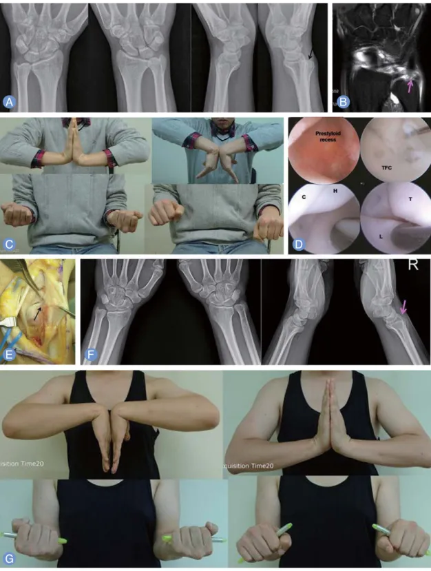
Fig. 2.
Case of patient presenting with persisted ulnar side wrist pain after the initial injury 5 years ago.
(A)Dorsal subluxation of the ulnar head
(black arrow) on the lateral view of simple radiographs was found.
(B)The detachment of deep fiber of triangular fibrocartilage complex (TFCC)
from the ulnar fovea (pink arrow) was observed in coronal view of magnetic resonance imaging scans.
(C)Slight restriction of the pronation was
seen.
(D)During the arthroscopic examination, synovitis around the prestyloid recess was found with the loss of the tension of the TFCC (positive
trampoline test) and traction induced inward folding of the TFCC from the ulnar fovea (positive Hook test). Other combined injuries did not accom-
pany. TFC, triangular fibrocartilage; C, capitate; H, hamate; L, lunate; T, triquetrum.
(E)Open repair of the deep fiber of TFCC to the original ulnar
fovea (black arrow) was done using the transosseous suture technique.
(F)The reduced state of the preoperatively subluxated ulnar (pink arrow)
head was seen on the follow-up radiographs taken at five years after operation.
(G)Functional outcomes measured by Mayo modified wrist score
and disability of the arm, shoulder and arm score improved from 55 preoperatively to 100 postoperatively and 35 to 3.3, separately.
J Korean Soc Surg Hand Vol. 19, No. 2, June 2014
J
OURNAL OF THEK
OREANS
OCIETY FORS
URGERY OF THEH
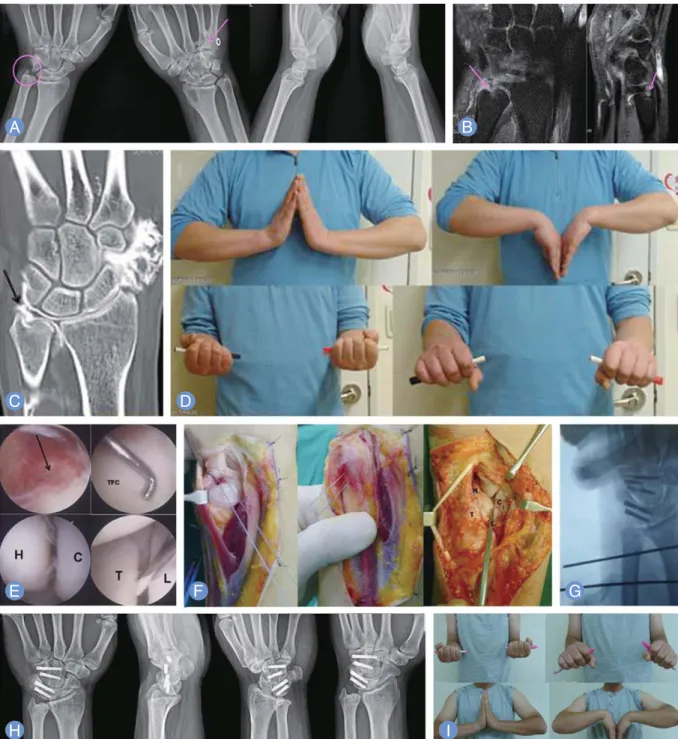
ANDFig. 3.
Case of patient presenting with persisted ulnar side wrist pain after the initial injury 6 months ago despite of the initial conservative man- agement using long arm cast for 4 weeks.
(A)Except the finding of the interpositional arthroplasty of fourth and fourth carpometacarpal joint of the right side (pink arrow), specific finding was not found around the ulnar head (pink circle). The detachment of deep fiber of triangular fibrocartilage complex (TFCC) from the ulnar fovea (pink arrow) was observed in coronal view of magnetic resonance imaging scans
(B)and computed tomogra- phy arthrogam (black arrow)
(C).
(D)Slight restriction of the volar and dorsiflexion was seen preoperatively.
(E)During the arthroscopic examination (black arrow), synovitis (black arrow) around the prestyloid recess was found with the loss of the tension of the TFCC (positive trampoline test) and traction induced inward folding of the TFCC from the ulnar fovea (positive Hook test). Other injuries of the capitohamate instability and lunotrique- tral instability accompanied. TFC, triangular fibrocartilage; C, capitate; H, hamate; L, lunate; T, triquetrum.
(F)Open repair of the deep fiber of TFCC to the original ulnar fovea was done using the transosseous suture technique.
(G)On the intraoperative fluoroscopic examination, well-posi- tioned screws at the lunotriqetral and capitohamate joints was observed with the temoporary fixation of the distal radioulnar joint using two 2.0 mm K-wires.
(H)Solid union of the lunotriquetral and capitohamate joints was seen on the simple radiographs taken at seven months after operation.
(G)
Functional outcomes measured by Mayo modified wrist score and disability of the arm, shoulder and hand score improved from 55 preopera-
tively to 99 postoperatively and 28.3 to 15, separately. (I) Near full range of motion except the slight limitation of the volar flexion was recovered.
수 있을 것으로 생각된다.
REFERENCES
1. Stuart PR, Berger RA, Linscheid RL, An KN. The dor- sopalmar stability of the distal radioulnar joint. J Hand Surg Am. 2000;25:689-99.
2. Gofton WT, Gordon KD, Dunning CE, Johnson JA, King GJ. Soft-tissue stabilizers of the distal radioulnar joint:
an in vitro kinematic study. J Hand Surg Am. 2004;29:
423-31.
3. Palmer AK. Triangular fibrocartilage disorders: injury patterns and treatment. Arthroscopy. 1990;6:125-32.
4. Hagert E, Hagert CG. Understanding stability of the dis- tal radioulnar joint through an understanding of its anatomy. Hand Clin. 2010;26:459-66.
5. Estrella EP, Hung LK, Ho PC, Tse WL. Arthroscopic repair of triangular fibrocartilage complex tears.
Arthroscopy. 2007;23:729-37, 37 e1.
6. Hermansdorfer JD, Kleinman WB. Management of chronic peripheral tears of the triangular fibrocartilage complex. J Hand Surg Am. 1991;16:340-6.
7. Cooney WP, Linscheid RL, Dobyns JH. Triangular fibro- cartilage tears. J Hand Surg Am. 1994;19:143-54.
8. Nakamura T, Sato K, Okazaki M, Toyama Y, Ikegami H.
Repair of foveal detachment of the triangular fibrocarti- lage complex: open and arthroscopic transosseous techniques. Hand Clin. 2011;27:281-90.
9. Garcia-Elias M, Smith DE, Llusa M. Surgical approach
to the triangular fibrocartilage complex. Tech Hand Up Extrem Surg. 2003;7:134-40.
10. Pederzini LA, Tosi M, Prandini M, Botticella C. All- inside suture technique for Palmer class 1B triangular fibrocartilage repair. Arthroscopy. 2007;23:1130.e1-4.
11. Atzei A. New trends in arthroscopic management of type 1-B TFCC injuries with DRUJ instability. J Hand Surg Eur Vol. 2009;34:582-91.
12. Bain GI, McGuire D, Lee YC, Eng K, Zumstein M.
Anatomic foveal reconstruction of the triangular fibro- cartilage complex with a tendon graft. Tech Hand Up Extrem Surg. 2014;18:92-7.
13. Adams BD, Divelbiss BJ. Reconstruction of the post- traumatic unstable distal radioulnar joint. Orthop Clin North Am. 2001;32:353-63.
14. Moritomo H. Advantages of open repair of a foveal tear of the triangular fibrocartilage complex via a palmar surgical approach. Tech Hand Up Extrem Surg. 2009;13:
176-81.
15. Moritomo H, Masatomi T, Murase T, Miyake J, Okada K, Yoshikawa H. Open repair of foveal avulsion of the triangular fibrocartilage complex and comparison by types of injury mechanism. J Hand Surg Am. 2010;35:
1955-63.
16. Anderson ML, Larson AN, Moran SL, Cooney WP, Amrami KK, Berger RA. Clinical comparison of arthro- scopic versus open repair of triangular fibrocartilage complex tears. J Hand Surg Am. 2008;33:675-82.
J Korean Soc Surg Hand Vol. 19, No. 2, June 2014
J
OURNAL OF THEK
OREANS
OCIETY FORS
URGERY OF THEH
AND삼각섬유연골 복합체 손상 제1B형의 관혈적 봉합술
류인혁
포항세명기독병원 정형외과
외상성 삼각섬유연골 복합체 손상의 가장 흔한 형태인 plamer 분류 제1B형의 경우 원위 요척골 관절의 안정성이 소실 되고 지속적인 완관절 척측부 통증의 원인이 되고 있다. 최근에 삼각섬유연골 복합체의 해부학적 구조가 알려지고 ulnar fovea에 부착하는 원위 요척골 관절 인대의 심부 섬유가 중요한 역할을 하고 있다는 사실이 인정받으며 파열된 심부 섬유를 ulnar fovea에 부착하고자 하는 노력이 있어 왔으며 좋은 결과를 보고하고 있다. 가장 먼저 시도된 관혈적 봉합술 후 관절경적 술기의 발달과 함께 여러 술기가 소개되고 있다. 가장 기본적인 관혈적 봉합술에 대해 여러 논문 고 찰과 함께 수술 시 유의해야 할 사항에 대해 논문 고찰과 함께 고찰해보고자 한다.
색인단어:관혈적 봉합술, 제1B형 파열, 삼각섬유연골 복합체
접수일2014년 5월 15일 게재확정일2014년 6월 5일 교신저자류인혁
경북 포항시 남구 포스코대로 351 포항세명기독병원 정형외과
TEL054-289-1765 FAX054-289-1766 [email protected]