INTRODUCTION
Kikuchi disease (KD) or histiocytic necrotizing lymphade- nitis is a self-limiting disorder of unknown origin; KD is char- acterized by painful cervical lymphadenopathy with prolonged fever (1). Therefore, although rare, KD should be part of the differential diagnosis of posterior cervical lymphadenopathy in children, especially in Asian pediatric patients (1, 2).
As most KD patients recover spontaneously and are sub- sequently followed in outpatient clinics, accurate diagnosis can avoid both unnecessary procedures and treatment. Com- puted tomography (CT) findings as well as the clinical char- acteristics of KD have already been reported, however, only a few published studies have analyzed the characteristics of KD in pediatric patients. Therefore, the authors retrospec- tively reviewed the clinical manifestations and imaging fea- tures of KD in a pediatric cohort.
MATERIALS AND METHODS
Between March 2001 and December 2007, a total of 15 children with a pathologic diagnosis of KD and who under- went neck CT, were included in this study. Specimens from the excisional biopsies of the cervical lymph nodes at level V showed necrotizing lymphadenitis and confirmed the diag- nosis of KD in all patients. Indications for biopsy included
lymph nodes in the supraclavicular region of the neck, pro- longed fever more than 7 days and increase in the size of nodes which failed to respond to antibiotic therapy.
We retrospectively analyzed both the medical records and the imaging findings of these patients. The medical records of all 15 patients were retrospectively reviewed in order to identify the clinical and laboratory features including the age/gender, duration of fever before admission, leukocyte count, hemoglobin level, platelet count, lactate dehydroge- nase (LDH), the erythrocyte segmentation rate (ESR), the C-reactive protein (CRP), treatment response, and associat- ed diseases (Table 1).
A total of 15 neck CT scans, 13 neck ultrasound scans (US), three abdominal US, three abdominal CT, and two 2-[fluo- rine 18]-fluoro-2-deoxy-D-glucose (FDG)-Positron Emission Tomography (PET) scans were performed on these fifteen children.
CT examinations (Somatom Plus, Siemens Medical Sys- tems, Erlangen, Germany) were performed following intra- venous administration of non-ionic contrast medium (Ultra- vist: Schering, Berlin, Germany). Neck CT was performed using axial scans from the skull base to the upper mediasti- num. The ultrasound exam was performed on an ATL HDI Ultramark 9 scanner (Bothell, WA, U.S.A.). We analyzed the sites, size (greatest dimension), shape, perinodal infiltra- tion, and contrast enhancement pattern of the affected lymph nodes (Table 2). The sites of lymph nodes identified by imag-
1105
Hye Jeong Han1, Gye-Yeon Lim1, Dong-Myung Yeo1, and Nak-Gyun Chung2
Departments of Radiology1and Pediatrics2, St. Mary’s Hospital, The Catholic University of Korea, College of Medicine, Seoul, Korea
Address for correspondence Gye-Yeon Lim, M.D.
Department of Radiology, St. Mary’s Hospital, The Catholic University of Korea, 62 Yeouido-dong, Yeongdeungpo-gu, Seoul 150-713, Korea Tel : +82.2-3779-2168, Fax : +82.2-783-5188 E-mail : [email protected]
DOI: 10.3346/jkms.2009.24.6.1105
Kikuchi’ s Disease in Children: Clinical Manifestations and Imaging Features
Previously published studies on Kikuchi disease (KD) have frequently addressed the computed tomography (CT) findings in the adult population, however, only a few studies have been reported for the pediatric age group. The purpose of this study is to analyze the clinical characteristics and imaging features of KD in children. Fif- teen children (2-14 yr) who had a neck CT and pathology diagnosis of KD were includ- ed in this study. Clinical features, including the duration of lymphadenopathy and fever, prognosis, and laboratory values, were evaluated. We analyzed the sites, size, and lymph node pattern as seen on their CT scans. The median duration of fever was 10 days. Fourteen patients experienced improvement in their condition, although four of these patients experienced recurrent episodes of KD. All patients had affected cervical nodes at level V. Perinodal infiltrates were observed in the affected cervical nodes in 14 cases (93%), and non-enhancing necrosis was also noted within the affected cervical nodes in 10 cases (63%). In conclusion, the com- bination of imaging findings in conjunction with clinical findings of KD may help to determine whether or not to perform pathology analysis and follow-up studies.
Key Words : Lymphatic Diseases; Histiocytic Necrotizing Lymphadenitis; Child
Received : 19 August 2008 Accepted : 23 February 2009
ing were analyzed using Som’s classification (3). The diag- nostic CT criteria for abnormal lymphadenopathy included 1) increased size (compared with the expected normal size of the respective region), 2) lymph nodes of any size with cen- tral necrosis, perinodal infiltrates, and increased contrast en- hancement, and 3) nodal grouping and the presence of three or more contiguous and confluent lymph nodes. On CT scan- ning, perinodal infiltration was visualized as obliteration or increased attenuation of the adjacent fat planes.
US and FDG-PET scans were also analyzed in patients with available data. On gray-scale US images, we analyzed the presence of the echogenic nodal hilum or internal ane- choic area and the sharpness of the border. We did not ana-
lyze color Doppler images as they were only obtained in some of our study patients. The FDG-PET data were evaluated qualitatively for regions of focally increased glucose meta- bolism. This study was reviewed and approved by our hos- pital’s human rights committee.
RESULTS Clinical findings
Our clinical and laboratory findings are detailed in Table 1.
There were 11 boys and 4 girls with a mean age of 10.5 yr (age range: 2-14 yr) and a male-female ratio of 2.8:1. The median duration of fever before admission was 10 days (6- 60 days). Laboratory findings included leukopenia (80%), leukocytosis (13%), anemia (67%), and elevated LDH (93%).
ESR and CRP levels were elevated in all 15 patients. Five patients had KD associated hemophagocytic lymphohistio- cytosis (HLH). Fourteen improved within five months (8 weeks-5 months), although four patients had recurrent epi- sodes of KD occurred 11-26 months after their initial epi- sodes. The follow-up period averaged 3.2 yr.
Ten patients recovered without specific therapy after exci- sional biopsy, and five patients were treated with predniso- lone. All patients treated with a steroid had both KD and HLH. In three of the five patients with both KD and HLH who failed to respond to corticosteroid treatment, chemo- therapy, i.e. etoposide, dexamethasone or cyclosporine-A, was started. The condition of other two children improved. One child treated with chemotherapy died from secondary dis- seminated coagulopathy caused by a buccal abscess.
KD, Kikuchi disease; Hb, hemoglobin; LDH, lactate dehydrogenase; ESR, erythrocyte segmentation rate; CRP, C-reactive protein; HLH, hemophago- cytic lymphohistiocytosis.
Leukocyte (/μL) 4,500-13,500 Days of
fever
Hb (g/dL) 12-16
Platelet (×103/μL)
150-450
LDH (IU/L) 218-472
ESR (mm/hr)
0-20
CRP (mg/dL)
<0.5
Associated disease Recurrency
Patients Age (yr)/
gender
1 9/M 8 3,540 12.2 139 662 21 28.32 + -
2 14/M 9 3,200 13 169 449 46 27.1 + HLH
3 10/M 6 4,820 13.8 348 546 29.6 59 - -
4 12/M 8 1,800 11.3 126 1,241 43 27.09 - HLH
5 12/M 6 4,100 13.7 220 686 39 13.47 - -
6 10/M 18 2,930 9 291 816 48 4.5 - -
7 2/F 10 20,630 10 472 521 120 63.87 + -
8 10/M 9 3,630 12.9 198 831 46 37 - -
9 12/M 60 3,320 11.1 250 872 47 15.61 - -
10 5/F 26 21,350 9.9 414 1,540 71 11.61 - HLH
11 12/M 10 3,140 11.8 403 1,467 77 12.33 - -
12 14/M 7 2,850 14 168 1,384 28 16.54 - -
13 14/F 12 2,000 11.6 120 849 87 47.2 + HLH
14 14/F 24 1,940 9.2 178 807 92 15.54 - -
15 8/M 13 2,110 11.1 152 1,308 52 39.36 - HLH
Table 1. Clinical and laboratory findings of 15 children with KD
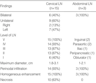
Abdominal LN (n=3) Cervical LN
(n=15) Findings
Bilateral 6 (40%) 3 (100%)
Unilateral 9 (60%)
Right 2 (13%)
Left 7 (47%)
Level of LN
V 15 (100%) Inguinal (2)
IV 14 (93%) Paraaortic (2)
III 13 (87%) Iliac (1)
II 13 (87%) Peripancreatic (1)
I 6 (40%) Obturator (1)
Maximum diameter, cm 1.8-3.1 1-2.1
Perinodal infiltration 14 (93%) 3 (100%) Homogeneous enhancement 15 (100%) 3 (100%)
Necrosis 10 (63%) 0
Table 2. Summary of CT findings in 15 children with KD
CT, Computed tomography; KD, Kikuchi disease; LN, lymph node.
Imaging findings
The CT findings are summarized in Table 2. All patients had affected cervical nodes at level V. Other frequently affect- ed nodes were at levels IV (n=14), II (n=13), III (n=13), and I (n=6); the supraclavicular nodes (n=6) were also affected.
Two cases were also noted to have parotid (n=1) and retroau- ricular (n=1) nodes. Six patients (40%) had bilateral neck involvement. Nine (60%) patients presented with unilateral
nodal involvement, mostly occurring on the left side (7/9).
In our series, all affected lymph nodes appeared as clusters of small- to medium-sized (1.8 to 3.1 cm, mean: 2.3 cm) nodes with oval to round shape (Figs. 1-3). Perinodal infiltrates were seen in the affected cervical nodes in 14 (93%) of our patients (Fig. 1-3). On enhanced CT, non-enhancing necrosis within the affected cervical nodes was also noted in ten patients (63
%) (Fig. 1).
In five patients (33%), all affected nodes showed uniform
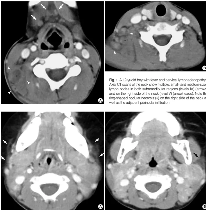
Fig. 2. A two-year-old girl with fever and bilateral cervical lymphadenopathy. Axial CT scans show multiple, medium-dimension lymph nodes with uniform enhancement on both sides of the neck at levels II and V. Note the increased attenuation in the adjacent superficial space (arrows).
A B
Fig. 1. A 12-yr-old boy with fever and cervical lymphadenopathy.
Axial CT scans of the neck show multiple, small- and medium-sized lymph nodes in both submandibular regions (levels IA) (arrows) and on the right side of the neck (level V) (arrowheads). Note the ring-shaped nodular necrosis (*) on the right side of the neck as well as the adjacent perinodal infiltration.
A
B
*
*
*
enhancement without necrosis (Fig. 2). Although the lymph node attenuation was less intense than that of the vascular structure, it was higher than that of the adjacent muscula- ture (Fig. 2).
Abdominal imaging was only performed in three patients (Fig. 3), all of whom demonstrated extensive lymphadenopa- thy involving many nodal groups in the inguinal (n=2), para- aortic (n=2), iliac (n=1), peripancreatic (n=1), and obturator (n=1) regions. Whole-body FDG PET was performed on two of these three patients and revealed multiple nodes demon- strating FDG-avid nodular masses in right cervical and supr- aclavicular nodes (standardized uptake value, SUV, 10.4) and abdominal nodes (SUV, 10) (Fig. 3C). Features of the perin- odal infiltrates were well-visualized on both CT and US, but regions of internal necrosis were not visualized on either modality (Fig. 3). The imaging features in our patients with KD associated with HLH did not differ from those in the patients with KD only.
DISCUSSION
KD is characterized by fever and self-limiting tender cervi- cal lymphadenopathy as well as by the histological character- istics of histiocytic necrotizing lymphadenitis (1, 2). Although rare, KD should be included in the differential diagnosis of posterior cervical lymphadenopathy in children, especially in Asian pediatric patients (1, 2).
Regarding the age at onset, a previous study with an adult population found that KD has a female predilection with a 3-4:1 ratio (4). However, the present study which was per- formed in a pediatric population, showed a completely dif-
ferent gender predominance, i.e. KD was found to be more prevalent in boys than in girls (Male/Female, 2.8:1). A pre- vious study with a pediatric population reported a similar gen- der predilection, i.e. M/F of 1.9:1 (2). Therefore, it is unclear why KD predominantly affects women in adult populations, but boys in pediatric populations younger than 16 yr of age.
In the present study, the youngest child was two years old, and three patients (20%) were less than 10 yr of age.
Laboratory tests are helpful in ruling out causes of cervical lymphadenopathy, but no specific tests are available for detect- ing KD. KD patients may have mild leukopenia, elevated ESR and CRP or rarely, leukocytosis. Leukopenia has been reported to occur in 16.6% to 58.3% of KD patients and was observed in 80% of our 15 pediatric patients. In a pre- vious study of pediatric KD patients, elevated ESR and CRP were noted in 72.7% and 72.2%, respectively, but in our study, all patients demonstrated elevated ESR and CRP lev- els (2). The LDH level in our series was generally increased (93%). Moreover, as found in adults in other studies, the lab- oratory indices of almost all of our study patients examined during the febrile period, showed leukopenia, anemia, and elevated ESR and LDH values.
In the present study, the imaging features of KD did not differ markedly from those in some general series (5, 6). Cer- vical lymph nodes in KD have a tendency to be located in the posterior triangle nodes, as seen in 48% to 77% of the cases in previous literature reports (5, 6), however, this ten- dency was more obvious in the present study in which all patients had affected nodes at level V. Moreover, in our study, all affected lymph nodes appeared as clusters of small- to medi- um-sized lymph nodes and no nodes exceeded 3 cm in great- est dimension. Therefore, the nodes appeared abnormal not
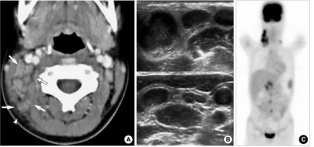
Fig. 3. A 10-yr-old girl with intermittent fever and right cervical lymphadenopathy. (A) CT scan shows multiple, small- and medium-sized lymph nodes on the right side of the neck (arrows) (levels IIB and V). Note the perinodal fat obliteration in the adjacent superficial space (arrowhead). (B) US obtained at the same time show somewhat matted, hypoechoic, nodal masses with irregular perinodal borders, sug- gestive of perinodal infiltrates. (C) MIP FDG-PET scan image shows multiple areas of FDG uptake in the right cervical, supraclavicular, right para-aortic and peripancreatic regions.
A B C
because of their sizes but rather because of their numbers and enhancement features.
Cervical nodes are most commonly affected by KD, but virtually any nodal chain can be involved in KD and axillary, supraclavicular, mediastinal, peripancreatic, iliac, and inguinal locations of nodes have been reported, as was observed in three of our study cases (6, 7). Isolated intra-abdominal adenopa- thy has only rarely been reported, however, as imaging of the thorax and abdomen is not usually performed, it is possible that to date thoracic and abdominal disease involvement has not been detected (7).
US is generally used as the modality of choice for evaluat- ing cervical lymphadenopathy in children because it has no radiation hazard. In the present study, US was also performed but was found to be of limited use in terms of evaluating inter- nal necrosis of affected nodes. Unfortunately, only a limited number of our study patients were evaluated on color Doppler US and this number was too small to allow us to analyze the significance of the imaging findings. In all four patients who underwent color Doppler US, it showed hilar vascularity, however, CT demonstrated eccentric necrosis in these cases.
A previous report of the Korean population mentioned that color Doppler US findings of KD include symmetrical cen- tral hilar vascularity with some flow signals in the peripher- al soft tissue which are similar to those of benign lymphadeni- tis (8). Further studies of the utility of color Doppler US in diagnosing KD, are needed in the pediatric population.
Although CT poses a radiation hazard, it is the preferred modality for evaluating perinodal infiltrates (93%) and nodal necrosis (63%), both of which were frequently observed in our patients. The cause of the more frequent nodal necrosis in this pediatric series than in previous adult series, is unclear.
However, the difference may be attributable to the fact that the clinical course and immune response of KD differ accord- ing to individual patients and their ages (1). Another factor contributing to this difference is thought to be stage of the histopathologic changes in KD associated with its clinical course. Although the early proliferative stage of KD with its lymphohistiocytic proliferation and numerous atypical mo- nonuclear cells, can cause it to be confused with malignant lymphoma, cortical and paracortical necrosis are the chief histopathologic features of KD (1, 7), and some previous lit- erature reports have indicated the rare occurrence of exten- sive necrosis (7). However, in our series, full-blown macro- scopic necrosis of affected nodes was not unusual on CT, espe- cially in febrile children. Clinically and radiologically, KD could lead to a misdiagnosis of tuberculosis or even malig- nant lymphoma (5, 7). In one of our patients, multiple homo- genously enhancing nodes without nodal necrosis were seen on CT, and multiple FDG-avid nodular masses in both cervi- cal and abdominal lymph nodes was seen on FDG PET scans, thus mimicking lymphoma. Previous PET studies have indi- cated that affected nodes in KD demonstrate FDG uptake (standardized uptake value, SUV) compatible with malig-
nant adenopathy (9). Although qualitative and quantitative analysis of regions of focally increased glucose metabolism seen on PET may falsely indicate malignant lymphoma, a finding of enlarged nodes <3 cm in greatest dimension and with perinodal infiltrates, might be helpful in differentiat- ing pediatric KD from lymphoma.
Most previous reports have suggested that nodes affected be KD resolve spontaneously without specific treatment with- in six months of biopsy; this was also observed in most of our pediatric patients (1, 2). The rate of KD recurrence has been reported to range from 1.3-4%, however, 27% (n=4) of our 15 patients experienced recurrent episodes at least one year after the initial resolution (2, 4). Furthermore, two of four of these patients had recurrent KD-associated HLH.
In conclusion, the present study demonstrates that KD in children has somewhat different clinical manifestations and imaging features compared with previously reported results in adult populations. Our study demonstrates that KD shows a male predominance in children which contrasts to the female predominance among young adults. Furthermore, in the pre- sent study, KD was found to have higher rates of recurrence and HLH development than have been previously reported.
In our pediatric series, the most common CT feature of KD was clusters of multiple, small- to medium-sized ovoid to round lymph nodes with internal necrosis and perinodal infil- trates at level V. The authors advise that awareness of the CT features and clinical characteristics of KD in children may help to determine whether or not to perform pathology anal- ysis and follow- up studies.
REFERENCES
1. Lee KY, Yeon YH, Lee BC. Kikuchi-Fujimoto disease with pro- longed fever in children. Pediatrics 2004; 114: e752-6.
2. Lin HC, Su CY, Huang SC. Kikuchi’s disease in Asian children.
Pediatrics 2005; 115: e92-6.
3. Som PM, Curtin HD, Mancuso AA. Imaging-based nodal classifi- cation for evaluation of neck metastatic adenopathy. AJR Am J Ro- entgenol 2000; 174: 837-44.
4. Tsang WY, Chan JK, Ng CS. Kikuchi’s lymphadenitis. A morpho- logic analysis of 75 cases with special reference to unusual features.
Am J Surg Pathol 1994; 18: 219-31.
5. Kwon SY, Kim TK, Kim YS, Lee KY, Lee NJ, Seol HY. CT find- ings in Kikuchi disease: analysis of 96 cases. AJNR Am J Neurora- diol 2004; 25: 1099-102.
6. Turner RR, Martin J, Dorfman RF. Necrotizing lymphadenitis. A study of 30 cases. Am J Surg Pathol 1983; 7: 115-23.
7. Miller WT Jr, Perez-Jaffe LA. Cross-sectional imaging of Kikuchi disease. J Comput Assist Tomogr 1999; 23: 548-51.
8. Pyeun YS, Lee SW, Rho MH. Ultrasonographic evaluation of Kikuchi disease in the neck. J Korean Soc Med Ultrasound 2001; 20: 221-6.
9. Kaicker S, Gerard PS, Kalburgi S, Geller MD, Hailoo D. PET-CT scan in a patient with Kikuchi disease. Pediatr Radiol 2008; 38: 596-7.