INTRODUCTION
Lung cancer is the most common fatal cancer worldwide and has become the leading cause of cancer deaths in Korea.
The number of new cases will continue to rise (1). The risk of human cancer can be associated with environmental, occu- pational, and recreational exposures to carcinogens (2). Onco- genesis is related with epigenetic changes, oncogenes, tumor suppressor genes, apoptosis, and genetic changes associated with DNA repair. There have been many investigations on the prognostic role of p53, bcl-2, and Ki-67 expression in non-small cell lung cancer (NSCLC). Early reports of p53 mutation suggested a variable relationship to survival (3, 4).
Another study showed that p53 mutations detectable in tumor tissues had been shown to be an independent marker for the poor prognosis in resectable stage I NSCLC (5). Data on bcl- 2 expression from a study on NSCLC patients showed posi- tive correlations with longer survival (6). There were reports on an inverse relationship between bcl-2 and p53 in NSCLC (7, 8). However, another studies did not support a relevant prognositic role for p53, bcl-2, or Ki-67 immunohistochem- ical markers in NSCLC regardless of stage (9, 10). No rela- tionship was observed between the expression of Ki-67 and that of bcl-2. The relationship between a positive rate for
Ki-67 and prognosis remains unclear (11). Small cell lung cancer (SCLC) has a poor prognosis. Most of the patients carry a large burden at the time of diagnosis. SCLC has been stud- ied less than NSCLC. There was a report that bcl-2 expres- sion did not influence survival in SCLC (12). We conducted a retrospective study on the value of mutant p53, bcl-2, and Ki-67 expressions in SCLC patients from Korea Cancer Cen- ter Hospital.
MATERIALS AND METHODS
All of the patients were primarily diagnosed as SCLC at the Department of Internal Medicine of Korean Cancer Cen- ter Hospital between February 1997 and December 2002.
Seventy-five of 107 SCLC patients were treated. Immuno- histochemical (IHC) stainings for mutant p53, bcl-2 and Ki- 67 expressions were performed in the 66 paraffin-embedded biopsy samples among the 75 member treatment group. The study group included patients (57 males and 9 females, 61 yr mean age) with cytologically or histopathologically diagnosed SCLC. Histopathological diagnoses were done by broncho- scopic biopsy, lymph node biopsy, and percutaneous lung gun biopsy. Staging procedures included physical examination, chest
Kwang Hyun Paik, Yeon Hee Park, Baek-Yeol Ryoo, Sung Hyun Yang, Jae Cheol Lee, Cheol Hyun Kim, Seung Seog Ki, Jung Min Kim, Myung Joon Park, Heui June Ahn, Won Choi, Jin Haeng Chung*
Departments of Internal Medicine and Pathology*, Korea Institute of Radiological and Medical Sciences, Seoul, Korea
Address for correspondence Yeon Hee Park, M.D.
Department of Internal Medicine, Korea Institute of Radiological and Medical Sciences, 215-4 Gongneung-dong, Nowon-gu, Seoul 139-706, Korea Tel : +82.2-970-1229, Fax : +82.2-970-2401 E-mail : [email protected]
35
Prognostic Value of Immunohistochemical Staining of p53, bcl-2, and Ki-67 in Small Cell Lung Cancer
Small cell lung cancer (SCLC) is one of the most fatal cancers in humans and many factors are known to be related to its poor prognosis. Immunohistochemical (IHC) stainings were done on SCLC specimens in order to investigate the prognostic value of the apoptosis-related gene expression and the tumor proliferative maker, and the relationships among these IHC results and patients clinical characteristics, chemoresponsiveness, and survival were analyzed. The medical records of 107 patients were reviewed retrospectively. IHC stainings for p53, bcl-2 and Ki-67 expres- sions were performed in the 66 paraffin-embedded biopsy samples. Sixty-six out of the 107 patients were evaluable for response rate and survival. The overall response rate was 75% (95% Confidence Interval=74-76%) and the median survival time was 14 months. The median survival time of limited stage was 16 months and that of extensive stage was 10 months. The prevalence of p53, bcl-2 and Ki-67 expression was 62%, 70%, and 49%, respectively. There were no correlations among the im- munoreactivities of p53, bcl-2 and Ki-67 with clinical stage, chemoresponsiveness or overall survival. The clinical stage was the only prognostic factor influencing sur- vival. The expression rates of p53, bcl-2, and Ki-67 were relatively high in SCLC without any prognostic significance. The exact clinical role of these markers should be defined through further investigations.
Key Words :Tumor Suppressor Protein p53; bcl-2-Associated X Protein; Ki-67 Antigen; Carcinoma, Small Cell; Tumor Marker, Biological; Prognosis
Received : 8 April 2005 Accepted : 5 September 2005
36 K.H. Paik, Y.H. Park, B.-Y. Ryoo, et al.
radiography, chest computed tomography (CT) scan, and bone scintigraphy. Brain CT/magnetic resonance image (MRI), bone marrow biopsy and other studies were optional in asymp- tomatic patients but mandatory in those with symptoms suggesting disseminations. Patients were staged as either limited, with disease confined to one hemithorax, or exten- sive, with disease beyond one hemithorax. All patients were administered one of the three chemotherapy regimens: cis- platin and etoposide (EP); cyclophosphamide, adriamycin, and vincristine (CAV); etoposide and carboplatin (EC). We established the principle that all limited disease patients would be treated by radiotherapy if the chemotherapy were effec- tive. And then 40 patients out of all limited disease patient were treated by radiotherapy. Response categories i.e. com- plete response (CR), partial response (PR), stable disease (SD), and progressive disease (PD) were evaluated according to new response evaluation criteria in solid tumor (RECIST) guidelines (13). The CR and PR patients were considered responsive. Survival was defined as time lapse from the day of first chemotherapy course until the day of death.
Five-micrometer thickness, paraffin-embedded, tissue sec- tions were deparaffinized in xylene and hydrated in a graded ethanol series. Endogenous peroxidase activity was blocked with 3% hydrogen peroxide in methanol. Tissue sections were heated in 10 mM sodium citrate, pH 6.0, in a microwave oven for 10 min to expose the antigens. Sections were then washed with Tris-buffered saline (TBS). The streptavidin- biotin-peroxidase complex technique (universal LSAB kit, DAKO, Glostrup, Denmark) was used for immunohisto- chemical stain. The sections were incubated overnight at 4℃ with the three monoclonal antibodies: p53 (1:50, DAKO, Glostrup, Denmark), bcl-2 (1:40, DAKO) and Ki-67 (1:50, Zymed, San Francisco, CA, U.S.A.). Sections were then washed with TBS and incubated with biotinylated secondary anti- body for 30 min at room temperature. After washing, the sections were incubated with peroxidase-labelled streptavidin at room temperature for 30 min. Diaminobenzidine/hydro- gen peroxidase was used as a chromogen and sections were counterstained with hematoxylin. When the cell nuclei were stained strongly with dark brown color, the cells were con- sidered to be positive for p53 and Ki-67. Bcl-2 expression was noted in the cytoplasm. If more than 10% of the tumor cells were positively stained, it was considered as positive for p53 and bcl-2. In the case of Ki-67, the labeling index was determined by scanning areas with uniformly stained cells at a low magnification, followed by counting of the cells at high power field (×400). Appropriate positive and negative controls were used for all the procedures.
Median values and ranges were used to calculate patient characteristics and to compare the factors. To examine factors affecting survival, the following variables were analyzed: age, sex, disease extent, performance status, and Ki-67, p53, and bcl-2 expression. Each of these variables was divided into two categories as follows: age was divided by mean age, i.e., less
than 60 yr vs. more than or equal to 60 yr; sex, male vs. female;
disease extent, limited disease (LD) vs. extended disease (ED);
performance status (PS), good (PS 0-1) vs. poor (PS 2-4); Ki- 67, equal to or greater than 50% vs. less than 50%; p53 and bcl-2, positive vs. negative. To compare the response rate, chi- square test was used. Survival was assessed by the Kaplan- Meier method and compared with log-rank test. Hazard ratio and its 95% confidence interval for each variable were esti- mated by Cox proportional hazard model. A p-value lower than 0.05 was considered statistically significant. Chi-square test with Fisher exact test were used as appropriate. SPSS for Windows, 11.0 standard version, was used for all analysis.
RESULTS
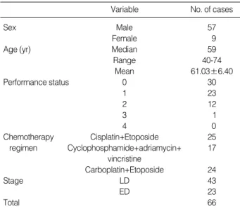
Patient characteristics are shown in Table 1. Overall response rate for evaluable 66 patients were 72%. There was no dif- ference of the response rate among three regimens (EP, 69%;
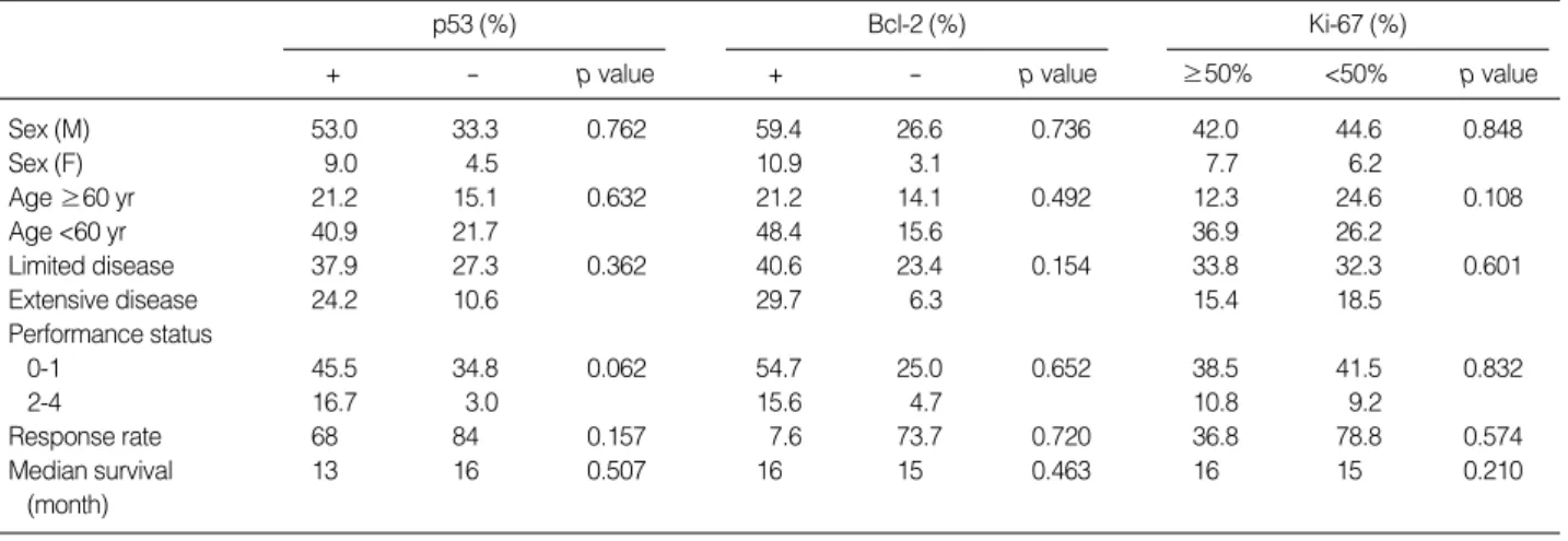
CAV, 65%; EC, 75%; p=0.83). Forty-one (62%) out of the 66 patients were positive for p53 antibodies; 25 (40%) with limited disease (LD) and 16 (24%) with extensive disease (ED) (p=0.36). The response rate for the positive p53 group was 68%, that for the negative group was 84% (p=0.15). Sur- vival data were available for 66 patients. Median survival was 13 months for the positive p53 group, and 16 months for the negative group (p=0.50). There were no significant differences in sex, age, disease extent, performance status, and survival between patients with and without mutant p53 antibodies (Table 2). According to the analysis of prognostic factors which influenced chemotherapeutic response rate and survival, only the extent of the disease was significant for patient survival by univariate and multivariate analysis (Table 3) (Fig. 1). Median survival for the entire group was 15 mon-
Variable No. of cases
Sex Male 57
Female 9
Age (yr) Median 59
Range 40-74
Mean 61.03±6.40
Performance status 0 30
1 23
2 12
3 1
4 0
Chemotherapy Cisplatin+Etoposide 25
regimen Cyclophosphamide+adriamycin+ 17 vincristine
Carboplatin+Etoposide 24
Stage LD 43
ED 23
Total 66
Table 1.Patient characteristics
LD, limited disease; ED, extensive disease.
ths; 16 months for LD patients, and 10 months for ED pati- ents (p=0.04). We also analyzed the survival between posi- tive and negative p53 groups in LD patients or ED patients.
In LD patients, median survival was 15 months for the pos- itive p53 group, and 18 months for the negative group (p=
0.37). In ED patients, median survival was 10 months for positive p53 and 12 months for negative (p=0.87). No cor- relation was found between survival and p53 expression in either LD or ED patients. Therefore p53 was not a useful prognostic indicator of SCLC.
For bcl-2, 45 (70%) out of 64 patients were positive; 26 (40%) with LD and 19 (30%) with ED (p=0.15) (Table 2).
The response rate was 75% for the positive bcl-2 group, and 73% for the negative group (p=0.72). Median survival was 16 months for positive bcl-2, and 15 months for the nega- tive group (p=0.46). In LD patients, median survival was 15 months for the positive bcl-2 group and 19 months for the negative group 19 months (p=0.37). In ED patients, median
survival was 9 months for positive bcl-2, and 12 months for negative (p=0.16). No correlation was found between sur- vival and bcl-2 expression in either LD or ED patients. Like mutant p53, there were no significant differences in sex, gen- der, disease extent, performance status, and survival between patients with and without bcl-2 expression.
We separately analyzed Ki-67 antigen expression both above and below 50%. Thirty-two (49%) out of 65 were in the above 50% group, 22 (34%) with LD and 10 (15%) with ED (p=0.60) (Table 2). The response rate was 69% for the above 50% group, and 79% for the below group (p=0.55).
Median survival was 16 months for the above 50% group, and 15 months for the below group (p=0.21). In LD patients, median survival was 16 months for the above 50% group, and 16 months for the below group (p=0.37). In ED patients, median survival was 12 months for the above 50% group and 10 months for the below group (p=0.65). No correlation was found between survival and Ki-67 expression in either LD or ED patients. There were no significant differences in sex, gender, disease extent, performance status, and survival accord- ing to Ki-67 expression.
Univariate analysis p-value
Multivariate analysis
p-value
Performance status 0-1 0.049 0.058
2-4
Sex M 0.732 0.853
F
Age (yr) ≥60 0.094 0.234
<60
Stage LD 0.032 0.047
ED
Radiation therapy Done 0.252 0.153
Not done Table 3.Prognostic factor analysis
LD, limited disease; ED, extensive disease.
p53 (%)
+ - p value
Bcl-2 (%)
+ - p value
Ki-67 (%)
≥50% <50% p value
Sex (M) 53.0 33.3 0.762 59.4 26.6 0.736 42.0 44.6 0.848
Sex (F) 9.0 4.5 10.9 3.1 7.7 6.2
Age ≥60 yr 21.2 15.1 0.632 21.2 14.1 0.492 12.3 24.6 0.108
Age <60 yr 40.9 21.7 48.4 15.6 36.9 26.2
Limited disease 37.9 27.3 0.362 40.6 23.4 0.154 33.8 32.3 0.601
Extensive disease 24.2 10.6 29.7 6.3 15.4 18.5
Performance status
0-1 45.5 34.8 0.062 54.7 25.0 0.652 38.5 41.5 0.832
2-4 16.7 3.0 15.6 4.7 10.8 9.2
Response rate 68 84 0.157 7.6 73.7 0.720 36.8 78.8 0.574
Median survival 13 16 0.507 16 15 0.463 16 15 0.210
(month)
Table 2.Response rate and median survival according to p53, bcl-2 and Ki-67
Percent surviving
100
80
60
40
20
0
0 12 24 36 48 60 72
Month Fig. 1.Survival according to stage.
Total LD ED
p=0.0407
38 K.H. Paik, Y.H. Park, B.-Y. Ryoo, et al.
DISCUSSION
The p53 protein is recognized as an important cell regu- latory factor that arrests the growth of cells containing dam- aged DNA. A reversible arrest in the G1 phase of the cell cycle enables DNA repair before DNA synthesis. When appropriate repair is not possible, p53 expression may trig- ger apoptosis, a reversible process culminating in cell death.
If normal, wild type p53 function is lost, the treatment is relatively resistant, as a result of deficiency of p53-dependent apoptosis (14, 15). The p53 mutation is the most common genetic mutation in cancer. Thus, the mutated gene loses its natural tumor suppressor function allowing damaged cells to divide unchecked and finally to become malignant cells.
There are still controversies concerning the prognosis and survival in lung cancer patients. Some studies reported that p53 mutation had been associated with poor prognosis and shorter survival in NSCLC (5, 8, 16, 17). However, some others reported no such correlation in NSCLC (3, 9, 10, 18).
Others reported a favorable prognosis in NSCLC (4, 19). There have been fewer studies in SCLC than in NSCLC. Some studies showed that bcl-2 expression and p53 mutation had no rela- tion to survival in SCLC (9, 10, 12, 20). Another study divid- ed their SCLC patients into limited and extensive-stage dis- ease, after which p53-antibody positivity emerged as an inde- pendent marker of poor prognosis in LD but had no rela- tion to survival (21). One study indicated that p53 played an important role as a determinant of chemosensitivity in SCLC and that p53 immunostaining could be used in clini- cal practice to determine the presence of tumor-chemoresis- tance (22). Kenichi et al. reported that patients with expres- sion of mutant p53 protein showed lower response rate than those having p53-negative tumors and were less sensitive to anticancer drugs (23). Our study, however, indicated that p53 antibody had no relation to survival.
The bcl-2 proto-oncogene is encoded by a 230-kb gene that gives rise to a 24- to 26-kDa protein that is localized in the inner mitochondrial membrane, and, to a lesser extent, in the cell membrane (24). The major function of bcl-2 appears to be the inhibition of apoptosis or programmed cell death, whereas bax, bad, bak, and others promote cell death (25). It is well documented that bcl-2 becomes deregulated in tumor cells as a result of translocation into the immunoglobulin heavy-chain locus, and is therefore constitutively activated in follicular lymphoma (26). In epithelial tumors, no genetic change of bcl-2 has been demonstrated, in contrast to lym- phocytic neoplasia. However, bcl-2 expression has been des- cribed in a series of solid tumors, particularly in NSCLC and in breast cancer (27). An in vitro study showed that bcl-2 expression may be related to chemoresistance due to inhibi- tion of drug-induced apoptosis (28). Thus multidrug resis- tance is probably linked, at least in part, to high levels of bcl-2 expression. Bcl-2 blocks the cell death pathway (apop- tosis) and is not directly associated with cell proliferation
(29). Studies examining the association of bcl-2 expression with survival in NSCLC have been contradictory, with some reporting bcl-2 as an indicator of better survival (6, 7), while other showed no survival differences according to bcl-2 sta- tus (30, 31). There have been few studies in SCLC. From our results, bcl-2 expression was not an independent predictor of survival in SCLC.
BrdU (bromodeoxyuridine), PCNA (proliferating cell nu- clear antigen), Ki-67, and others have been used as cell pro- liferation markers. PCNA, a 36-kilodalton, nuclear polypep- tide that is related to cell proliferation, is identical to cyclin, which is a protein that appears in the proliferative phase of cells (32). Being synthesized during the late G1 to S phase, PCNA is an auxillary protein for DNA polymerase . Ki- 67, a more reliable proliferating marker, reacts with nuclear antigen that is present only in proliferating cells (G1, S, G2, and M phase), not in resting (G0) cells (33-35), and thereby provides a reliable method for evaluating tumor growth frac- tion in many malignant tumors, including lung cancer (36).
It appears to be a useful prognostic marker of NSCLC, espe- cially in the early stage (37). Compared to p53 and bcl-2, the relationship between Ki-67 and survival has been studied less extensively in NSCLC, and especially so in SCLC. Our results showed that Ki-67 expression had no relation to sur- vival in SCLC.
From our results, the apoptosis-related genes, mutant p53 and bcl-2, and the proliferative marker, Ki-67, were found to be useful in the diagnosis of SCLC patients because of their relatively high prevalence (38, 39). However, as they did not show clinical significance for prognosis or survival, further investigation will be required to confirm the clinical role of these markers.
REFERENCES
1. Ministry of Health and Welfare. 2002 Annual reports of the Korea Central Cancer Center Registry (Published in 2003); 29.
2. Harris CC. Chemical and physical carcinogenesis: advances and perspectives for the 1990s. Cancer Res 1991; 51: 5023-44.
3. McLaren R, Kuzu I, Dunnill M, Harris A, Lane D, Gatter KC. The relationship of p53 immunostaining to survival in carcinoma of the lung. Br J Cancer 1992; 66: 735-8.
4. Lee JS, Yoon A, Kalapurakal SK, Ro JY, Lee JJ, Tu N, Hittleman WN, Hong WK. Expression of p53 oncoprotein in non-small cell lung cancer: a favorable prognostic factor. J Clin Oncol 1995; 13:
1893-903.
5. Harpole DH, Herndon JE 2nd, Wolf WG, Iglehart JD, Marks JR. A prognostic model of recurrence and death in stage I non-small cell lung cancer utilizing presentation, histopathology and oncoprotein expression. Cancer Res 1995; 55: 51-6.
6. Pezzella F, Turley H, Kuzu I, Tungerkar MF, Dunnil MS, Pierce CB, Harris A, Gatter KC, Mason DY. Bcl-2 protein in non-small cell lung carcinoma. N Engl J Med 1993; 329: 690-4.
7. Fontanini G, Vignati S, Bigini D, Mussi A, Lucchi M, Angeletti CA, Basolo F, Bevilacqua G. Bcl-2 protein: a prognostic factor inversely correlated to p53 in non-small cell lung cancer. Br J Cancer 1995;
71: 1003-7.
8. Ishida H, Irie K, Itoh T, Furukawa T, Tokunaga O. The prognostic significance of p53 and bcl-2 expression in lung adenocarcinoma and its correlation with Ki-67 growth fraction. Cancer 1997; 80:
1034-45.
9. van de Vaart PJ, Belderbos J, de Jong D, Sneeuw KC, Majoor D, Bartelink H, Begg AC. DNA-adduct levels as a predictor of outcome for NSCLC patients receiving daily cisplatin and radiotherapy. Int J Cancer 2000; 89: 160-6.
10. Cagini L, Monacelli M, Giustozzi G, Moggi L, Bellezza G, Sidoni A, Bucciarelli E, Darwish S, Ludovini V, Pistola L, Gregorc V, Tonato M. Biological prognostic factors for early stage completely resected non-small cell lung cancer. J Surg Oncol 2000; 74: 53-60.
11. Tungekar MF, Gatter KC, Dunnil MS, Mason DY. Ki-67 immuno- staining and survival in operable lung cancer. Histopathology 1991;
19: 545-50.
12. Maitra A, Amirkhan RH, Saboorian MH, Frawley WH, Ashfaq R.
Survival in small cell lung carcinoma is independent of Bcl-2 expres- sion. Hum Pathol 1999; 30: 712-7.
13. Therasse P, Arbuck SG, Eisenhauer EA, Wanders J, Kaplan RS, Rubin- stein L, Verweij G, Van Glabbeke M, van Oosterom AT, Christian MC, Gwyther SG. New guidelines to evaluate the response to treat- ment in solid tumors. J Natl Cancer Inst 2000; 92: 205-16.
14. Kastan MB, Canman CE, Leonard CJ. P53, cell cycle control and apo- ptosis: implications for cancer. Cancer Metastasis Rev 1995; 14: 3-15.
15. Greenblatt MS, Bennett WP, Hollstein M, Harris CC. Mutations in the p53 tumor suppressor gene: clues to cancer etiology and molec- ular pathogenesis. Cancer Res 1994; 54: 4855-78.
16. Quinlan DC, Davidson AG, Summers CL, Warden HE, Doshi HM.
Accumulation of p53 protein correlates with a poor prognosis in human lung cancer. Cancer Res 1992; 52: 4828-31.
17. Laudanski J, Burzykowski T, Niklinska W, Chyczewski K, Furman M, Niklinski J. Prognostic value of serum p53 antibodies in patients with resected non-small cell lung cancer. Lung Cancer 1998; 22:
191-200.
18. Mitsudomi T, Suzuki S, Yatabe Y, Nishio M, Kuwabara M, Gotoh K, Hatooka S, Shinoda M, Suyama M, Ogawa M, Takahashi T, Ari- yoshi Y, Takahashi T. Clinical implications of p53 autoantibodies in the sera of patients with non-small cell lung cancer. J Natl Cancer Inst 1998; 90: 1563-8.
19. Bergqvist M, Brattstrom D, Larsson A, Holmertz J, Hesselius P, Losen- berg L, Wagenius G, Brodin O. P53 auto-antibodies in non-small cell lung cancer patients can predict increased life expectancy after radiotherapy; Anticancer Res 1998; 18: 1999-2002.
20. Rosenfeld MR, Malats N, Schramm L, Graus F, Cardenal F, Vinolas N, Rosell R, Tora M, Real FX, Posner JB, Dalmau J. Serum anti-p53 antibodies and prognosis of patients with small cell lung cancer. J Natl Cancer Inst 1997; 89: 381-5.
21. Zalcman G, Tredaniel J, Schlichtholz B, Urban T, Milleron B, Lubin R, Meignin V, Couderc LJ, Hirsch A, Soussi T. Prognostic signifi- cance of serum p53 antibodies in patients with limited stage small
cell lung cancer. Int J Cancer 2000; 89: 81-6.
22. Rodriguez-Salas N, Palacios J, Moreno G, de Castro J, Gonzalez- Baron M, Gamallo C. Correlation of p53 oncoprotein expression with chemotherapy response in small cell lung carcinomas. Lung Cancer 2001; 34: 67-74.
23. Gemba K, Ueoka H, Kiura K, Tabata M, Harada M. Immunohisto- chemical detection of mutant p53 protein in small cell lung cancer:
relationship to treatment outcome. Lung Cancer 2000; 29: 23-31.
24. de Jong D, Prins FA, Mason DY, Reed JC, van Ommen GB, Kluin PM. Subcellular localization of the bcl-2 protein in malignant and normal lymphoid cells. Cancer Res 1994; 54: 256-60.
25. Reed JC. Bcl-2 family proteins. Oncogene 1998; 17: 3225-36.
26. Cleary ML, Sklar J. Nucleotide sequence of a t (14;18) chromosomal breakpoint in follicular lymphoma and demonstration of a breakpoint- cluster region near a transcriptionally active locus on chromosome 18. Proc Natl Acad Sci USA 1985; 82: 7439-43.
27. Harris AL. What does bcl-2 mean in solid tumors-friend or foe? Ann Oncol 1994; 5: 388-90.
28. Reed JC, Kitada S, Takayama S, Miyashita T. Regulation of chemore- sistance by the bcl-2 oncoprotein in non-Hodgkin’s lymphoma and lymphocytic leukemia cell lines. Ann Oncol 1994; 5 (Suppl 1): 61-5.
29. Korsmeyer SJ. Bcl-2 initiates a new category of oncogenes: regula- tors of cell death. Blood 1992; 80: 879-86.
30. Gaffney EF, O’Neil AJ, Staunton MJ. Bcl-2 and prognosis in non- small-cell lung carcinoma. N Engl J Med 1994; 330: 1757-8.
31. Kim YC, Park KO, Kim HJ, Choi IS, Park CS, Juhng SW. DNA ploidy and proliferative activity in bcl-2 expressed non-small cell lung cancer. Korean J Intern Med 1996; 11: 101-7.
32. Mathews MB, Bernstein RM, Franza BR Jr, Garrels JI. Identity of the proliferating cell nuclear antigen and cyclin. Nature 1984; 309:
374-6.
33. Landberg G, Ross G. Proliferating cell nuclear antigen and Ki-67 antigen expression in human hematopoietic cells during growth stimu- lation and differentiation. Cell Prolif 1993; 26: 427-37.
34. Wilson GD, Saunders MI, Dische S, Daley FM, Robinson BM, Mar- tindale CA, Joiner B, Richman PI. Direct comparison of bromo- deoxyuridine and Ki-67 labelling indices in human tumours. Cell Prolif 1996; 29: 141-52.
35. Gerdes J, Lemke H, Baisch H, Wacker HH, Schwab U, Stein H. Cell cycle analysis of a cell proliferation-associated human nuclear anti- gen defined by the monoclonal antibody Ki-67. J Immunol 1984;
133: 1710-5.
36. Tubiana M, Courdi A. Cell proliferation kinetics in human solid tumors: relation to probability of metastatic dissemination and long- term survival. Radiother Oncol 1989; 15: 1-18.
37. Scagliotti GV, Micela M, Gubetta L, Leonardo E, Cappia S, Borasio P, Pozzi E. Prognostic significance of Ki-67 labelling in resected non small cell lung cancer. Eur J Cancer 1993; 29: 363-5.
38. Wang DG, Johnston CF, Sloan JM, Buchanan KD. Expression of Bcl-2 in lung neuroendocrine tumors: comparison with p53. J Pathol 1998; 184: 247-51.
39. Mizera-Nyczak E, Dyszkiewicz W, Heider KH, Zeromski J. Isoform expression of CD44 adhesion molecules, Bcl-2, p53 and Ki-67 pro- teins in lung cancer. Tumour Biol 2001; 22: 45-53.