INTRODUCTION
Intracranial teratomas are rare, constituting less than 1%
of all intracranial tumors (1), and predominant in childhood and adolescence (2, 3). Most of them have been reported to involve pineal region and their occurrence in the third ven- tricle has been reported less frequently (4-6). Moreover, the development of mature teratoma in the third ventricle in post- adolescent age is extremely unusual. Despite some controver- sies, total extirpation of the tumor is thought to be the gold standard for treatment of mature teratoma and generally leads no recurrence (7, 8). We present a rare case of intraventricu- lar germinoma occurring 30 months after a total resection of mature teratoma in the third ventricle.
CASE REPORT The first admission
In January 1996, a 26-yr-old man was admitted with head- ache and vomiting. On admission, he was lethargic and showed a mild degree of motor weakness on both lower ex- tremities. No other abnormal neurological deficits were noted.
Laboratory studies including intradermal test for Parago- nimus and Clonorchis disclosed no abnormalities. Magnetic
resonance (MR) images showed strongly enhancing small mass lesion in the third ventricle accompanied by severe ob- structive hydrocephalus (Fig. 1). A ventriculo-peritoneal shunting procedure was performed to relieve the hydroce- phalic crisis.
The second admission
He was re-admitted in May 1996 for management of head- ache that aggravated with positional change and evaluation of mass lesion previously noted in the third ventricle. Gadolin- ium-enhanced MR images depicted a homogeneously en- hanced rapid growing oval-shaped mass with a large cyst in the third ventricle (Fig. 2). Results of hormonal tumor marker studies, including -fetoprotein ( -FP), human chorionic gonadotropin (hCG), and carcinoembryonic antigen (CEA), were in normal ranges. On May 21, 1996, the tumor was totally resected via subfrontal-interhemispheric translamina terminalis approach with division of anterior communicat- ing artery. The tumor consisted of pink flesh solid portion and a cyst filled with clear yellowish fluid (Fig. 3). Although it was attached to the floor of the third ventricle, total extir- pation of the tumor was done easily.
Histopathological examination confirmed a diagnosis of a mature cystic teratoma containing well-differentiated three distinct germ cell layers (Fig. 4). No malignant features were
Jae Min Kim, Jin Hwan Cheong, Hyeong Joong Yi*, Koang Hum Bak, Choong Hyun Kim, Suck Jun Oh*
Department of Neurosurgery, Hanyang University Kuri Hospital, Kuri and Hanyang University Medical Center*, College of Medicine, Seoul, Korea
Address for correspondence Jae Min Kim, M.D.
Department of Neurosurgery, Hanyang University Kuri Hospital, 249-1 Kyomun-dong, Kuri 471-701, Korea
Tel : +82.31-560-2323, Fax : +82.31-560-2327 E-mail : [email protected]
287
Metachronous Germinoma After Total Removal of Mature Teratoma in the Third Ventricle
: A Case Report
A rare case of intraventricular germinoma in the third ventricle, which occurred 30 months after total removal of mature teratoma on the same location in a 29- yr-old man is presented. Recurrence is supposed to represent an acceleration of localized dysplastic processes of totipotent germ cells present in the midline neuraxis or a growth of unidentified microscopic residue of germinoma compo- nent in mature teratoma. Although the radiation therapy after total removal of mature teratoma is still controversial, careful follow-up is warranted for evaluat- ing a possible recurrence of other germ cell tumors.
Key Words : Germinoma; Third ventricle; Teratoma; Recurrence
Received : 7 March 2001 Accepted : 7 May 2001
observed. During the several postoperative days, patient showed transient polyuria and confusion. No further post- operative adjuvant therapy was provided. At the time of hos- pital discharge, follow-up computerized tomography (CT) scan was performed and there was no evidence of residual tumor. Eventually, he was discharged without any neurologi- cal deficits.
The third admission
In December 1998, about 30 months after the total removal of tumor, he was re-admitted due to generalized weakness and intractable vomiting. He had lost his body weight 5 kg during the previous three weeks. Detailed evaluation with contrast-enhanced CT scan demonstrated a homogeneously
Fig. 1.Pretreatment gadolinium-enhanced axial (A) and coronal (B) T1-weighted MR images showing strongly enhancing intraventric- ular mass in the third ventricle with ventriculomegaly.
PI
R 1 0 5
A B
Fig. 2.Gadolinium-enhanced T1-weighted coronal (A) and sagittal (B) MR images showing decreased dimension of the lateral ventri- cle 4 months after ventriculo-peritoneal shunt surgery and size-increased homogeneously enhancing oval-shaped mass, which has a large cystic portion (arrows) within the third ventricle.
A 1 2 0
A B
R 1 0 5
P 1 2 0
5). No serum or cerebrospinal fluid (CSF) tumor marker studies showed abnormal findings.
On December 8, 1998, an anterior transcallosal transfora- minal approach via a left foramen of Monro was performed due to adhesion of fornix with recurrent tumor tissue. A
the medial side of the choroid plexus and thalamostriate vein in the third ventricle. Due to its dense adhesion and gliosis around the tumor, the tumor was partially resected. Surgical specimen proved to be a germinoma, which was composed of large epithelioid cells with large, round, and vesicular nuclei and lymphocytic infiltration. Teratomatous compo- nents were not found at all (Fig. 6). Postoperative course was uneventful. He received 54 Gy of localized irradiation for 6 weeks. Now then, he has been well and back to his previous job without any evidence of tumor recurrence (Fig. 7).
DISCUSSION
Based on histology, intracranial germ cell tumors (GCTs) are largely classified into germinomas, non-germinomatous germ cell tumors (teratoma, embryonal carcinoma, endoder- mal sinus tumor, and choriocarcinoma), and mixed germ- cell tumors (1, 9, 10). Among these GCTs, teratomas repre- sent less than 1% of all intracranial tumors (1), while the incidence of intracranial GCTs varied according to the pop- ulation studied (9, 11, 12). The age distribution of intracra- nial teratomas showed a peak incidence during the first two decades of life and mostly in children (2, 3), whereas the germinoma in the early pubertal years (13). Jenning et al.
(13) postulated that the neuroendocrine events of puberty might be a “triggering" effect on the abrupt rise of GCTs in the puberty.
Teratomas usually occur in the pineal, suprasellar region
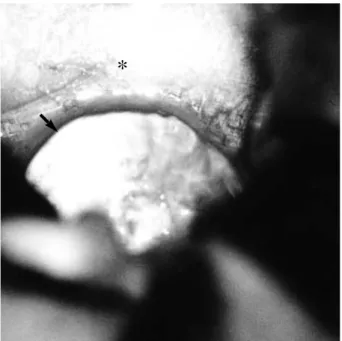
Fig. 3.Intraoperative photograph showing a translucent cystic wall (arrow), which contains clear fluid after dissection of lamina terminalis with a distended optic chiasm (asterisk).
Fig. 4.Photomicrographs from different parts of mature teratoma specimens showing skin with appendages, salivary glandular compo- nent, fibrofatty tissue, and lymphocytes (A) (H&E,×40) and a single layer of columnar epithelium mimicking intestine, cartilage islands, skeletal muscle, and gastric body glandular components (B) (H&E,×100).
A B
*
(1, 6), and rarely in the cerebellar vermis (1) and lateral ven- tricles (8, 14), whereas most germinomas arise in suprasellar cistern, the third ventricle, and less frequently, in the lateral ventricles (6, 13, 15). There have hitherto been only six cases of metachronous GCTs in which a germinoma occurred after total removal of a pineal (16-20) or intrasellar teratomas (21) in the literature. Moreover, several cases of the primary intra- ventricular teratomas have been reported (4-6), but there is no report on a germinoma developing 30 months after total
extirpation of an intraventricular cystic mature teratoma in adults.
It is very difficult to draw a conclusion about the correla- tion between the primary and secondary GCTs in a same patient with a long time interval. Some pathogeneses have been postulated on the development of metachronous GCTs.
Firstly, although the recurrence of nongerminomatous GCT usually occurs within 1 yr after treatment (22), there is a good possibility of tumor recurrence or dissemination especially in the mixed tumors that are composed of various combina- tions of two or more types of GCTs. Secondly, although it is uncertain whether the antecedent presence of germ cells is due to germinal aberrant migration of germ cells, embryon- ic “cell rest", or localized harmatomatous or dysplastic pro- cesses (23), the pre-existing germ cells have been implicated.
Thirdly, it may develop due to genetic alterations that lead to mutational inactivation of tumor suppressor genes or acti- vation of oncogenes (24). In our case, the second germinoma, which occurred in the same location with the primary tumor is most plausibly considered as a recurrence from a microsco- pic residue of germinoma component in previously resected mature cystic teratoma despite the long silent period. Other possibilities include the sporadic acceleration of a localized hamartomatous or dysplastic process of pre-existing germ cells.
Although the treatment strategy and prognosis of intracra- nial GCTs generally correlate with the major contributing and the most malignant element of the tumors, it is gener- ally accepted that mature teratomas are radioresistant and that total removal of tumor is the treatment of choice (7, 8).
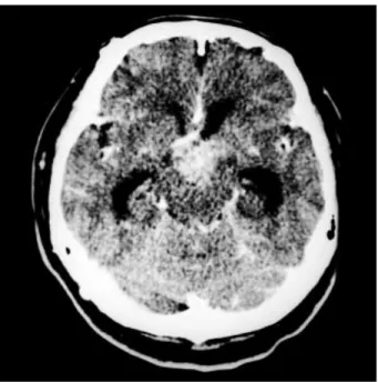
Fig. 5.Postcontrast CT scan obtained 30 months after total re- moval of the cystic mature teratoma via subfrontal-interhemispher- ic translaminal approach showing homogeneously enhanced, ill-defined round shaped mass on the suprasellar region.
Fig. 6.Photomicrograph of the recurrent germinoma specimen showing two distinguishable cell types: large polygonal cells and lymphocytes with adjacent normal brain tissue (H&E,×200).
Fig. 7.Postcontrast CT scan obtained 20 months after a subto- tal resection of intraventricular germinoma via anterior transcal- losal transforaminal approach and completion of adjuvant radio- therapy showing no evidence of tumor recurrence with a metal- lic artifact on the anterior communicating artery.
5 C M
minoma that recurred in the third ventricle after total removal of mature cystic teratoma in a post-adolescent patient. Care- ful follow-up CT/MRI scans are indispensable to detect the evidence of recurrence even after total removal of mature ter- atomas.
REFERENCES
1. Russell DS, Rubinstein LJ. Tumours and tumour-like lesion of malde- velopmental origin. In Russell, DS, & Rubinstein LJ (Eds), Patholo- gy of Tumours of the Nerve System, ed 5, Baltimore, Williams &
Wilkins, 1989; 681-7.
2. Hunt SJ, Johnson PC, Coons SW, Pittman HW. Neonatal intracra- nial teratoma. Surg Neurol 1990; 34: 336-42.
3. Ventureyna R, Herder S. Neonatal intracranial teratoma. J Neuro- surg 1983; 59: 879-83.
4. Fiser Z. A teratoma of the third ventricle of the brain. Acta Neurochir 1979; 50: 343-7.
5. Lin CL, Wang CJ, Yin HL, Howng SL. Successful resection of a ter- atoma of the third ventricle in a 3-yr-old boy. J Formos Med Assoc 1998; 97: 217-9.
6. Tekeuchi J, Mori K, Moritake K, Tani F, Waga S. Teratomas in the suprasellar region: report of five cases. Surg Neurol 1975; 3: 247- 55.
7. Edwards MSB, Hudgins RJ, Winson CB, Levin VA, Wara WM.
Pineal region tumors in children. J Neurosurg 1988; 68: 689-97.
8. Sel uki M, Attar A, Yuceer N, akiroglu E. Mature teratoma of the lateral ventricle: report of two cases. Acta Neurochir 1998; 140:
171-4.
9. Jellinger K. Primary intracranial germ cell tumours. Acta Neuropathol 1973; 25: 291-306.
10. Sano K. So-called intracranial germ cell tumours: are they really of germ cell origin? Br J Neurosurg 1995; 9: 391-401.
11. Araki C, Matsumoto S. Statistical reevaluation of pinealoma and related tumors in Japan. J Neurosurg 1969; 53: 146-9.
germinoma and pinealoma in Japan. Cancer 1980; 45: 2119-30.
13. Jennings MT, Gelman R, Hochberg F. Intracranial germ cell tumors:
natural history and pathogenesis. J Neurosurg 1985; 63: 155-67.
14. Jelinek J, Smirniotopoulos JG, Parisi JE, Kanzar M. Lateral ventric- ular neoplasms of the brain: differential diagnosis based on clinic, CT and MR findings. Am J Neuroradiol 1990; 11: 567-74.
15. Sano K, Matsutani M. Pinealoma (germinoma) treated by direct surgery and postoperative irradiation: a long-term follow-up. Child's Brain 1981; 8: 81-97.
16. Carrillo R, Ricoy JR, Del Pozo JM, Garcia-Uria J, Herrero J. Dissem- ination with malignant changes from a pineal tumor through the corpus callosum after total removal. Child's Brain 1977; 3: 230-7.
17. Ikeda J, Sawamura Y, Kato T, Abe H. Metachronous neurohypophy- seal germinoma occurring 8 yr after total resection of pineal mature teratoma. Surg Neurol 1998; 49: 205-9.
18. Matsuda K, Maeda M, Kodama S, Kanemaru R, Sugata I, Asakura T, Mihara T. Problems of recurrence after removal of pineal region - an experience of recurrence of pineal teratoma 4 yr after tumor removal. No To Shinkei 1982; 34: 1107-15.
19. Nagasawa S, Handa H, Yamashita J, Yamamura K. Suprasellar ger- minoma occurring two-years after total removal of pineal teratoma.
Surg Neurol 1984; 22: 481-5.
20. Tsuchida T, Tanaka R, Kobayashi K, Ueki K, Koizumi R. Develop- ment of 2 cell pattern pinealoma 15 years after total removal of pineal teratoma. No To Shinkei 1976; 28: 893-9.
21. Czirjak S, Pasztor E, Slowik F, Szeifert G. Third ventricle germino- ma after total removal of intrasellar teratoma: case report. J Neu- rosurg 1992; 77: 643-7.
22. DeLeo MJ, Greco FA, Hainsworth JD, Johnson DH. Late recurrences in long-term survivors of germ cell neoplasms. Cancer 1988; 62:
985-8.
23. Grote E, Lorenz R, Vuia O. Clinical and endocrinological findings in ectopic pinealoma and spongioblastoma of the hypothalamus. Acta Neurochir 1980; 53: 87-98.
24. Hollstein M, Sidransky D, Vogelstein B, Harris CC. p53 mutations in human cancers. Science 1991; 253: 49-53.
∨
. . ∨