Induction of Apoptosis by Hwangheuk-san in AGS Human Gastric Carcinoma Cells through the Generation of Reactive Oxygen Species and Activation of Caspases
Su Hyun Hong
1*, Cheol Park
2, Kyoung Min Kim
4and Yung Hyun Choi
1,3*
1Department of Biochemistry, Dongeui University College of Korean Medicine, Busan 614-052, Korea
2Department of Molecular Biology, College of Natural Sciences and Human Ecology, Dongeui University, Busan 614-714, Korea
3Anti-Aging Research Center & Blue-Bio Industry RIC, Dongeui University, Busan 614-714, Korea
4Department of Caridovascular & Neurologic Disease, College of
Korean Medicine, Dongeui University, Busan 614-052, Korea Received June 25, 2015 /Revised July 22, 2015 /Accepted July 23, 2015
Hwangheuk-san (HHS) is a Korean multi-herb formula comprising four medicinal herbs. HHS, which was recorded in “Dongeuibogam,” has been used to treat patients with inflammation syndromes and digestive tract cancer for hundreds of years. However, little is known about its anti-tumor efficacy.
The present study investigated the pro-apoptotic effect and mode of action of HHS against AGS hu- man gastric carcinoma cells. HHS inhibited the cell growth of AGS cells in a dose-dependent manner, which was associated with the induction of apoptotic cell death, as evidenced by the formation of apoptotic bodies, chromatin condensation, and an accumulation of cells in the sub-G1 phase. HHS-in- duced apoptotic cell death was associated with the up-regulation of pro-apoptotic Bax protein ex- pression, down-regulation of antiapoptotic Bcl-2 protein, and the release of cytochrome c from mi- tochondria to the cytosol. The treatment of AGS cells with HHS significantly elevated the generation of reactive oxygen species (ROS). Additionally, apoptosis-inducing concentrations of HHS induced the activation of both caspase-9 and -8, initiator caspases of the mitochondrial-mediated intrinsic and death receptor-mediated extrinsic pathways, respectively, and caspase-3, accompanied by proteolytic degradation of poly (ADP-ribose)-polymerase. However, ROS scavenger and pan-caspases inhibitor significantly blocked HHS-induced growth inhibition and apoptosis. Taken together, these findings suggest that HHS induces apoptosis through ROS- and caspase-dependent mechanisms and that HHS may be a potential chemotherapeutic agent for the control of human gastric cancer.
Key words :
AGS cells, apoptosis, caspase, Hwangheuk-san, ROS (reactive oxygen species)
*Corresponding authors
*Tel : +82-51-850-7413, Fax : +82-51-853-4036
*E-mail : [email protected] (Yung Hyun Choi)
*Tel : +82-51-850-8664, Fax : +82-51-853-4036
*E-mail : [email protected] (Su Hyun Hong)
This is an Open-Access article distributed under the terms of the Creative Commons Attribution Non-Commercial License (http://creativecommons.org/licenses/by-nc/3.0) which permits unrestricted non-commercial use, distribution, and reproduction in any medium, provided the original work is properly cited.
Journal of Life Science 2015 Vol. 25. No. 11. 1235~1243 DOI : http://dx.doi.org/10.5352/JLS.2015.25.11.1235
서 론
항암치료의 가장 중요한 전략은 다양한 기전을 통하여 암세 포의 분열과 증식을 억제하고 선택적으로 암세포를 제거하는 데 있다. 이러한 측면에서 특정 약물의 처리에 의한 암세포 특이적 프로그램화된 세포 사멸로 알려진 apoptosis 유발은 항암제 개발에 가장 중요한 영역으로 인식되고 있다[7, 8].
Apoptosis는 intrinsic (mitochondrial-mediated) 및 extrinsic (death receptor-mediated) apoptosis 두 가지의 경로로 크게 구별된다[1, 5, 17]. Extrinsic pathway는 세포막에 존재하는
death receptor에 죽음의 신호 ligand가 결합하여 특정 cas- pase (caspase-8)의 활성을 유도하여 이루어진다. 반면, in- trinsic pathway는 미토콘드리아의 기능과 연관된 다양한 유 전자 산물들의 조절 하에 해당 caspase (caspase-9)의 활성 증 가를 통하여 유도된다. 이러한 두 가지 경로는 미토콘드리아 에서 세포질로의 cytochrome c 방출과 연계될 수 있으며, cas- pase cascade를 유발하여 최종 effector caspase (caspase-3/-7) 의 활성을 통하여 apoptosis를 일으킨다[3, 29].
한편 오랫동안 다양한 약초들이 특정 질환의 치료와 예방의
목적으로 전세계적으로 널리 사용되어 왔다. 최근 우리나라를
포함한 동아시아 지역에서 널리 사용되어온 전통 처방(tradi-
tional multi-herb formulas)들에 대한 약리학적 관심이 증대
되고 있으며, 현대 약물로 치료가 어려운 질환의 제어 목적으
로 처방 사용 가능성 및 그들의 약리 효능의 기전에 대한 연구
가 이루어지고 있다[28, 30]. 한의학에서 사용되어 온 처방들은
각 본초의 기미(氣味)를 근거로 하여 부작용을 경감시키면서
의학적 효능은 증대시키고 환자 삶의 질적 향상을 증대시키기
위한 목적으로 발전되어 왔다[12, 19]. 그 중 황흑산(黃黑散)은
동의보감(東醫寶鑑)에 기재된 장옹(腸癰)과 복옹(腹癰)을 치
Table 1. Herbal components and amounts of Hwangheuk-san Herbal medicine
(pharmacognostic nomenclature)
Raw material amount (g/%) Rheum palmatum L. (Rhei Radix et
Rhizoma)
Psoralea corylifolia L. (Psoraleae Fructus) Pharbitis nil Chois. (Pharbitidis Semen) Arctium lappa L. (Arctii Fructus) Total amounts
36.0 (47.4) 16.0 (21.0) 16.0 (21.0) 8.0 (10.5) 76 ( 100)
료하는 처방으로 대황(大黃, Rheum palmatum L.), 보골지(補骨 脂, Psoralea corylifolia Linné), 견우자(牽牛子, Pharbitis nil Chois.) 및 우방자(牛蒡子, Arctium lappa L.) 등 4가지 약초 (Table 1)로 구성되어 있다. 비록 최근 들어 이들 각 한약재인 대황[15, 16], 보골지[17, 22], 우방자[4], 현호색[29]에 대한 항 암, 항산화 및 면역활성 등의 효능이 보고되었으나, 이들로 구 성된 복합처방전인 황흑산에 대한 연구는 이루어진 바 없다.
본 연구에서는 황흑산의 항암 효능을 알아보기 위하여, 인체 위암세포(AGS)의 증식에 미치는 영향을 평가하고 세포 증식 억제효과가 apoptosis 유도와 연관성이 있는지를 조사하였다.
재료 및 방법
시료 준비
본 실험에서 사용한 황흑산(Hwangheuk-san, HHS; Table 1)의 구성 한약재는 대한생약(주)(부산, 한국)에서 건조된 상태 로 구입하여, 추출에 적합하도록 세절 한 후 약재 무게의 10배 에 해당하는 증류수로 100℃에서 3시간 동안 추출하여 사용하 였다. 이후 열수 추출액을 Whatsman No.2 filter paper (Sigma-Aldrich Co., St. Louis, MO, USA)로 거른 후 동결건조 기를 사용하여 분말로 만들어 증류수에 100 mg/ml의 농도로 녹인 후 고압증기멸균기에서 멸균 처리하였다. 이 준비된 황 흑산 추출물은 무균실험대 안에서 Minisart® Syringe filter (0.2 μm, Sartorius AG Weender Landstr, Germany)를 사용하 여 거른 후 배지에 적정 농도로 희석하여 처리하였다.
세포배양
실험에 사용한 AGS 인체 위암세포는 생명공학연구소(대 전, 한국)에서 분양 받았으며, 세포의 배양을 위해 10%의 우태 아혈청(fetal bovine serum, FBS, Gibco BRL, NY, USA) 및 1%의 penicillin 및 streptomycin이 포함된 RPMI-1640 배지 (Gibco BRL)를 사용하였으며, 37℃, 5% CO
2조건 하에서 배양하 였다. 세포수의 증식에 따른 과밀도 현상과 적정수의 세포를 유 지하기 위하여 성장배지의 교환을 매 48 시간마다 실시하였다.
MTT assay를 이용한 세포 생존율의 측정
세포 배양용 6 well plate에 1×10
5cells/ml로 AGS 세포를
분주하고 다양한 황흑산 추출물을 각 well에 처리하여 배양하 였다. 48시간 후 배지를 제거하고 3-(4,5-dimethyl-2 thiazolyl)- 2,5-diphnyl-2H-tetrazolium bromide (MTT, Sigma-Aldrich Co.)를 0.5 mg/ml 농도로 희석하여 200 μl씩 분주하고 37℃에 서 2시간 동안 다시 배양하였다. 배양이 끝난 다음 MTT 시약 을 제거하고 dimethyl sulfoxide (DMSO, Sigma-Aldrich Co.) 를 1 ml씩 각 well에 분주하여 생성된 formazan을 모두 녹인 후 96 well plate에 200 μl씩 옮겨서 enzyme-linked immun- osorbent assay (ELISA) reader (Molecular Devices, Sunny- vale, CA, USA)로 540 nm에서 흡광도를 측정하였다.
DAPI staining에 의한 세포 핵의 형태 관찰
Apoptosis가 유발되었을 경우 특이적으로 나타나는 핵의 형태적 변화를 관찰하기 위하여 황흑산이 처리된 세포를 모은 다음 37% formaldehyde 용액과 phosphate buffer saline (PBS)를 1:9의 비율로 섞은 fixing solution을 모아진 세포에 500 μl 첨가하여 충분히 섞은 후, 상온에서 10분 동안 고정하였 다. 고정된 세포를 2,000 rpm으로 5분간 원심 분리하여 fixing solution을 제거하고 cytospin을 이용하여 slide glass에 부착 하였다. 세포가 부착된 slide glass를 PBS로 2~3 회 정도 세척 하고 0.2%의 Triton X-100 (Amresco, Solon, OH, USA)를 첨가 하여 상온에서 10 분간 고정한 후 2.5 μg/ml 농도의 4', 6-dia- midino-2-phenylindole (DAPI, Sigma-Aldrich Co.) 용액을 처 리하여 상온에서 15분간 염색하였다. 염색이 끝난 후 DAPI 용액을 충분하게 세척하고 mounting solution을 처리한 후 형광현미경(Carl Zeiss, Oberkochen, Germany)을 이용하여 400배의 배율로 핵의 형태 변화를 관찰하였다.
Flow cytometry 분석
황흑산 추출물이 유발하는 apoptosis 정도를 정량적으로 분 석하기 위하여 정상 및 황흑산 추출물이 처리된 세포들을 모 은 다음 2,000 rpm으로 5분간 원심 분리하여 상층액을 제거한 후 PBS를 이용하여 2~3회 정도 세척하였다. 준비된 세포는 Cycle TEST PLUS DNA REAGENT Kit (Becton Dickinson, San Jose, CA, USA)를 이용하여 고정 및 염색을 하여 4℃, 암실에서 30분 동안 반응을 시켰다. 반응시킨 세포를 35-mm mesh를 이용하여 단일세포로 분리한 후 flow cytometer (FACS Calibur, Becton Dickinson)를 적용시켜 형광반응에 따 른 Cellular DNA content 및 histogram을 CellQuest software 및 ModiFit LT (Becton Dickinson) 프로그램을 이용하여 분석 하였다.
Western blot analysis에 의한 단백질 발현의 분석
준비된 세포들을 모은 다음 적당량의 lysis buffer [25 mM
Tris-Cl (pH 7.5), 250 mM NaCl, 5 mM ethylenediaminetetra-
acetic acid (EDTA), 1% Nonidet-P40 (NP-40), 1 mM phe-
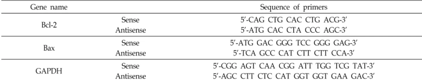
Table 2. Sequences of the primer pairs employed in the RT-PCR reactions
Gene name Sequence of primers
Bcl-2 Sense
Antisense
5'-CAG CTG CAC CTG ACG-3' 5'-ATG CAC CTA CCC AGC-3'
Bax Sense
Antisense
5'-ATG GAC GGG TCC GGG GAG-3' 5'-TCA GCC CAT CTT CTT CCA-3'
GAPDH Sense
Antisense
5'-CGG AGT CAA CGG ATT TGG TCG TAT-3' 5'-AGC CTT CTC CAT GGT GGT GAA GAC-3'
ny-methylsulfonyl fluoride (PMSF) 및 5 mM dithiothreitol
(DTT)]를 첨가하여 4℃에서 1시간 동안 반응시킨 후, 14,000 rpm으로 30 분간 원심분리하여 상층액에 있는 단백질을 분리 하여 Bio-Rad 단백질 정량 시약(Bio-Rad, Herculs, CA, USA) 을 이용하여 정량하였다. 세포질과 미토콘드리아 단백질의 분 리를 위해서는 mitochondria/cytosol fractionation kit (Biovi- sion, Milpitas, CA, USA)를 사용하였다. 적정 양의 단백질을 동량의 Laemilni sample buffer (Bio-Rad)와 혼합하여 sodium dodecyl sulphate (SDS)-polyacrylamide gel을 이용하여 전기 영동으로 분리한 후, nitrocellulose membrane (Schleicher and Schuell, Keene, NH, USA)으로 전이시켰다. 분리된 단백 질이 전이된 nitrocellulose membrane을 5% skim milk를 1 시간 처리하여 비특이적인 단백질들에 대한 blocking을 실시 하고 1 차 항체(Santa Cruz Biotechnology Inc., Santa Cruz, CA, USA)를 처리하여 상온에서 2시간 이상 반응시킨 다음 PBS-T (PBS with Tween 20)로 세척(10분간 3번)하고 적정 2차 항체(Amersham Life Science Co., Arlington Heights, IL, USA)를 사용하여 상온에서 1시간 정도 반응시켰다. 반응이 끝난 후 암실에서 enhanced chemiluminoesence solution (Amersham Life Science Corp.)을 적용시킨 다음 X-ray film 에 감광시켜 특정 단백질의 발현 양을 분석하였다.
RNA의 분리 및 reverse transcription-polymerase chain reaction (PCR)
전사수준에서 Bax 및 Bcl-2 유전자의 발현 변화를 조사하기 위하여 적정 조건에서 배양된 세포들의 RNA를 RNeasy kit (Qiagen, La Jolla, CA, USA)를 이용하여 분리한 후, AMV re- verse transcriptase (Amersham Life Science Corp.)를 이용하 여 cDNA를 합성하였다. Bax 및 Bcl-2 유전자에 적절한 primer (Table 2)를 이용한 PCR은 Mastercycler (Eppendorf, Ham- burg, Germany)를 사용하여 수행하였으며, PCR 반응 산물을 1% agarose을 이용하여 분리한 후, ethidium bromide (EtBr, Sigma-Aldrich Co.) 염색을 실시한 후 발 현의 정도를 비교하 였다.
ROS 생성 변화 측정
세포 내 reactive oxygen species (ROS, 활성산소종) 생성
변화를 확인하기 위하여 준비된 세포들을 fluorescent probe 인 2`, 7`-di-chlorodihydrofluorescein diacetate (H
2DCFDA, Molecular Probes, Leiden, Netherlands) 10 μM로 20 분간 염 색 후 flow cytometer를 사용하여 분석을 하였다. ROS 억제를 위한 ROS scavenger인 N-ace-tylcysteine (NAC, Sigma- Aldrich) 처리는 황흑산 추출물을 처리하기 1시간 전에 처리하 였다.
Caspase 활성 측정
Caspase-3, -8 및 -9의 in vitro 활성 측정을 위한 colorimetric assay kits는 R&D Systems (Minneapolis, MN, USA)에서 구 입하여 제시된 방법에 준하여 활성의 증감여유를 조사하였다.
이를 위하여 정상 및 황흑산이 처리된 배지에서 48시간 배양 된 세포를 모은 뒤 단백질을 추출하고 정량하여 각각 20 μg의 단백질을 fluorogenic peptide 기질 100 μM이 함유된 50 μl의 extraction buffer [40 mM 4-(2-hydroxyethyl)-1-piperazinee- thanesulfonic acid (pH 7.4), 20% glycerol (v/v), 1 mM EDTA, 0.2% NP-40 및 10 mM DTT]에 혼합하였으며, micro- plate에 다시 extraction buffer에 희석하여 각 시료당 총 vol- ume이 100 μl가 되게 하였다. 준비된 plate를 37℃에서 2시간 동안 반응시킨 후 ELISA reader를 이용하여 405 nm의 흡광도 를 이용하여 반응의 정도를 측정하였다. 실험에 사용된 기질 은 caspase-3의 경우에는 Asp-Clu-Val-Asp (DEVD)-p-nitro- aniline (pNA), caspase-8의 경우에는 Ile-Glu-Thr-Asp (IETD)- pNA이었으며, caspase-9는 Leu-Glu-His-Asp (LEHD)-pNA 였다.
통계처리
실험 결과들의 유의성을 검정하기 위하여 분산분석을 실시 한 후 p<0.05 수준에서 Duncan's multiple range tests를 실시 하였다. 이때 사용한 모든 통계분석은 SPSS 17.0 (IBM SPSS Inc, New York, USA) 통계 프로그램을 이용하여 처리하였다.
결과 및 고찰
황흑산 추출물 처리에 의한 AGS 세포의 증식 억제 및
apoptosis 유발
A
Fig. 1. Inhibition of cell viability and induction of apoptosis by Hwangheuk-san in human gastric cancer AGS cells. AGS cells were seeded in 6 well plate at 1×10⁵ cells/ml and treated with the indicated concentrations of Hwangheuk-san (HHS) for 48 hr. (A) Cell viability was estimated by the metabolic-dye-based MTT assay. Each point represents the mean ± SD of three independent experiments. The significance was determined by the Student’s t-test (*p<0.05 vs. untreated control). (B) Cell morphology was visualized by an inverted light microscope (Magnification, ×200). (C) The cells were fixed and stained with DAPI solution. The stained nuclei were observed under a fluorescent microscope (Original magnification, 400×). (D) To quantify the degree of apoptosis induced by HHS, the cells grown under the same conditions as (A) were evaluated for sub-G1 DNA content, which represents the fractions undergoing apoptotic DNA degradation, using a DNA flow cytometer.
Each point represents the mean of two independent experiments.
황흑산 추출물 처리에 따른 AGS 위암세포의 증식억제 정 도를 알아보기 위하여 다양한 농도의 황흑산 추출물을 48 시 간 동안 처리한 후 MTT assay를 실시하였다. Fig. 1A에 나타 낸 결과에서 알 수 있듯이 황흑산 추출물의 처리 농도 의존적 으로 AGS 세포의 증식이 유의적으로 감소되었다. 아울러 이 러한 증식 억제는 처리 농도 증가에 따른 세포 밀도의 감소와 다양한 형태적 변형을 동반하였다(Fig. 1B). 이러한 변화가 apoptosis 유발과 관련성이 있는지의 여부를 조사하기 위하여 핵의 형태적 변화와 flow cytometry 분석을 실시하였다. 먼저 apoptosis 유발의 직접적인 증거를 제시하기 위하여 정상 및 황흑산이 처리된 배지에서 배양한 AGS 세포를 대상으로 핵산 에 특이적으로 결합하는 DAPI 염색을 통하여 핵의 형태학적 변화를 관찰하였다. Fig. 1C의 결과에서 알 수 있듯이 정상 조건에서 배양된 세포와는 다르게 황흑산이 처리된 배지에서 배양된 세포에서는 apoptosis가 일어난 세포에서 전형적으로 관찰되는 DNA 단편화에 의한 apoptotic body의 형성[21, 27]
이 처리 농도 의존적으로 증가되었다. 다음은 propidium io- dide (PI) 염색을 통한 세포주기 분포도를 조사한 결과, Fig.
1D에 나타낸 바와 같이 apoptosis 유발 집단에 해당 하는 sub- G1기의 세포 빈도가 황흑산 추출물 처리 농도 의존적으로 증 가하였음을 알 수 있었다. 따라서 황흑산 추출물 처리에 의한 AGS 위암세포의 증식억제가 apoptosis 유도와 연관성이 있음 을 알 수 있었다.
Bcl-2 family 인자와 cytochrome c의 발현에 미치는 황 흑산 추출물의 영향
이상의 결과들을 바탕으로 하여 황흑산 처리에 의한 apop-
tosis 유발 현상과 연관된 몇 가지 기본적인 기전 해석의 일환
으로 Bcl-2 family에 속하는 주요 인자들의 발현 변화를 먼저
조사하였다. Bcl-2 family에 속하는 단백질은 미토콘드리아 막
투과성을 조절하는 단백질로, Bax와 같은 pro-apoptotic 단백
질은 미토콘드리아 막으로 이동하여 막 투과성을 증가시킴으
Fig. 2. Effects of Hwangheuk-san on the expression of Bcl-2, Bax and cytochrome c in human gastric cancer AGS cells. (A and C) Cells were treated with HHS for 48 hr, and aliquots containing total (A), cytosolic, and mi- tochondrial (C) proteins were subjected to SDS-poly- acrylamide gels followed by immunoblot analysis with specific antibodies against Bcl-2, Bax and cytochrome c proteins, and an ECL detection system. Actin and cyto- chrome c oxidase IV (COX IV) were used as internal controls of cytosolic (CF) and mitochondrial fractions (NF), respectively. (B) Total RNAs were isolated from cells grown under the same conditions as (A) and re- verse-transcribed. The resulting cDNAs were then sub- jected to PCR with indicated primers and the reaction products were separated in 1.0% agarose gel and vi- sualized by EtBr staining. Glyceraldehyde-3'-phosphate dehydrogenase (GAPDH) was used as an internal con- trols for the RT-PCR.
로써 미토콘드리아의 내막과 외막 사이에 존재하는 cyto- chrome c의 방출을 촉진하여 apoptosis를 유도한다. 반면 Bcl- 2와 같은 anti-apoptotic Bcl-2 family 단백질은 미토콘드리아 막의 탈분극을 억제하여 막 투과성을 감소시켜 cytochrome
c의 방출을 억제하여 apoptosis를 억제한다[14, 26].Fig. 2A에 나타낸 바와 같이, 황흑산 추출물 처리 농도 증가에 따라 Bcl-2 의 단백질 발현은 감소한 반면, Bax의 발현은 황흑산 추출물 처리에 의하여 처리 농도 증가에 따라 증가되었다. 아울러 이 러한 변화는 전사 수준에서 조절되고 있음을 RT-PCR 결과를
통하여 확인할 수 있었다(Fig. 2B). 이러한 결과는 미토콘드리 아 막 투과성의 증가에 기인하여 cytochrome c의 방출을 촉진 할 가능성이 높기 때문에[9-11] 미토콘드리아와 세포질 단백 질을 분리하여 cytochrome c의 발현을 비교하였다. 그 결과, Fig. 2C에 나타낸 바와 같이 황흑산 추출물 처리 농도 증가에 따라 미토콘드리아 내 cytochrome c의 발현은 감소된 반면, 세포질에서의 발현이 증가되었음을 확인하였다. 이상의 결과 는 황흑산 추출물이 AGS 세포에서 Bcl-2 단백질 발현 감소와 Bax의 발현 증가에 기인한 미토콘드리아 막의 투과성을 증가 시켜 cytochrome c의 방출을 촉진하였음을 의미하는 것이다.
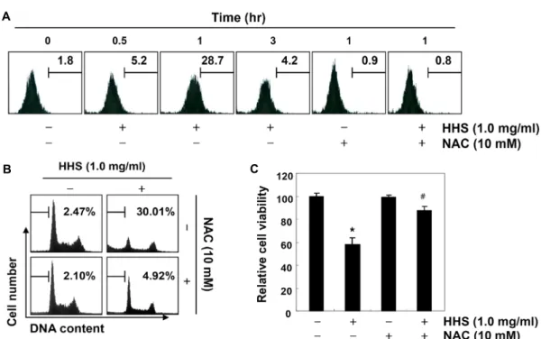
ROS의 생성에 미치는 황흑산 추출물의 영향
다양한 세포 내․외 신호에 의하여 발생하는 ROS는 DNA 및 미토콘드리아의 손상을 야기하여 apoptosis 유발 촉진 신 호로 작용한다[2, 13, 23]. 따라서 황흑산 추출물 처리에 의한 AGS 위암세포의 apoptosis 유발이 ROS 생성과 연관성이 있 는지의 여부를 조사하였다. 이를 위하여 황흑산 추출물이 처 리된 세포들을 H
2-DCFDA 용액에 반응시킨 후 flow cy- tometry 분석을 실시하였다. Fig. 3A의 결과에서 알 수 있듯이 황흑산 추출물의 처리 30분부터 ROS의 생성이 증가되었고, 1시간 처리 군에서 ROS의 생성이 가장 높게 나왔으며, 그 이 후부터는 점차적인 감소 경향성을 보여주어 황흑산 추출물 처리 초기에 ROS의 생성이 동반되었음을 알 수 있었다. 따라 서 황흑산 추출물 처리에 따른 ROS의 생성이 AGS 위암세포 의 증식 억제 및 apoptosis 유발에 직접적으로 관여하는지를 조사한 결과, Fig. 3B에서 알 수 있듯이 ROS scavenger인 NAC 의 전처리로 ROS의 생성을 억제한 경우 황흑산 추출물 처리 에 의한 apoptosis 유발이 현저하게 억제되었으며, 이는 AGS 세포의 생존율 회복과 연관성이 있었다. 이상의 결과는 황흑 산 추출물 처리에 의한 ROS의 생성이 AGS 위암세포의 증식 억제 및 apoptosis 유발에 직접적인 인자로 관여함을 의미하 는 것이다.
Caspase의 활성에 미치는 황흑산 추출물의 영향
Apoptosis 유발에 핵심적인 효소로 작용하는 caspase fam-
ily 단백질은 initiator caspases (caspase-8 및 -9)와 effector
caspases (caspase-3 및 -7)로 구분되며, initiator caspase는
death-inducing signal에 의해 활성화되어 effector caspase를
활성화하고 활성화된 effector caspase는 poly (ADP-ribose)-
polymerase (PARP) 등의 단백질을 분해하여 apoptosis를 유
도한다[6, 20, 24]. 특히 PARP는 정상세포의 DNA 수복이나
유전자 안정성 유지에 중요한 역할을 하며, apoptosis 유발
시 caspase-3에 의해 분해가 일어나면 이러한 회복 기능이 상
실된다[25]. 본 연구에서는 황흑산 추출물 처리에 의한 AGS
위암세포의 apoptosis 유발에 caspase 활성이 관여하는지의
여부를 부가적으로 조사하였다. 먼저 caspase의 발현 정도를
A
B C
Fig. 3. Effects of ROS scavenger on the generation of ROS, apoptosis and cell viability in Hwangheuk-san-treated AGS cells. AGS cells were treated with HHS (1.0 mg/ml) for the indicated times (A) or 48 hr (B and C) after 1 hr pre-treatment with or without NAC (10 mM), a ROS scavenger. (A) The medium was discarded and cells were incubated at 37°C in the dark for 20 min with new culture medium containing 10 μM H2DCFDA and then used to evaluate ROS generation by flow cytometry. Each point represents the mean of two independent experiments. (B) The cells were evaluated for sub-G1 DNA content using a DNA flow cytometer. Each point represents the mean of two independent experiments. (C) Cell viability assay was done using an MTT assay. Each point represents the mean ± SD of three independent experiments. The significance was determined by Student’s t-test (*p<0.05 versus untreated control; #p<0.05 versus HHS-treated cells).
A B
Fig. 4. Activation of caspases by Hwangheuk-san treatment in human gastric cancer AGS cells. (A) Cells were treated with HHS for 48 hr, and aliquots containing total proteins were subjected to SDS-polyacrylamide gels followed by immunoblot analysis with specific antibodies and an ECL detection system. Actin was used as an internal control. (B) The cells were lysed, and aliquots (20 μg protein) were assayed for in vitro caspase-3, -8 and -9 activity using DEVD-pNA, IETD-pNA and LEHD- pNA as substrates, respectively, at 37°C for 1 hr. The released fluorescent products were measured. The data are expressed as the mean ± SD of three independent experiments. The significance was determined by the Student’s t-test (*p<0.05 vs.
untreated control).
확인한 결과, Fig. 4A에서 나타난 바와 같이 황흑산 추출물 처리시 caspase-3, -8 및 -9의 불활성형 단백질의 발현이 모두 처리 농도 의존적으로 감소하는 것으로 나타났다. 이는 상대 적으로 활성형 caspase의 발현이 증가될 수 있음을 의미하기
에, caspase의 활성 여부를 정량적으로 확인하기 위하여 in vi-
tro caspase 활성을 분석한 결과, 황흑산 추출물 처리에 따라caspase-3, -8 및 -9의 활성 정도가 대조군과 비교하였을 때
처리 농도 의존적으로 유의적으로 증가되었으며(Fig. 4B), 황
B A
Fig. 5. Effects of a pan-caspase inhibitor, z-VAD-fmk, on the Hwangheuk-san-induced apoptosis and growth in- hibition in human gastric cancer AGS cells. Cells were treated with or without z-VAD-fmk (50 M) for 1 hr before challenge with HHS (1.0 mg/ml) for 48 hr. (A) The cells were evaluated for sub-G1 DNA content using a DNA flow cytometer. Each point represents the mean of two independent experiments. (B) Cell viability assay was done using an MTT assay. Each point represents the mean ± SD of three independent experiments. The significance was determined by Student’s t-test (*p<0.05 versus untreated control; #p<0.05 versus HHS-treated cells).
흑산 추출물 처리 농도의 증가에 따라 PARP 단백질의 단편화 가 증가하였다(Fig. 4A).
따라서 황흑산 추출물 처리에 의한 apoptosis 유발이 cas- pase 활성 의존적으로 이루어지는지의 여부를 조사하기 위하 여 pan-caspase의 억제제인 z- Val- Ala- Asp (VAD)-fm를 전처 리하여 황흑산 추출물에 의한 apoptosis 유발이 억제될 수 있 는지의 여부를 조사하였다. Fig. 5에 나타낸 바와 같이 황흑산 추출물에 의한 apoptosis는 z-VAD-fmk를 전처리에 의하여 거 의 완벽하게 차단되었으며, 이에 따른 생존율의 회복 현상이 관찰되었다. 이상의 결과는 황흑산 추출물 처리에 따른 AGS 세포의 apoptosis 유발이 intrinsic 및 extrinsic apoptosis 유발 경로를 모두 활성화시켜 이루어지고 있음을 의미하며 황흑산 추출물 처리에 의한 caspase의 활성이 중요한 역할을 하고 있 음을 시사하는 것이다.
결론적으로 황흑산 추출물 처리에 의한 AGS 인체 위암세 포의 증식억제는 ROS의 생성에 의한 미토콘드리아의 기능
손상과 caspase 활성 의존적 intrinsic 및 extrinsic apoptosis 경로의 활성을 통한 apoptosis 유발과 밀접한 연관성이 있음 을 알 수 있었다. 따라서 황흑산의 황암 활성에 대한 in vivo 동물 실험에 의한 효능의 검증과 생리활성 성분 분석 등에 대한 추가적인 연구가 수행되어야 할 것이다.
감사의 글
본 연구는 한국연구재단의 기초과학연구 프로그램 지원 사 업(No. 2012046358 및 2013R1A1A2065537)으로 수행된 연구 결과이며 이에 감사드립니다.
References
1. Brenner, D. and Mak, T. W. 2009. Mitochondrial cell death effectors. Curr. Opin. Cell Biol. 21, 871877.
2. Circu, M. L. and Aw, T. Y. 2001. Reactive oxygen species, cellular redox systems, and apoptosis. Free Radic. Biol. Med.
48, 749-762.
3. de Oliveira, J. R., de Aguiar Almeida, R. B., das Graças Figueiredo Vilela, P., de Oliveira, F. E., da Rocha, R. F., Jorge, A. O. and de Oliveira, L. D. 2014. Control of micro- organisms of oral health interest with Arctium lappa L.
(burdock) extract non-cytotoxic to cell culture of macro- phages (RAW 264.7). Arch. Oral Biol. 59, 808-814.
4. Jeong, S. Y. and Seol, D. W. 2008. The role of mitochondria in apoptosis. BMB Rep. 41, 1122.
5. Jin, Z. and El-Deiry, W. S. 2005. Overview of cell death sig- naling pathways. Cancer Biol. Ther. 4, 139-163.
6. Jung, J. I., Lim, S. S., Choi, H. J., Cho, H. J., Shin, H. K., Kim, E. J., Chung, W. Y., Park, K. K. and Park, J. H. 2006.
Isoliquiritigenin induces apoptosis by depolarizing mi- tochondrial membranes in prostate cancer cells. J. Nutr.
Biochem. 17, 689-696.
7. Kasibhatla, S. and Tseng, B. 2003. Why target apoptosis in cancer treatment. Mol. Cancer Ther. 2, 573-580.
8. Kerr, J. F., Wyllie, A. H. and Currie, A. R. 1972. Apoptosis:
a basic biological phenomenon with wide-ranging im- plications in tissue kinetics. Br. J. Cancer 26, 239-257.
9. Kim, E. J., Park, H., Lim, S. S., Kim, J. S., Shin, H. K. and Park, J. H. Y. 2008. Effect of the hexane extracts of Saussure lappa on the growth of HT-29 human colon cancer cells. Kor.
J. Food Sci. Technol. 40, 207-214.
10. Kim, E. J., Park, S. Y., Hong, J., Shin, M., Lim, S. S., Shin, H. K. and Park, J. H. Y. 2007. Inhibitory effect of the meth- anolic extract of Symphyocladia latiuscula on the growth of HT-29 human colon cancer cells. J. Kor. Soc. Food Sci. Nutr.
36, 431-438.
11. Kim, E. J., Park, S. Y., Shin, H. K., Kwon, D. Y., Surh, Y.
J. and Park, J. H. Y. 2007. Activation of caspase-8 contributes to 3,3'-diindolymethane-induced apoptosis in colon cancer cells. J. Nutr. 137, 1-6.
12. Kumar, H., Song, S. Y., More, S. V., Kang, S. M., Kim, B.
W., Kim, I. S. and Choi, D. K. 2013. Traditional Korean east asian medicines and herbal formulations for cognitive impairment. Molecules 18, 14670-14693.
13. Li, Z. Y., Yang, Y., Ming, M. and Liu, B. 2011. Mitochondrial ROS generation for regulation of autophagic pathways in cancer. Biochem. Biophys. Res. Commun. 414, 5-8.
14. Luo, X., Budihardjo, I., Zou, H., Slaughter, C. and Wang, X. 1998. Bid, a Bcl2 interacting protein, mediates cytochrome c release from mitochondria in response to activation of cell surface death receptors. Cell 94, 481-490.
15. Ma, Y. S., Hsiao, Y. P., Lin, J. H., Hsu, S. C., Chueh, F. S., Weng, S. W., Lai, K. C., Lin, J. G. and Chung, J. G. 2014.
Crude extract of Rheum palmatum L inhibits migration and invasion of LS1034 human colon cancer cells acts through the inhibition of matrix metalloproteinase-2/-9 by MAPK signaling. Environ. Toxicol. 30, 852-863.
16. Ma, Y. S., Hsu, S. C., Weng, S. W., Yu, C. C., Yang, J. S., Lai, K. C., Lin, J. P., Lin, J. G. and Chung, J. G. 2014. Crude extract of Rheum palmatum L induced cell death in LS1034 human colon cancer cells acts through the caspase-depend- ent and -independent pathways. Environ. Toxicol. 29, 969-980.
17. Mohammadparast, B., Rustaiee, A. R., Rasouli, M., Zardari, S. and Agrawal, V. 2014. In vitro enhancement of psoralen as an important anticancer compound in Psoralea corylifolia through precursor feeding. Pharm. Biol. 21, 1-4.
18. Park, D. G. 2009. The Changes of expression of surviving by butyrate in HCT116 colon cancer cells. J. Kor. Surg. Soc.
77, 297-305.
19. Park, H. and Kim, H. S. 2014. Korean traditional natural herbs and plants as immune enhancing, antidiabetic, chemo- preventive, and antioxidative agents: a narrative review and perspective. J. Med. Food 17, 21-27.
20. Park, S. Y., Kim, I. S., Lee, S. H., Lee, S. H., Jung, D. W., Park, O. J. and Kim, Y. M. 2012. Anti-proliferative effects
of selenium in HT-29 colon cancer cells via inhibition of Akt. J. Life Sci. 22, 55-61.
21. Park, S. Y., Lee, S. H., Park, O. J. and Kim, Y. M. 2011.
Apoptotic effects of curcumin and EGCG via Akt-p53 sig- naling pathway in HCT116 colon cancer cells. J. Life Sci. 21, 89-95.
22. Rajan, V., Tripathi, J., Variyar, P. and Pandey, B. N. 2014.
Mechanism of cytotoxicity by Psoralea corylifolia extract in human breast carcinoma cells. J. Environ. Pathol. Toxicol.
Oncol. 33, 265-77.
23. Ray, P. D., Huang, B. W. and Tsuji, Y. 2012. Reactive oxygen species (ROS) homeostasis and redox regulation in cellular signaling. Cell. Signal. 24, 981-90.
24. Schreiber, V., Dantzer, F., Ame, J. C. and de Murcia, G. 2006.
Poly (ADP-ribose): novel functions for an old molecule. Nat.
Rev. Mol. Cell Biol. 7, 517-528.
25. Schulze-Osthoff, K., Ferrari, D., Los, M., Wesselbrorg, S. and Peter, M. E. 1998. Apoptosis signaling by death receptors.
Eur. J. Biochem. 254, 439-459.
26. Scorrano, L. and Korsmeyer, S. J. 2003. Mechanisms of cyto- chrome c release by proapoptotic BCL-2 family members.
Biochem. Biophys. Res. Commun. 9, 437-444.
27. Siegel, R., Ma, J., Zou, Z. and Jemal, A. 2014. Cancer statistics. CA Cancer J. Clin. 64, 9-29.
28. Sucher, N. J. 2013. The application of Chinese medicine to novel drug discovery. Expert Opin. Drug Discov. 8, 21-34.
29. Sun, Q., Liu, K., Shen, X., Jin, W., Jiang, L., Saeed Sheikh, M., Hu, Y. and Huang, Y. 2014. Lappaol F, a novel anti- cancer agent isolated from plant arctium Lappa L. Mol.
Cancer Ther. 13, 49-59.
30. Teschke, R., Wolff, A., Frenzel, C. and Schulze, J. 2014.
Review article: Herbal hepatotoxicity--an update on tradi- tional Chinese medicine preparations. Aliment. Pharmacol.
Ther. 40, 32-50.
초록:AGS 인체 위암세포에서 황흑산에 의한 ROS 생성 및 caspase 활성 의존적 apoptosis 유발
홍수현
1*․박 철
2․김경민
4․최영현
1,3*
(1동의대학교 한의과대학 생화학교실, 2동의대학교 자연생활과학대학 분자생물학과, 3동의대학교 항노화연구소
및 Blue-Bio Industry RIC, 4동의대학교 한의과대학 순환신경내과학교실)