The Sanguinarine Apoptosis Induction of Hep3B Human Hepatocellular Carcinoma Cells is Dependent on the Activation of Caspase
Min Ho Han
1, Sung Hyun Choi
2, Su Hyun Hong
3, Dong Il Park
4and Yung Hyun Choi
3,5*
1Department of Applied Research, National Marine Biodiversity Institute of Korea, Seocheon 33662, Korea
2Department of System Management, Korea Lift College, Geochang 50141, Korea
3Open Laboratory for Muscular and Skeletal Disease, and Department of Biochemistry, Dongeui University College of Korean Medicine, Busan 47227, Korea
4Department of Internal Medicine, Dongeui University College of Korean Medicine, Busan 47227, Korea
5Anti-Aging Research Center, Dongeui University, Busan 47340, Korea
Received July 21, 2017 /Revised November 15, 2017 /Accepted November 15, 2017
Sanguinarine is a benzophenanthridine alkaloid derived from the roots of Sanguinaria canadensis L., which is used for the purpose of treating various diseases. Although studies of anticancer activities have been performed using various cancer cell lines, the phenomenon of inducing apoptosis in cancer cells by using sanguinarine requires more research. Therefore, this study investigated the anti-cancer activities and related mechanisms of sanguinarine used with Hep3B human hepatocellular carcinoma cells in terms of the regulation of apoptosis. Sanguinarine inhibited the proliferation of Hep3B cells in a concentration-dependent manner, which was associated with the induction of apoptosis.
Sanguinarine also increased the activity of caspase-3, which is a typical effector caspase, and the activ- ities of caspase-8 and caspase-9, which are key when initiating extrinsic and intrinsic apoptosis path- ways, respectively. In addition, sanguinarine increased the expression of death receptor-related genes and pro-apoptotic BAX, which belongs to the Bcl-2 family, while suppressing the expression of an- ti-apoptotic Bcl-2. Sanguinarine promoted the truncation of Bid and enhanced the release of cyto- chrome c from the mitochondria to the cytoplasm due to a loss of mitochondrial membrane potential.
Furthermore, the reduction of a survival rate that was induced by sanguinarine and the induction of apoptosis disappeared with the inhibition of artificial caspase activity. Therefore, the results of the study indicated that sanguinarine-induced apoptosis in Hep3B cells involves both extrinsic and in- trinsic pathways; such apoptosis is a caspase-dependent phenomenon.
Key words : Apoptosis, caspase, HepG2 cells, sanguinarine, tBid
*Corresponding author
*Tel : +82-51-850-7413, Fax : +82-51-853-4036
*E-mail : [email protected]
This is an Open-Access article distributed under the terms of the Creative Commons Attribution Non-Commercial License (http://creativecommons.org/licenses/by-nc/3.0) which permits unrestricted non-commercial use, distribution, and reproduction in any medium, provided the original work is properly cited.
Journal of Life Science 2017 Vol. 27. No. 11. 1340~1348 DOI : https://doi.org/10.5352/JLS.2017.27.11.1340
서 론
최근 새로운 항암제의 탐색을 위하여 전통의약 자원에 대한 관심이 증가되고 있다. 특히 식물 유래 천연물은 화학합성 약 물에 비하여 부작용이 적으며, 오랜 경험적 자료가 충분한 장 점을 가진다[8, 13]. 그중 혈근초(血根草, bloodroot)로 알려진
Sanguinaria canadensis L.의 뿌리는 기관지염과 천식을 포함한다양한 질병 치료의 목적으로 사용되어 왔다[38]. 혈근초에서 추출된 sanguinarine은 benzophenanthridine alkaloid 계열 물질로서 치약과 구강 세정제의 성분[12, 31] 뿐만 아니라 재상 염 치료제 또는 항생제로도 널리 사용되고 있다[23, 30, 38].
Sanguinarine에 대한 연구에 의하면, 이 화합물은 강력한 항균 효과[30, 41]와, 항염증[28, 40], 항산화[11, 39], 면역 증강 [35, 38] 및 혈관 신생억제[2, 10] 등을 함유하고 있는 것으로 보고되고 있다. 아울러 sanguinarine은 정상 세포의 증식에는 큰 영향을 주지 않으면서, 암세포의 증식을 억제하거나 암세 포 유형별로 세포주기 정지(cell cycle arrest) 및 세포사멸 (apoptosis)의 유도[1, 4, 15, 16, 27]와 전이(metastasis)를 억제 [34, 42]하는 등 강력한 항암효과가 있음을 알려졌다. 특히 sanguinarine에 의한 다양한 암세포에서 apoptosis 유도 과정 에는 활성산소종(reactive oxygen species, ROS)의 생성 증가 가 동반된다는 결과들에서 미토콘드리아의 기능 손상이 주요 apoptosis 유도 기전 중의 하나로 인식되고 있으며[5, 29, 33], 본 연구실에서도 그동안 다양한 암세포를 대상으로 유사한 연구를 수행하여 왔다[6, 7, 18-21, 26].
한편 가장 전형적인 세포죽음 기전임 apoptosis는 caspase
cascade의 활성을 특징으로 한다[14, 32]. Apoptosis는 외인성
(extrinsic) 및 내인성(intrinsic) 경로로 대별되는데, 전자는
death receptor (DR) 분자에 death ligand가 결합하여 cas-
pase-8을 활성화시키고 이는 다시 caspase-3 및 caspase-7과 같은 effect caspase의 활성을 통하여 다양한 세포 내 기질 단 백질의 분해를 유도한다[25, 37]. 반면에 미토콘드리아가 관여 하는 intrinsic 경로는 apoptosis를 촉진하거나 억제할 수 있는 단백질로 구성된 Bcl-2 family의 발현 변화에 의해 조절된다 [14, 17]. Bcl-2 family 중, Bax를 포함한 pro-apoptotic 단백질 이 Bcl-2와 같은 anti-apoptotic 단백질과 비교하여 상대적으로 발현이 증가하면 Bax가 미토콘드리아로 이동하여 미토콘드리 아 막 잠재력(mitochondrial membrane potential, MMP, Δψ m)의 소실과 세포질로의 cytochrome c 방출을 촉진한다[17, 37]. 세포질에서 cytochrome c는 apoptosome을 형성하여 cas- pase-9와 caspase-3을 활성화시키며 그 결과 poly(ADP-ribose) polymerase (PARP)를 포함한 기질 단백질의 절단을 촉진하는 공동 경로로 유입된다[9, 17]. 그리고 이 두 경로는 pro-apop- totic BH3-interacting domain death agonist (Bid)를 통해 신 호 전달에 연결될 수 있는데, 활성화된 caspase-8은 Bid를 절단 하여 활성형 tBid (truncated form of Bid)로 전환시켜 미토콘 드리아에서 세포질로의 cytochrome c 방출을 촉진한다[3, 24].
비록 sanguinarine에 의한 apoptosis 유도와 연관된 두 가지 apoptosis 경로에 대한 해석이 부분적으로 이루어져 왔으나, 항암 활성의 정확한 분자 표적과 기전은 아직 명확하지 않다.
따라서 본 연구에서는 인간 간암세포(Hep3B)를 대상으로 sanguinarine이 현재까지 알려진 두 가지 apoptosis 유도 경로 를 모두 활성화 시키는지의 여부와, 이러한 과정이 caspase 활성 의존적인 현상인지를 조사하였다.
재료 및 방법
세포배양 및 sanguinarine의 처리
본 연구에 사용된 Hep3B 간암세포는 American Type Cul- ture Collection (Manassas, VA, USA)에서 구입하였으며, 10%
의 fetal bovine serum과 1%의 penicillin–streptomycin이 함 유된 Dulbecco’s modified Eagle’s medium (DMEM, Welgene, Daegu, Republic of Korea)을 이용하여 37
oC, 5% CO
2조건에 서 배양하였다. Sanguinarine은 Sigma-Aldrich Chemical Co.
(St. Louis, MO, USA)에서 구입하였으며, ethanol에 녹여 10 mM의 stock solution으로 만든 후, 적정 농도로 DMEM에 희 석하여 처리하였다.
세포 생존율의 측정
세포 생존에 미치는 sanguinarine의 영향을 조사하기 위하 여 6-well plate에 well 당 1×10
5개의 세포를 분주하고 24시간 안정화시켰다. 적정 농도의 sanguinarine이 함유된 배지로 교 체한 후 48시간 추가 배양하고 sanguinarine 처리에 따른 형태 적 변화를 관찰한 후, 3시간 동안 0.5 mg/ml의 3-(4,5-dimethylthiazol-2-yl)-2,5-diphenyltetrazolium bromide
(MTT, Sigma- Aldrich Chemical Co.) 용액을 처리하였다, 그 후 well에 형성된 formazan을 dimethyl sulfoxide (DMSO, Sigma- Aldrich Chemical Co.)에 용해시킨 후, enzyme-linked immunosorbent assay (ELISA) reader (Molecular Devices, Sunnyvale, CA, USA)를 이용하여 540 nm에서 흡광도 변화를 측정하여 대조군에 대한 세포 생존율을 백분율로 표시하였다.
Apoptosis 유도 확인을 위한 핵의 형태 변화 관찰 Sanguinarine 처리에 따른 Hep3B 세포의 apoptosis 유도 여부를 확인하기 위하여 48시간 동안 다양한 농도의 sangui- narine이 처리된 세포들을 모아 phosphate-buffered saline (PBS) 용액으로 세척하고, 실온에서 10분 동안 3.7%의 paraf- ormaldehyde가 함유된 PBS 용액으로 고정시켰다. 고정된 세 포를 PBS로 세척하고 실온에서 10분 동안 4,6-diamidino-2- phenylindole (DAPI, Sigma-Aldrich Chemical Co.) 용액(2.5 g/ml)으로 염색 한 후, 세척하고 형광 현미경(fluorescene mi- croscope, Carl Zeiss, Germany)을 사용하여 세포의 핵 형태 변화를 분석하였다.
세포주기 분석에 따른 apoptosis 유발의 정량화
Sanguinarine 처리에 따른 Hep3B 세포의 세포주기 분포도 에 따른 apoptosis 유발 빈도의 정량화를 위해, 준비된 세포를 PBS로 세척하고, 냉각시킨 70% 에탄올에 고정시켰다. 이들 세 포를 PBS로 다시 세척하고, 100 mg/ml RNase A, 50 mg/ml 의 propidium iodide (PI), 0.1%의 sodium citrate 및 0.1%의 NP-40이 함유된 PI 용액을 이용하여 암하에서 30분간 처리하 였다. 각 실험군당 최소 10,000개 이상의 세포를 flow cytometer (Becton Dickinson, San Jose, CA, USA)에 적용시켜 세포 내 DNA 함량에 따른 histogram을 대상으로 sub-G1기에 속하는 세포를 apoptosis가 유발된 세포로 산출하였다.
Annexin V 염색에 의한 apoptosis 유발의 정량화 Sanguinarine 처리에 따른 apoptosis 정량화의 또 다른 방 법으로 Annexin V 염색법을 적용하였다. 이를 위하여 준비된 세포들을 PBS를 이용하여 2~3회 정도 세척하고 Annexin V binding buffer (Becton Dickinson)에 부유시킨 다음 Annexin V-fluorescein isothiocyanate (FITC) 및 PI를 처리하여 20분 동안 암실에서 반응시켰다. 반응이 끝난 후 단일 세포로 분리 한 다음 flow cytometer에 적용해 apoptosis가 유발된 세포 (V
+/PI
–)를 형광반응에 따라 분석하였다.
RNA의 분리 및 reverse transcriptase-polymerase chain reaction (RT-PCR)의 분석
Sanguinarine이 함유된 배지에서 배양된 세포들에서 전사
수준의 특정 유전자 발현 변화를 조사하기 위하여, 준비된 세
포들을 대상으로 Tryzol Reagent (Invitrogen Co., Carlsbad,
Table 1. Oligonucleotides used in RT-PCR
Gene name Sequence
Fas Sense
Antisense
5'-TCT AAC TTG GGG TGG CTT TGT CTT C-3' 5'-GTG TCA TAC GCT TTC TTT CCA T-3'
FasL Sense
Antisense
5'-GGA TTG GGC CTG GGG ATG TTT CA-3' 5'-AGC CCA GTT TCA TTG ATC ACA AGG-3'
DR4 Sense
Antisense
5‘-CAG AAC GTC CTG GAG CCT GTA AC-3’
5‘-ATG TCC ATT GCC TGA TTC TTT GTG-3’
DR5 Sense
Antisense
5‘-GGG AAG AAG ATT CTC CTG AGA TGT G-3’
5‘-ACA TTG TCC TCA GCC CCA GGT CG-3’
TRAIL Sense
Antisense
5‘-ATG GCT ATG ATG GAG TCC AG-3' 5‘-TTG TCC TGC ATC TGC TTC AGC-3'
Bax Sense
Antisense
5'-ATG GAC GGG TCC GGG GAG-3' 5'-TCA GCC CAT CTT CTT CCA-3'
Bcl-2 Sense
Antisense
5'-CAG CTG CAC CTG ACG-3' 5'-ATG CAC CTA CCC AGC-3'
GAPDH Sense
Antisense
5'-CGG AGT CAA CGG ATT TGG TCG TAT-3' 5'-AGC CTT CTC CAT GGT GGT GAA GAC-3'
Table 2. Primary antibodies used in the present study
Antibody Dilution Product no. Species of origin and supplier
Fas FasL DR4 DR5 TRAIL
Bax Bcl-2 Bid PARP Cytochrome c
COX VI Actin
1:1000 1:1000 1:1000 1:1000 1:1000 1:1000 1:1000 1:500 1:1000 1:1000 1:500 1:1000
sc-715 sc-957 sc-7863 sc-65314 sc-7877 sc-493 sc-783 sc-11423
sc-7150 sc-7159 20E8C12 BS6007M
Rabbit polyclonal, Santa Cruz Biotechnology, Inc.
Rabbit polyclonal, Santa Cruz Biotechnology, Inc.
Rabbit polyclonal, Santa Cruz Biotechnology, Inc.
Mouse polyclonal, Santa Cruz Biotechnology, Inc.
Rabbit polyclonal, Santa Cruz Biotechnology, Inc.
Rabbit polyclonal, Santa Cruz Biotechnology, Inc.
Rabbit polyclonal, Santa Cruz Biotechnology, Inc.
Rabbit polyclonal, Santa Cruz Biotechnology, Inc.
Rabbit polyclonal, Santa Cruz Biotechnology, Inc.
Rabbit polyclonal, Santa Cruz Biotechnology, Inc.
Mouse monoclonal, Abcam
Mouse polyclonal, Bioworld Technology, Inc.
CA, USA)를 이용하여 RNA를 추출하였으며, 해당 유전자들 의 primer (Table 1)와 ONE-STEP RT-PCR PreMix kit (iNtRON Biotechnology, Republic of Korea)를 이용하여 PCR 을 수행하였다. 이후 증폭된 DNA 산물을 1.5% agarose gel를 사용하여 100 volt에서 20분간 전기영동하고 ethidium bro- mide (EtBr, Sigma-Aldrich Chemical Co.)로 염색 후 UV 하에 서 관찰하였다. 아울러 PCR 반응의 대조군으로 glyceraldehyde- 3-phosphate dehydrogenase (GAPDH)를 사용하였다.
단백질의 분리 및 Western blot analysis
Sanguinarine 처리에 따른 Hep3B 세포의 apoptosis 유발 관련 유전자들의 번역수준에서 발현 변화 관찰을 위하여 준비 된 세포에 적당량의 lysis buffer를 첨가하여 4℃에서 1시간 이상 용해시켰다. 상층액에 있는 단백질을 분리한 후 동량의 단백질을 sdium dodecyl sulfate (SDS)-polyacrylamide gel을
이용하여 분리하고, gel에 함유된 단백질을 polyvinylidene difluoride membrane (Schleicher and Schuell, Keene, NH, USA)에 전이시켰다. Membrane에 5% skim milk 용액을 1시 간 처리하여 비특이적인 단백질들에 대한 blocking을 실시하 였다. 그리고 적정 1차 항체(Table 2)를 처리하여 상온에서 2시 간 이상 반응시킨 후, PBS-T 용액(PBS with Tween 20)으로 세척하고 2차 항체를 상온에서 1시간 정도 반응시켰다. 반응 이 끝난 후 암실에서 enhanced chemiluminesence (ECL) sol- ution (Santa Cruz Biotechnology Inc.)을 적용시킨 다음 X-ray film에 감광시켜 특정 단백질의 발현 변화를 분석하였다.
Caspase 활성의 측정
Sanguinarine에 노출된 세포의 caspase 활성 증가에 대한
정량적인 평가를 위하여 R&D Systems (Minneapolis, MI,
USA)에서 구입한 kit를 이용하였다. 이를 위하여 다양한 농도
A
B
Fig. 1. Inhibition of cell viability and morphological changes by sanguinarine in Hep3B human hepatocellular carcinoma cells. Hep3B cells were seeded into 6-well plates at 1×10⁵ per well and treated with the indicated concentrations of sanguinarine for 48 hr. (A) Cell viability was de- termined by an MTT assay. Results are expressed as per- centages of the vehicle-treated control ± SD of three sep- arate experiments (*p<0.05 vs. untreated control). (B) After treatment with sanguinarine for 48 h, cellular mor- phology was visualized by an inverted microscope (Magnification, ×200). And, nuclei stained with DAPI solution were photographed with a fluorescence micro- scope using a blue filter (Magnification, ×400). After treatment with sanguinarine, the cells were harvested, washed with PBS, and fixed with 3.7% paraformalde- hyde in PBS at 25°C. After 10 min, the cells were washed with PBS, stained with DAPI solution for 10 min and then washed twice with PBS. The morphology changes in the nucleus were examined using a fluorescence mi- croscope using a blue filter (Magnification, ×400).
의 sanguinarine이 처리된 세포에서 추출한 동량의 단백질 (150 μg)을 50 μl의 sample에 50 μl의 reaction buffer [40 mM HEPES (pH 7.4), 20% glycerol (v/v), 1 mM ethylenedi- aminetetraacetic acid, 0.2% Nonidet P-40 및 10 mM dithio- threitol]를 혼합한 다음 5 μl caspases 기질[caspase-3, Asp- Glu-Val-Asp (DEVD)-p-nitroaniline (pNA); caspase-8, Ile- Glu-Thr-Asp (IETD)-pNA; caspase-9, Leu-Glu-His-Asp (LEHD)- pNA]을 첨가하여 37
oC, 암실에서 3시간 동안 반응시켰다. 반 응이 끝난 후 ELISA reader를 이용하여 405 nm 파장에서 각 caspase 활성 정도에 따른 변화를 측정하였다.
MMP (Δψm) 값 변화의 측정
세포 내 미토콘드리아 기능 손상의 여부를 확인하기 위하여 MMP 값 변화 정도를 측정하기 위해서 5,5‘,6,6’-tetrachloro- 1,1‘,3,3’-tetraethyl-imidacarbocyanine iodide (JC-1, Sigma- Aldrich Chemical Co.)를 사용하였다. 이를 위해 적정 농도의 sanguinarine이 처리된 세포를 수집하고 37℃ 암하에서 20분 동안 10 μM의 JC-1 용액으로 염색하였다. 이어서, 세포를 PBS 로 세척하고 부유시킨 후 flow cytometer에 적용시켜 MMP의 변화를 측정하였다.
통계 분석
실험 결과들의 유의성을 검정하기 위하여 분산분석(ANOVA) 을 실시한 후 p<0.05 수준에서 Duncan's multiple range tests 를 실시하였으며, 그 결과는 평균(mean)±표준편차(standard deviation, SD)로 표시하였다.
결과 및 고찰
Sanguinarine에 의한 Hep3B 간암세포의 apoptosis 유도 Hep3B 간암세포의 증식에 미치는 sanguinarine의 영향을 조사하기 위한 MTT assay를 실시하기 위하여 다양한 농도의 sanguinarine이 함유된 배지에서 Hep3B 세포를 48시간 동안 배양하였다. Fig. 1A에 나타낸 바와 같이, sanguinarine에 노출 된 Hep3B 세포는 sanguinarine의 처리 농도 증가에 따라 생존 율이 유의적으로 억제되었음을 알 수 있었다. 예를 들어, 0.6 μM의 처리군에서는 대존군에 비하여 약 65% 정도의 생존율 을 보였고, 이 보다 두 배 높은 1.2 μM 처리군에서는 약 25%
정도의 생존율을 나타내었다. 이와 같은 수치는 암세포의 종 류에 따라 약간의 차이는 있으나 대체로 대장암(HCT-116), 신 경교종(U87MG 및 U118MG), 위암(SGC-7901 and HGC-27) 등의 다른 고형암 세포들에서의 결과와 유사하였지만[18, 33, 42], 구강편평세포암종세포(KB), 방광암세포(T24), 신경아세 포종(SH-SY5Y) 등에 비해서는 다소 감수성이 높게 나타났다 [4, 20, 27].
아울러 sanguinarine이 함유된 배지에서 배양된 Hep3B 세
포는 정상 배지에서 배양된 세포에 비하여 부착력이 약해지면 서 부유되거나 세포의 크기도 수축되는 등의 형태적 변화를 동반하였다(Fig. 1B). 그리고 전향적인 apoptosis가 유도된 세 포에서 관찰되는 핵의 염색질 응축(chromatin condensation) 에 의한 apoptotic body의 형성이 증가되었다. 따라서 sangui- narine 처리에 따른 apoptosis 유도의 정도를 정량적으로 평가 한 결과 세포주기 분석에 따른 sub-G1기에 해당되는 세포의 빈도 및 annexin-V 염색에 따른 apoptosis 유도 세포 빈도의 상승이 sanguinarine 처리 농도 의존적으로 증가하였다(Fig.
2). 이상의 결과들은 sanguinarine에 의한 Hep3B 세포의 증식 억제가 apoptosis 유도와 연관성이 있음을 의미한다.
DR 및 Bcl-2 family 관련 유전자들의 발현에 미치는 san-
guinarine의 영향
A
B
C
Fig. 2. Induction of apoptosis by sanguinarine in Hep3B cells.
(A) Hep3B cells were treated with the indicated concen- trations of sanguinarine for 48 hr and the cells were eval- uated for sub-G1 DNA content, which represents the fractions undergoing apoptotic DNA degradation using a flow cytometer. Data are the mean of the two different experiments. (B) To quantify the degree of apoptosis in- duced by sanguinarine, the cells were stained with an- nexin V-FITC and PI, and the percentages of the apop- totic cells (annexin V+ cells) were then analyzed by a flow cytometer. (C) Data are reported as means ± SD of three independent experiments (*p<0.05 vs. untreated control).
A B
Fig. 3. Effects of sanguinarine on levels of DR-related genes in Hep3B cells. (A) Hep3B cells were treated with various concentrations of sanguinarine for 48 hr, total RNA was isolated and RT-PCR was performed using the indicated primers. The amplified PCR products were run in 1.5%
agarose gel and visualized by EtBr staining. GAPDH was used as a housekeeping control gene. (B) Cells grown under the same conditions as (A) were lysed, and then equal amounts of cell lysates were separated on SDS-pol- yacrylamide gels and transferred to nitrocellulose membranes. Membranes were probed with the indicated antibodies, and the proteins were visualized using an ECL detection system. Actin was used as an internal control.
이상에서 관찰된 Hep3B 세포에서 sanguinarine에 의한 apoptosis 유도에 대표적인 두 가지 apoptosis 경로 중의 하나 인 extrinsic 경로에 미치는 영향을 조사하기 위하여, 상기와 동일한 조건에서 배양된 Hep3B 세포를 대상으로 DR 관련 유 전자들의 발현에 미치는 sanguinarine의 영향을 조사하였다.
Fig. 3의 결과에서 알 수 있듯이 세포막에 존재하는 수용체 중에서 Fas의 발현은 sanguinarine의 처리 농도 증가에 따라 농도 의존적으로 전사 및 번역 수준에서 모두 증가하였으나, Fas ligand (FasL)의 경우 큰 변화가 없었다. DRs의 경우, DR4 의 발현은 큰 변화가 없었으나, DR5와 tumor necrosis fac- tor-related apoptosis-inducing ligand (TRAIL)의 발현은 san- guinarine 처리에 따라 다소 증가되었다. 다음은 intrinsic 경로 의 핵심 조절 유전자에 해당하는 Bcl-2 family에 속하는 인자 들의 발현 변화를 조사하였다. Fig. 4A 및 Fig. 4B에 제시된 결과에서 알 수 있듯이, sanguinarine 처리 농도의 증가에 따
라 Bcl-2 family 중에서 대표적인 pro-apoptotic 유전자에 해당 되는 Bax의 발현은 sanguinarine에 노출된 세포에서 처리 농 도 증가에 따라 점진적으로 증가된 반면, 대표적인 anti-apop- totic 유전자에 해당되는 Bcl-2의 발현은 mRNA 및 단백질 수 준에서 모두 감소되었다.
Apoptosis 유도에서 extrinsic 경로의 활성을 위해서는 해당 수용체에 적절한 ligand의 결합이 개시 신호에 중요하며[25, 37], extrinsic 경로 활성에서는 MMP의 소실에 Bcl-2 family 인자들 중, pro-apoptotic 단백질의 상대적인 발현 증가가 필 요하다는 점에서[14, 22], 본 연구의 결과는 sanguinarine에 의 한 Hep3B 세포의 apoptosis 유도 과정이 extrinsic 및 intrinsic 경로의 동시 활성화를 통해서 이루어 질 가능성이 있음을 보 여주는 것이다. 그러나 DR4와 FasL의 큰 증가 현상이 나지 않고, Fas, DR5 및 TRAIL의 발현 증가가 직접적인 extrinsic 경로 활성에 관여하는지의 여부에 대한 추가적인 실험이 요구 된다.
Caspase의 활성 및 PARP의 발현에 미치는 sanguinar- ine의 영향
다음은 apoptosis 유도의 핵심 기전이 caspase cascade의
활성인 점을 고려하여[14, 32], sanguinarine에 의한 Hep3B 세
포의 apoptosis 유도에도 이러한 현상이 동반되는지의 여부를
조사하였다. 이를 위하여 대표적인 extrinsic 및 intrinsic 경로
의 개시 caspase에 해당되는 caspase-8 및 caspase-9를 포함한,
두 caspase의 활성에 의하여 활성이 증가되는 effector caspase
A B
C
Fig. 4. Effects of sanguinarine on levels of Bcl-2 family members and activity of caspases in Hep3B cells. Hep3B cells were treated with the indicated concentrations of sanguinarine for 48 hr. (A) Total RNA was isolated and performed RT-PCR. The amplified PCR products were run in 1.5%
agarose gel and visualized by EtBr staining. GAPDH was used as a housekeeping control gene. (B) The cells were lysed, and then equal amounts of cell lysates were sepa- rated on SDS-polyacrylamide gels and transferred to ni- trocellulose membranes. Membranes were probed with the indicated antibodies. An ECL detection system was used to visualize proteins. Actin was used as an internal control. (C) Aliquots were incubated with DEVD-pNA, IETD-pNA, and LEHD-pNA for caspase-3, -8, and -9, in- dividually, at 37oC for 1 hr. Released fluorescence prod- ucts were measured. Data represent the mean ± SD of three independent experiments (*p<0.05 vs. untreated control).
에 해당되는 caspase-3의 활성 여부를 조사한 결과, sanguinar- ine의 처리 농도 증가에 따라 3가지 caspase의 활성이 모두 증가되었음을 확인하였다(Fig. 4C). 아울러 활성화된 caspase- 3의 기질 단백질로서 전형적인 apoptosis 유도 과정에서 분해 가 관찰되는 PARP의 발현[9, 17]을 조사해 본 결과, Fig. 4B에 서 알 수 있듯이 sanguinarine이 처리된 세포에서 PARP의 단 편화가 유도되어, sanguinarine에 의한 Hep3B 세포의 apop- tosis 유도에는 caspase의 활성 증가가 관여하고 있음을 유추 할 수 있다.
한편 두 가지 apoptosis 경로를 서로 연결하면서 intrinsic 경로의 증폭에 관여하는 대표적인 단백질이 Bid이며, Bcl-2 family에 속하는 세포질 단백질로서, caspase-8의 기질 단백질 에 해당한다[3, 24]. 본 연구의 결과에 의하면, sanguinarine이 함유된 배지에서 배양된 Hep3B 세포에서 Bid의 truncation 형태(tBid)의 발현이 다소 증가하였으며, 이는 아마도 sangui-
narine에 의하여 활성화된 caspase-8에 의한 것으로서 두 경로 의 연결에 부분적으로 기여했을 것으로 추정된다.
Sanguinarine에 의한 MMP의 소실과 apoptosis 유도에 서 caspase의 역할
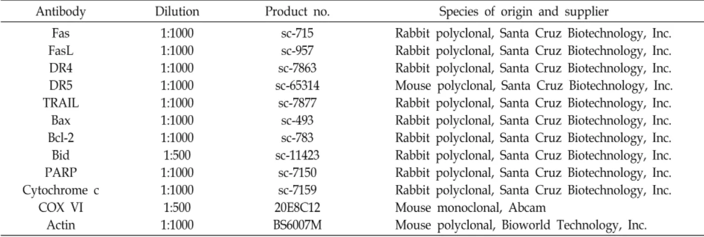
한편, 다양한 자극에 의한 intrinsic 경로의 활성은 미토콘드 리아의 기능 손상에 따른 미토콘드리아의 내막과 외막 사이에 존재하는 cytochrome c와 같은 apoptosis 유도 인자들의 세포 질 방출이 필수적이다[9, 17]. 따라서 sanguinarine에 의한 apoptosis 유도에 이러한 현상들이 나타나는지를 조사하기 위 하여 sanguinarine 처리에 따른 MMP 값의 변화를 측정하였 다. Fig. 5A 및 Fig. 5B에 나타낸 바와 같이, sanguinarine 처리 농도의 증가에 따른 미토콘드리아의 기능 손상을 의미하는 MMP의 소실이 유의적으로 증가하였다. 또한 세포질과 미토 콘드리아 분획 단백질을 이용한 cytochrome c의 발현에 대한 immunoblotting 결과에서는 sanguinarine 처리에 따라 미토 콘드리아에서 세포질로의 cytochrome c 유리 현상이 증가하 였음을 알 수 있었다. 이는 Hep3B 세포에서 sanguinarine에 의한 apoptosis 유도 과정에 intrinsic 경로가 활성화되었으며, 세포질로 유리된 cytochrome c는 intrinsic 경로의 initiator caspase인 caspase-9의 활성 증가에 기여하였음을 보여주는 결과이다.
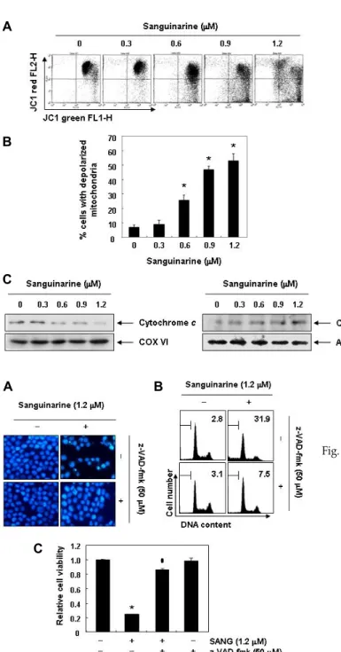
이상에서 제시한 결과들에서 sanguinarine은 아마도 cas- pase cascade의 활성에 따른 extrinsic 및 intrinsic 경로를 모두 활성화시킴으로서 apoptosis를 유도하였을 것이며, 이 과정에 caspase 활성이 필수적으로 관여되었을 것으로 추정된다. 따 라서 이를 재확인하기 하기 위하여 sanguinarine 처리에 따른 apoptosis가 caspase 활성 의존적인 현상인지의 여부를 조사 하였다. 이를 위하여 강력한 pan-caspase 활성 저해제인 ben- zyloxycarbonyl-Val-Ala-Asp (OMe) fluoromethylketone (z- VAD-fmk) [36]을 사용하였다. 즉 caspase의 인위적인 활성을 저해하기 위하여 z-VAD-fmk를 sanguinarine 처리 1시간 전 에 전처리를 한 후 sanguinarine을 24시간 처리 후, sanguinar- ine에 의한 apoptosis 유도 현상이 억제되는지의 여부를 조사 하였다. Fig. 6A의 결과에서 알 수 있듯이, z-VAD-fmk가 존재 하는 조건에서 sanguinarine에 노출된 Hep3B 세포에서는 sanguinarine 단독처리에 의한 핵의 형태적 변화가 거의 관찰 되지 않았다. 아울러 sanguinarine에 의하여 유도되는 apopto- sis 유발 빈도도 매우 감소되었으며, 이에 따라 sanguinarine 처리에 의한 생존율 감소 현상도 유의적으로 차단되어(Fig.
6C), sanguinarine 처리에 의한 Hep3B 세포의 apoptosis 유도 는 caspase 활성 의존적으로 일어나는 현상임을 알 수 있었다.
본 연구에서는 혈근초 유래 sanguinarine의 항암 활성을
Hep3B 간암세포를 대상으로 조사하였으며, sanguinarine에
의한 Hep3B 세포의 증식 억제가 apoptosis 유도에 의한 것임
을 알 수 있었다. 또한 sanguinarine은 extrinsic 및 intrinsic
A
B
C
Fig. 5. Effects of sanguinarine on the expression of cytochrome c and the MMP values in Hep3B cells. (A) After treatment with sanguinarine for 48 hr, the cells were col- lected and incubated with 10 μM JC-1 for 20 min at 37oC in the dark. The cells were then washed with PBS and analyzed us- ing a flow cytometer. (B) The data are ex- pressed as the means ± SD of three in- dependent experiments (*p<0.05 vs. un- treated control). (C) The cytosolic and mi- tochondrial proteins were extracted from the cells and analyzed by Western blot- ting using the indicated antibodies. Cyto- chrome oxidase IV (COX IV) and actin were used as internal controls for the mi- tochondrial and cytosolic fractions, re- spectively.
A B
C
Fig. 6. Inhibition of sanguinarine-induced apoptosis by pan-cas- pase inhibitor in Hep3B cells. Hep3B cells were pre-treated for 1 hr with or without z-VED-fmk, and then treated with sanguinarine for an additional 48 hr. (A) The cells were stained with DAPI and photographed with a fluorescence microscope using a blue filter (Magnification, ×400). (B) The cells were evaluated for sub-G1 DNA content using a flow cytometer. The data are the mean of the two different experiments. (C) Growth inhibition was measured by the metabolic-dye-based MTT assay. Results are expressed as percentages of the vehicle-treated control ± SD of three sep- arate experiments (*p<0.05 vs. untreated control; #p<0.05 vs.
sanguinarine-treated cells).
경로의 동시 활성화를 통하여 apoptosis를 유도하였으며, 이 과정이 caspase 활성 의존적임을 알 수 있었다.
감사의 글
이 논문은 2017학년도 동의대학교 교내연구비(201702660001) 의 지원에 의해 수행되었음.
References
1. Ahsan, H., Reagan-Shaw, S., Breur, J. and Ahmad, N. 2007.
Sanguinarine induces apoptosis of human pancreatic carci-
noma AsPC-1 and BxPC-3 cells via modulationsin Bcl-2 family proteins. Cancer Lett. 249, 198-208.
2. Basini, G., Bussolati, S., Santini, S. E. and Grasselli, F. 2007.
Sanguinarine inhibits VEGF-induced angiogenesis in a fi- brin gel matrix. Biofactors 29, 11-18.
3. Billen, L. P., Shamas-Din, A. and Andrews, D. W. 2008. Bid:
a Bax-like BH3 protein. Oncogene 27, S93-104.
4. Cecen, E., Altun, Z., Ercetin, P., Aktas, S. and Olgun, N.
2014. Promoting effects of sanguinarine on apoptotic gene expression in human neuroblastoma cells. Asian Pac. J.
Cancer Prev. 15, 9445-9451.
5. Chang, M. C., Chan, C. P., Wang, Y. J., Lee, P. H., Chen, L. I., Tsai, Y. L., Lin, B. R., Wang, Y. L. and Jeng, J. H.
2007. Induction of necrosis and apoptosis to KB cancer cells
by sanguinarine is associated with reactive oxygen species production and mitochondrial membrane depolarization.
Toxicol. Appl. Pharmacol. 218,143-151.
6. Choi, W. Y., Kim, G. Y., Lee, W. H. and Choi, Y. H. 2008.
Sanguinarine, a benzophenanthridine alkaloid, induces apoptosis in MDA-MB-231 human breast carcinoma cells through a reactive oxygen species-mediated mitochondrial pathway. Chemotherapy 54, 279-287.
7. Choi, Y. H., Choi, W. Y., Hong, S. H., Kim, S. O., Kim, G.
Y., Lee, W. H. and Yoo, Y. H. 2009. Anti-invasive activity of sanguinarine through modulation of tight junctions and matrix metalloproteinase activities in MDA-MB-231 human breast carcinoma cells. Chem. Biol. Interact. 179, 185-191.
8. Cragg, G. M. and Newman, D. J. 2005. Plants as a source of anti-cancer agents. J. Ethnopharmacol. 100, 72-79.
9. Decker, P. and Muller, S. 2002. Modulating poly (ADP-ri- bose) polymerase activity: potential for the prevention and therapy of pathogenic situations involving DNA damage and oxidative stress. Curr. Pharm. Biotechnol. 3, 275-283.
10. Eun, J. P. and Koh, G. Y. 2004. Suppression of angiogenesis by the plant alkaloid, sanguinarine. Biochem. Biophys. Res.
Commun. 317, 618-624.
11. Firatli, E., Unal, T., Onan, U. and Sandalli, P. 1994. Antiox- idative activities of some chemotherapeutics. A possible mechanism in reducing gingival inflammation. J. Clin.
Periodontol. 21, 680-683.
12. Frankos, V. H., Brusick, D. J., Johnson, E. M., Maibach, H.
I., Munro, I., Squire, R. A. and Weil, C. S. 1990. Safety of Sanguinaria extract as used in commercial toothpaste and oral rinse products. J. Can. Dent. Assoc. 56, S41-47.
13. Fridlender, M., Kapulnik, Y. and Koltai, H. 2015. Plant de- rived substances with anti-cancer activity: from folklore to practice. Front. Plant Sci. 6, 799.
14. Fulda, S. and Debatin, K. M. 2006. Extrinsic versus intrinsic apoptosis pathways in anticancer chemotherapy. Oncogene 25, 4798-4811.
15. Gaziano, R., Moroni, G., Bue, C., Miele, M. T., Sinibaldi- Vallebona, P. and Pica, F. 2016. Antitumor effects of the ben- zophenanthridine alkaloid sanguinarine: Evidence and per- spectives. World J. Gastrointest. Oncol. 8, 30-39.
16. Gupta, S. C., Kim, J. H., Prasad, S. and Aggarwal, B. B. 2010.
Regulation of survival, proliferation, invasion, angiogenesis, and metastasis of tumor cells through modulation of in- flammatory pathways by nutraceuticals. Cancer Metastasis Rev. 29, 405-434.
17. Hajra, K. M. and Liu, J. R. 2004. Apoptosome dysfunction in human cancer. Apoptosis 9, 691-704.
18. Han, M. H., Kim, G. Y., Yoo, Y. H. and Choi, Y. H. 2013.
Sanguinarine induces apoptosis in human colorectal cancer HCT-116 cells through ROS-mediated Egr-1 activation and mitochondrial dysfunction. Toxicol. Lett. 220, 157-166.
19. Han, M. H., Kim, S. O., Kim, G. Y., Kwon, T. K., Choi, B.
T., Lee, W. H. and Choi, Y. H. 2007. Induction of apoptosis by sanguinarine in C6 rat glioblastoma cells is associated with the modulation of the Bcl-2 family and activation of caspases through downregulation of extracellular sig-
nal-regulated kinase and Akt. Anticancer Drugs 18, 913-921.
20. Han, M. H., Park, C., Jin, C. Y., Kim, G. Y., Chang, Y. C., Moon, S. K., Kim, W. J. and Choi, Y. H. 2013. Apoptosis induction of human bladder cancer cells by sanguinarine through reactive oxygen species-mediated up-regulation of early growth response gene-1. PLoS One 8, e63425.
21. Han, M. H., Yoo, Y. H. and Choi, Y. H. 2008. Sanguinarine- induced apoptosis in human leukemia U937 cells via Bcl-2 downregulation and caspase-3 activation. Chemotherapy 54, 157-165.
22. Hata, A. N., Engelman, J. A. and Faber, A. C. 2015. The BCL2 family: Key mediators of the apoptotic response to targeted anticancer therapeutics. Cancer Discov. 5, 475-487.
23. Hong, S. J., Jeong, S. S. and Song, K. B. 2005. Effects of san- guinaria in fluoride-containing dentifrices on the reminerali- sation of subsurface carious lesion in vitro. Int. Dent. J. 55, 128-132.
24. Kantari, C. and Walczak, H. 2011. Caspase-8 and bid: caught in the act between death receptors and mitochondria.
Biochim. Biophys. Acta. 1813, 558-563.
25. Kaufmann, T., Strasser, A. and Jost, P. J. 2012. Fas death receptor signalling: roles of Bid and XIAP. Cell Death Differ.
19, 42-50.
26. Lee, T. K., Park, C., Jeong, S. J., Jeong, M. J., Kim, G. Y., Kim, W. J. and Choi, Y. H. 2016. Sanguinarine induces apop- tosis of human oral squamous cell carcinoma KB cells via inactivation of the PI3K/Akt signaling pathway. Drug Dev.
Res. 77, 227-240.
27. Lee, J. S., Jung, W. K., Jeong, M. H., Yoon, T. R. and Kim, H. K. 2012. Sanguinarine induces apoptosis of HT-29 human colon cancer cells via the regulation of Bax/Bcl-2 ratio and caspase-9-dependent pathway. Int. J. Toxicol. 31,70-77.
28. Li, W., Li, H., Mu, Q., Zhang, H., Yao, H., Li, J. and Niu, X. 2014. Protective effect of sanguinarine on LPS-induced endotoxic shock in mice and its effect on LPS-induced COX-2 expression and COX-2 associated PGE2 release from peritoneal macrophages. Int. Immunopharmacol. 22, 311-317.
29. Matkar, S. S., Wrischnik, L. A. and Hellmann-Blumberg, U.
2008. Sanguinarine causes DNA damage and p53-independ- ent cell death in human colon cancer cell lines. Chem. Biol.
Interact. 172, 63-71.
30. Miao, F., Yang, X. J., Zhou, L., Hu, H. J., Zheng, F., Ding, X. D., Sun, D. M., Zhou, C. D. and Sun, W. 2011. Structural modification of sanguinarine and chelerythrine and their an- tibacterial activity. Nat. Prod. Res. 25, 863-875.
31. Miller, R. A., McIver, J. E. and Gunsolley, J. C. 1988. Effects of sanguinaria extract on plaque retention and gingival health. J. Clin. Orthod. 22, 304-307.
32. Nakajima, Y. I. and Kuranaga, E. 2017. Caspase-dependent non-apoptotic processes in development. Cell Death Differ.
24, 1422-1430.
33. Pallichankandy, S., Rahman, A., Thayyullathil, F. and Gala- dari, S. 2015. ROS-dependent activation of autophagy is a critical mechanism for the induction of anti-glioma effect of sanguinarine. Free Radic. Biol. Med. 89, 708-720.
34. Park, S. Y., Jin, M. L., Kim, Y. H., Lee, S. J. and Park, G.
초록:Sanguinarine에 의한 Hep3B 인체 간암세포의 apoptosis 유도에 관한 연구
한민호
1․최성현
2․홍수현
3․박동일
4․최영현
3,5*
(1국립해양생물자원관, 2한국승강기대학교 승강기시스템관리과, 3동의대학교 한의과대학 생화학교실 및 근ˑ골격계
질환제어 융합연구실, 4동의대학교 한의과대학 내과학교실, 5동의대학교 항노화연구소)
Sanguinarine은 다양한 목적으로 사용되고 있는 Sanguinaria canadensis L.의 뿌리에서 유래된 benzophenan- thridine alkaloid 계열 물질중의 하나이다. 그동안 sanguinarine의 다양한 약리학적인 효능이 알려져 왔고, 항암활 성에 대한 연구도 여러 암세포들을 대상으로 수행되어 왔다. 그러나 sanguinarine에 의한 암세포의 apoptosis 유 도에 대한 현상은 여전히 많은 부분에서 연구의 대상으로 남아 있다. 본 연구는 Hep3B 인체 간암세포를 대상으로 sanguinarine의 항암활성에 대한 추가적인 자료를 제시하기 위하여 수행되었다. 본 논문의 결과에 의하면, san- guinarine은 처리 농도 의존적으로 Hep3B 세포의 증식을 억제하였으며, 이는 apoptosis 유도와 연관성이 있었다.
Sanguinarine은 두 가지 apoptosis 경로인 extrinsic 및 intrinsic 경로의 개시 initiator caspase인 caspase-8 및 cas- pase-9 뿐만 아니라 대표적인 effector caspase인 caspase-3의 활성을 증가시켰고, caspase-3의 기질인 PARP의 분 절을 유발하였다. 아울러 sanguinarine은 DR-related 유전자들의 발현을 부분적으로 증가시켰으며, Bcl-2 family에 속하는 pro-apoptotic Bax의 발현을 증가시킨 반면, anti-apoptotic Bcl-2의 발현은 억제시켰다. 또한 sanguinarine 은 Bid의 truncation을 촉진하였고, MMP의 소실에 따른 cytochrome c를 미토콘드리아에서 세포질로의 이동을 증가시켰다. 그리고 sanguinarine에 의한 apoptosis 유도 및 세포 증식율 억제 현상이 caspase의 활성을 인위적으 로 억제하였을 경우, 모두 사라졌다. 따라서 sanguinarine에 의하여 유도하는 Hep3B 세포의 apoptosis 유발에는 caspase 의존적으로 extrinsic 및 intrinsic 경로가 모두 관여하고 있음을 알 수 있었다.
2014. Sanguinarine inhibits invasiveness and the MMP-9 and COX-2 expression in TPA-induced breast cancer cells by inducing HO-1 expression. Oncol. Rep. 31, 497-504.
35. Senchina, D. S., Flinn, G. N., McCann, D. A., Kohut, M. L.
and Shearn, C. T. 2009. Bloodroot (Sanguinaria canadensis L., Papaveraceae) enhances proliferation and cytokine pro- duction by human peripheral blood mononuclear cells in an in vitro model. J. Herbs Spices Med. Plants 15, 45.
36. Slee, E. A., Zhu, H., Chow, S. C., MacFarlane, M., Nicholson, D. W. and Cohen, G. M. 1996. Benzyloxycarbonyl-Val- Ala-Asp (OMe) fluoromethylketone (Z-VAD.FMK) inhibits apoptosis by blocking the processing of CPP32. Biochem. J.
315, 21-24.
37. Tummers, B. and Green, D. R. 2017. Caspase-8: regulating life and death. Immunol. Rev. 277, 76-89.
38. Vlachojannis, C., Magora, F. and Chrubasik, S. 2012. Rise and fall of oral health products with Canadian bloodroot extract. Phytother. Res. 26, 1423-1426.
39. Vrba, J., Hrbac, J., Ulrichova, J. and Modriansky, M. 2004.
Sanguinarine is a potent inhibitor of oxidative burst in DMSO-differentiated HL-60 cells by a non-redox mechanism.
Chem. Biol. Interact. 147, 35-37.
40. Wang, Q., Dai, P., Bao, H., Liang, P., Wang, W., Xing, A.
and Sun, J. 2017. Anti-inflammatory and neuroprotective ef- fects of sanguinarine following cerebral ischemia in rats.
Exp. Ther. Med. 13, 263-268.
41. Yang, X. J., Miao, F., Yao, Y., Cao, F. J., Yang, R., Ma, Y.
N., Qin, B. F. and Zhou, L. 2012. In vitro antifungal activity of sanguinarine and chelerythrine derivatives against phyto- pathogenic fungi. Molecules 17, 13026-13035.
42. Zhang, R., Wang, G., Zhang, P. F., Zhang, J., Huang, Y. X., Lu, Y. M., Da, W., Sun, Q. and Zhu, J. S. 2017. Sanguinarine inhibits growth and invasion of gastric cancer cells via regu- lation of the DUSP4/ERK pathway. J. Cell. Mol. Med. 21, 1117-1127.