천화분이 MCF-7 유방암 세포주의 G2/M 세포주기 억제에 미치는 영향
정승민․정미경․고성규1․최유경․박종형․전찬용*
경원대학교 한의과대학 내과학교실, 1 : 경희대학교 한의과대학 예방의학교실
Effect of Arresting MCF-7 Human Breast Carcinoma Cell at G2/M Phase of Trichosanthes Kirilowii
Seung Min Jeong, Mi Kyung Jeong, Seong Gyu Ko1, You Kyung Choi, Jong Hyeong Park, Chan Yong Jun*
Department of Orinetal Internal Medicine, College of Oriental Medicine, Kyungwon University, 1 ; Preventive Medicine, College of Oriental Medicine, Kyung-Hee University
The purpose of this study is to investigate the anti-proliferative mechanism by Trichosanthes kirilowii (TCK) in MCF-7 human breast carcinoma cell. In this study, we used human breast cancer cell line, Michigan cancer foundation-7 cells (MCF-7 cells). They were co-incubated with 30∼200 ㎍/ml TCK for 48 hours, and cell viability was measured by Water-soluble tetrazolium salt-1 (WST-1) assay. After MCF-7 cells were exposed to 60 ㎍/ml of TCK for 0, 3, 6, 12, 24, 48 hours, We performed flow analysis cytometry sorting(FACS) and western blot analysis. We investigated the effect of dose-dependent cell growth inhibition by TCK, which could be proved by WST-1 assay. Also, flow cytometry analysis showed that TCK increased percentage of subG1 phase and G2/M phase cell cycle. In addition, TCK induced apoptosis through the expression of caspase-9, -3 and poly(ADP-ribose) polymerase(PARP) activation. Moreover, we showed that ATM-dependent G2/M phase arrest by DNA damage and phosphorylation of chk2, cdc25C, cdc2(Tyr15). Taken together, these results suggest that by G2/M phase arrest through DNA damage and inducing of apoptosis through intrinsic pathway, TCK may have potential tumor suppressor in breast cancer.
Key words : Trichosanthes kirilowii(TCK), MCF-7 cell, G2/M phase arrest, apoptosis
* 교신저자 : 전찬용, 인천시 남구 구월동 1200-1, 경원대학교 부속 길한방병원
․E-mail : [email protected], ․Tel : 070-7120-5007
․접수 : 2011/07/20 ․수정 : 2011/08/11 ․채택 : 2011/08/26
서 론
암에 의한 사망률은 국내에서 사망원인 1위로, 매년 그 수가 증가하고 있다 1) . 그 중 유방암은 산업화 사회가 진행될수록 현대 인들의 식습관이 고단백, 고지방식으로 변화함에 따라 발병률이 더욱 증가하고 있다 2) . 서양에서 여성들의 암에 의한 사망원인으 로 높은 순위를 차지하고 있는 유방암은 우리나라 여성들에게도 매년 증가하여 현재 여성 암 중 1위를 차지하고 있다 3,4) . 이러한 유방암을 치료하기 위한 항암제 중 식물 추출물 및 성분이 유방 암의 예방과 치료에 긍정적인 효과를 나타내고 있다고 보고되고 있다 5-8) .
천화분(Trichosanthes kirilowii, 이하 TCK)은 葫蘆科(박과,
Cucurbitaceae)에 속한 다년생 攀援性草質藤本인 하늘타리 및 동 속 근연식물의 괴근으로 淸熱生津, 淸肺化痰, 消腫排膿의 효능을 지닌 것으로 알려져 있으며 9) , 최근 천화분을 이용한 항암연구로 는 암세포의 증식억제 및 세포사멸 유발, 혈관신생억제 등에 관 한 일부 보고가 있다 10-13) .
이에 본 연구에서는 천화분의 에탄올 추출물을 MCF-7 human breast carcinoma cell에 처리하여 microscope, WST-1 assay, FACS, western blot analysis를 통해 관찰한 결과 세포 억 제에 대한 유의성 있는 결과를 얻었기에 보고하는 바이다.
재료 및 방법
1. 재료 1) 약재
본 실험에서 사용한 약재인 천화분은 대표적 한약공급업체
인 옴니허브(Korea)에서 구입하였으며, 100 g 정량 후 1 ℓ의 80% 에탄올에 30분 동안 sonication하였다. 3 mm 여과지 (Whatman, Maidstone, England)를 이용하여 감압 여과한 후, 여 과된 에탄올 추출물은 감압농축기(Eyela, Japan)를 이용하여 농축 한 후, 동결 건조하여(Freezedryer, Matsushita, Japan) 얻은 분말 을 3차 증류수에 200 ㎎/㎖로 녹인 후 stock으로 사용하였다.
2) 세포주
인간 유방암 세포주 MCF-7은 한국세포주은행을 통해 구입 하였으며, DMEM 배지에 10% (v/v) 혈청과 1% 항생제를 넣어 5% CO 2 가 든 37℃ humidified incubator에서 배양한다. 각 실험 에 사용된 세포는 70% 정도의 밀도로 준비하였다.
3) 시약 및 항체
WST-1 assay kit는 Roche에서 구입하였으며, Propidium Iodide 염색 시약은 Sigma-Aldrich (Michigan, USA)에서 구입하 였다. 실험에 사용한 antibody인 ATM, phospho-ATM, phospho-chk2, phospho-cdc25C, phospho-cdc2 (Thy15), cyclin B 는 cell signaling technology (MA, USA)에서 구입하였으며, cleaved-caspase-9, cleaved-caspase-8, procaspase-3, cleaved- caspase-3, actin, poly-(ADP-ribose) polymerase(이하 PARP)는 santa cruz biotechnology (CA, USA)에서 구입하였다. 세포 배양 에 사용한 DMEM 배지 및 fetal bovine serum (FBS)과 antibiotic- antimycotic은 Gibco BRL (Grand Island, NY)에서 구 입하였다.
2. 방법
1) WST-1 assay를 이용한 세포 생존율 측정
MCF-7 세포를 하루 전날 96 well plate에 1x10 5 /well로 6 well씩 8개의 群으로 깔아놓은 후 세포가 하루 동안 plate 바닥에 붙도록 배양한다. 다음날 100 ㎕의 천화분을 각각의 농도(0 ㎍/
㎖, 30 ㎍/㎖, 40 ㎍/㎖, 50 ㎍/㎖, 60 ㎍/㎖, 70 ㎍/㎖, 100 ㎍/
㎖, 200 ㎍/㎖)에 맞게 넣어준 후 배양기에서 48시간 동안 배양한 다. 48시간 후 WST-1 solution을 10 ㎕씩 각각의 well에 넣고 2시 간동안 배양한 후 물에 녹아져 나온 formazan (water-soluble formazan)을 ELISA reader 기기(Molecular devices, VersaMax microplate reader, USA)로 440 ㎚ 필터에서 값을 측정하였다.
2) FACS를 이용한 세포사멸 및 세포주기 분석
flow analysis cytometry sorting (FACS)를 통해 세포사멸을 보기 위해 4×10 5 의 세포를 60 ㎜ culture plate에 배양하고, 다음 날 60 ㎍/㎖의 천화분을 처리한 후 0, 3, 6, 12, 24, 48시간 동안 3 ㎖의 95% ethanol(0.5% Tween-20 첨가)에 세포를 고정시켰다.
48시간동안 샘플이 다 모아질 때까지 나머지 샘플은 -20℃에서 보관하였다. 샘플을 다 모은 후 에탄올에 고정시켜둔 세포는 차 가운 PBS로 두 번 washing한 후 暗상태에서 10 ㎎/㎖
propidium iodide(이하 PI)로 염색하여 FACStar flow cytometer (Becton Dickinson, San Jose, CA)와 ModFit LT V 2.0 software를 이용하여 분석하였다.
3) Western blot analysis
MCF-7 세포를 100 ㎜ culture plate에 7×10 5 /plate로 10%
FBS DMEM에 12시간 동안 안정화 하였다. 안정화 후 세포는 각 조건에 맞게 천화분을 처리한 후 시간에 맞게 샘플을 모았다. 샘 플을 모을 때는 plate에 붙어있는 세포뿐만 아니라 붙어있지 않 은 세포까지 튜브에 다 모은 후 원심분리(2000 RPM, 5 min)cell down한 후 그 상층액을 버리고, 차가운 PBS로 2번 washing하여 cell pellet만 남겼다. 각 시간별로 모아진 세포는 protein lysis buffer를 이용하여 단백질만 분리하고, 단백질의 정량은 bradford(bio-rad protein assay kit) 시약으로 하였으며, SDS-PAGE gel에 동량의 단백질을 loading하여 분리한 후, nitrocellulose membrane에 옮겼다. 단백질이 옮겨진 membrane 은 1% BAS+1% skim milk가 섞인 PBST(1×PBS in 0.1%
Tween-20)로 상온에서 1시간 동안 blocking한 후, 여러 가지 1차 항체를 4℃에서 over night(O/N)으로 반응시킨 후, PBST로 washing후 HRP-conjugated 2차 항체를 상온에서 1시간 동안 반 응시켰다. 역시 PBST를 사용하여 washing과정을 거친 다음에 Electrochemiluminescence detection system (Amersham- Pharmacia Biotech, Buckinghamshire, England)을 이용하여 X -ray film으로 결과를 확인하였다.
3. 통계처리
실험결과의 모든 분석은 각 그룹의 측정값을 Mean(평균) ± S.E.(표준오차)로 요약하였으며, Student t-test method로 분석하 여 p-value가 0.05미만일 경우 유의성을 인정하였다.
결 과
1. MCF-7 세포 모양에 미치는 효과
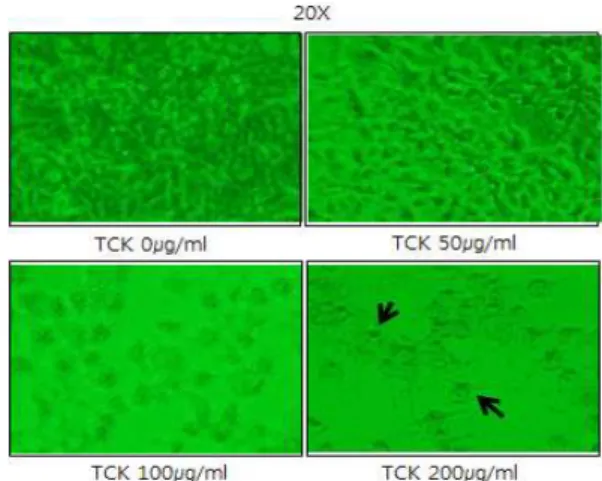
먼저 세포에 천화분을 농도별로 처리한 후 세포 모양에 어떤 영향을 미치는지 살펴보았다. 그 결과 천화분의 농도가 증가할수 록 세포의 밀도도 낮아지며, apoptosis의 전형적인 모양인 apoptotic body를 세포 표면에 형성하는 것을 관찰할 수 있었다 (Fig. 1).
Fig. 1. Effect of TCK on cell morphology in MCF-7 cell.
Cell density of MCF-7 cell decreased dose-dependently after TCK. Also, TCK increased apoptotic bodies(arrows) on MCF-7 cell surfaces. Cells were treated with 50∼200㎍/ml TCK for 48 hours. Cell morphology was observed with microscope.
2. MCF-7 세포 생존율에 미치는 효과
WST-1 assay 결과, MCF-7 세포에 30∼200 ㎍/ml의 천화분 을 처리한 후 48시간 후에 농도 의존적으로 세포생존율이 낮아지 는 것을 확인할 수 있었다(Fig. 2).
Fig. 2. Effect of TCK on cell proliferation in MCF-7 cell.
It shows growth inhibition of MCF-7 cell by TCK. Cells were treated with 30∼200 ㎍/ml TCK for 48 hours, and cell viability was measured by WST-1 assay. The shown data is representative of three independent experiments.(Error bars were shown in mean± standard error means(SEM)). *p<0.05, **p<0.01, ***p<0.001.
3. MCF-7 세포사멸에 미치는 효과
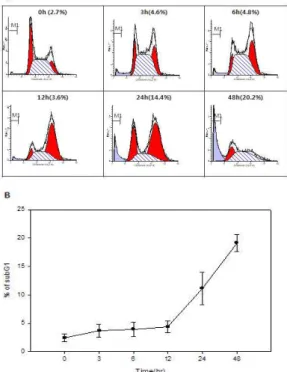
FACS 결과, 천화분(60 ㎍/ml)을 처리한 지 48시간째 약 20%
가량 subG1 값이 높아지는 것을 확인하였으며, 이로써 천화분에 의해 세포사멸이 유도됨을 확인하였다(Fig. 3).
Fig. 3. TCK increases subG1 phase in MCF-7 cell.
SubG1(M1) increased after TCK. MCF-7 cells were exposed to 60 ㎍/ml of TCK for indicated periods of time. DNA content distribution was analyzed using a FACStar flow cytometer and percentage of subG1 phase cells was determined based on a DNA content histogram(A) and graph(B).4. MCF-7 세포주기에 미치는 효과
FACS를 이용하여 MCF-7 세포에 천화분(60 ㎍/ml)을 처리
한 후, cell cycle의 변화를 관찰한 결과, G2/M phase arrest가 일 어나는 것을 확인하였다. G2/M phase arrest는 천화분 처리 12시 간째에 약 60%, 24시간째에 약 50%가량 증가하였다(Fig. 4).
Fig. 4. TCK induces G2/M phase arrest in MCF-7 cell.
TCK(60 ㎍/㎖) treatment significantly increased G2/M phase arrest time-dependently in MCF-7 cell. The percentage of cells in each phase of the cell cycle was obtained by MODFIT software. A and B are graphs of different type.5. Caspase 발현 및 활성에 미치는 효과
Western blot analysis 결과, MCF-7 cell에 천화분(60 ㎍/ml) 처리 후 caspase-8은 변화가 없었으나, caspase-9는 12시간 후부 터 활성이 미미하게 증가하는 것을 확인하였으며, caspase-3의 활 성은 24시간, 48시간 후에 높게 나타나는 것을 확인하였다. 직접 적인 세포사멸의 marker인 PARP의 활성도 확인하였다(Fig. 5).
Fig. 5. Effect of TCK on caspase expression and activation.
After 3∼46 hours of incubation with TCK(60 ㎍/ml), the cells were lysed, and cellular proteins were western blotted. Caspase-9 activity was up-regulated in TCK treatment time 12 hours to 48 hours. Cleaved procaspase-3, subsequently PARP in MCF-7 cell. Significant cleavage of PARP was observed at 12 hours after TCK treatment.
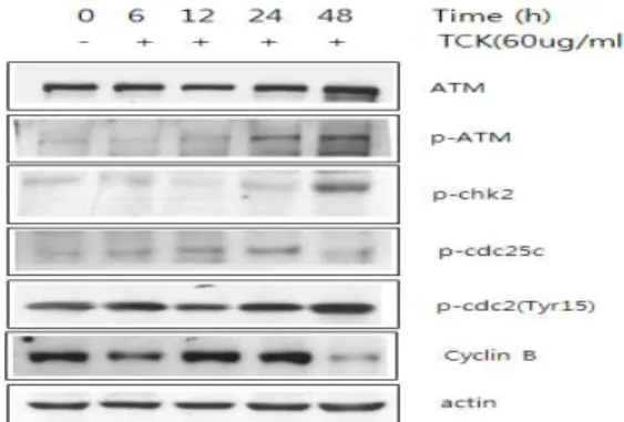
6. G2/M phase 조절단백질에 미치는 효과
Western blot analysis 결과, ATM 인산화 level이 천화분 처 리 후 24∼48시간까지 증가하였으며, ATM에 의해 조절되어지는 그 하위단계에 있는 chk2의 인산화 level 역시 48시간째 확연히 증가하는 것을 확인하였다. Chk2의 하위단계에 있는 cdc25C 역 시 24시간째 미미하게 증가하였고, cdc2(Tyr15)의 인산화 잔기가 천화분 처리 후 48시간째 증가하였으며, 결국 cyclin B의 level을 조절하였다(Fig. 6).
Fig. 6. G2/M phase arrest is dependent on ATM pathways.
After 6∼48 hours of incubation with TCK(60 ㎍/ml), the cells were lysed and cellular proteins were western blotted. TCK induced ATM, chk2, cdc25C phosphorylation in MCF-7 cell for indicated periods. In addition, cdc2 phosphorylation and cyclin B level were regulated by interaction with cdc25C.고 찰
Apoptosis와 세포주기 억제는 무수히 많은 연구에서 항암제 개발에 높은 빈도로 target이 되고 있다. Apoptosis는 조직의 항 상성이나 발생 과정에서 일어나는 것으로 세포가 이겨낼 수 없 는 자극이 가해질 때 개체를 보호하기 위해 진화적으로 생겨난 과정이라고 알려져 있다 14) . Apoptosis 메커니즘의 핵심요소는 caspase라는 proteolytic system으로 15) extrinsic pathway를 통한 caspase-8과 intrinsic pathway와 관련된 caspase-9는 다른 downstream effector caspase인 caspase-3, -6, -7들을 활성화시킨 다. 그 중 caspase-3의 활성은 apoptosis 과정에서 핵심적인 역할 을 하며 세포 내 많은 단백질 poly-(ADP-ribose) polymerase (PARP), gelsolin, retinoblastoma (Rb)와 b-catenin 등을 분해시 킨다 16) .
Ataxia telangiectasia mutated kinase(ATM)은 serine/threon ine kinase이며 세포주기와 DNA의 복구를 조절하는데 이것의 인산화는 두 가닥의 DNA사슬에 손상을 가져 온다 17) . 이 ATM에 의해 조절되어지는 protein들 중 세포주기와 관련된 protein들로 는 p53, Chk2, Chk1, CtIP, 4E-BP1, BRCA1, RPA3, H2AX, SMC1, FANCD2 및 Rad17과 같은 것들이 있다 18) .
Chk2가 포함하고 있는 amino-terminal domain으로는 7개의 serine 또는 threonine잔기(Ser19, Thr26, Ser28, Ser33, Ser35, Ser50 and Thr68)가 있는데, 이것은 ATM/ATR kinase에 의해 쉽 게 인산화 된다 19,20) .
Cdc25는 인산화효소에 반응하는 단백질로
dephosphorylating되거나 cdc2를 activation시킴으로써 진핵세포
의 체세포분열을 조절하는데 중요한 역할을 한다 21) . 또한 이것은 세포주기 간기에서 c-TAK1에 의해 Ser261잔기가 인산화 되며, 이것은 DNA 손상에 의한 G2/M phase arrest와 관련된다 22) .
Cdc2는 이것의 kinase 활성에 의해 진핵세포의 체세포분열 을 조절하는 단백질로 세포주기를 도는데 중요한 protein인 cyclin의 binding을 조절하며, cdc2의 두 가지 잔기인 Tyr14와 Tyr15의 인산화는 cell cycle을 조절하는데 중요한 것으로 나타난 다 23) . 이 두 잔기의 인산화는 Wee1과 Myt1과 같은 protein을 억 제할 수 있다 24,25) .
과루의 뿌리를 봄이나 가을에 채취하여 외피를 벗겨내고 작 게 토막내 건조시킨 것이 천화분으로 이것의 유효성분은 다종의 단백질 혼합성분인 trichosanthin이다. 천화분은 淸熱生津, 淸肺 化痰, 消腫排膿의 효과가 있어 口渴, 消渴, 黃疸, 咳血, 癰腫 등을 치료한다 26) .
천화분을 이용한 항암연구로는 HepA-H cell과 HeLa cell의 MTT assay와 현미경 분석 결과 HeLa cell만 세포증식억제효과가 있다는 연구 10) 와 ME-180 cell에서 caspase-3을 활성화시켜 세포 사멸을 일으킨다는 연구 11) 및 B16/F10 melanoma cell의 tyrosinase activity와 melanin synthesis를 억제한다는 연구 12) , B16-F10 melanoma cell, CT-26 cell, Hep-G2 cell, HT1080 cell, SNU-1 cell에서 HUVEC(혈관신생 내피세포)을 억제하고 Anchorage dependent colony assay를 통해 세포증식이 억제된 다는 연구 13) 가 있다.
본 연구에서는 인체 유방암 세포주인 MCF-7 세포를 이용하 여 세포증식억제 및 기전, 혈관신생억제에 대한 효과를 알아보았 다. 이에 세포증식억제와 관련된 apoptosis의 유발과 경로, 세포 주기의 변화 및 세포주기조절에 관련된 인자들의 단백질 변화 여 부를 조사하였다.
먼저 천화분이 MCF-7 세포에 어떤 영향을 미치는지 알아보 기 위해 WST-1 assay와 microscope 관찰을 하였다. 그 결과 천화 분을 처리한 그룹에서 농도 의존적으로 세포의 증식이 억제되는 것을 WST-1 assay로 확인하였고, 농도가 증가할수록 세포의 부 착력 상실이 늘어나고 apoptotic body가 나타나는 것을 현미경으 로 확인하였는데, 이는 세포질의 응축으로 인해 세포의 부착력을 상실한 것으로, 세포질의 응축변화는 천화분 처리 후 1시간 만에 도 눈으로 확인되었다.
이러한 차이를 수치로 나타내기 위해서 FACS 실험을 통한 PI 염색방법을 이용해 apoptosis 수치를 나타내는 subG1의 양의 변화를 확인해보았다. PI 염색결과 천화분 처리 48시간 후에 subG1이 약 20% 증가하는 것을 확인할 수 있었다. 또한 세포주 기의 변화를 관찰한 결과 G2/M phase arrest가 나타나는 것을 확인할 수 있었으며, 그 정도가 12시간째에 약 60%가량 증가하였 고, 24시간째에는 50%가량을 유지하는 것으로 봐서 시간이 지나 면서 효과가 떨어지는 것을 짐작케 하였다.
FACS 결과를 바탕으로 천화분에 의한 MCF-7 세포의 감소
는 G2/M phase arrest가 apoptosis를 유발하여 감소된다는 사실
을 알 수 있었으며, 이를 확인하기 위해 apoptosis 및 G2/M
phase 관련 인자들을 western blot analysis를 통해 살펴보았다.
Apoptosis pathway는 일단 미토콘드리아를 통한 intrinsic pathway와 death receptor를 통해 caspase-8의 활성을 일으키는 extrinsic pathway가 있다 20) . 이러한 apoptosis pathway가 활성화 되면, 최종적으로 caspase의 최하위 단계이며, 세포사멸 과정에서 핵심적인 역할을 하는 것으로 알려진 caspase-3이 활성화 되고, 세포내에서 PARP나 retinoblastoma (Rb)와 같은 단백질들을 분 해시킨다 21) . 이에 먼저 caspase-3과 PARP의 발현 및 활성 정도를 확인하였는데 천화분에 의해 이 두 단백질의 활성이 증가하는 것 을 관찰할 수 있었다. 그리고 천화분에 의한 apoptosis의 활성이 intrinsic pathway를 통한 것인지 또는 extrinsic pathway를 통한 것인지 알아보기 위해 caspase-8과 caspase-9의 활성을 관찰한 결 과 caspase-9의 활성은 증가한 반면 caspase-8은 변하지 않는 것 으로 보아 천화분이 intrinsic pathway를 통해 세포사멸을 유도하 는 것을 확인하였다.
또한 ATM, chk2 인산화 level을 천화분이 조절한다는 결과 를 바탕으로, DNA damage에 의해 G2/M phase arrest를 유발시 킨다는 사실을 확인할 수 있었으며, 이는 결국 cdc25C 인산화와 cdc2/cyclin B의 interaction에 의해 세포주기를 arrest시킨다고 할 수 있겠다.
이상의 결과로, 천화분을 MCF-7 세포에 처리하였을 경우 세 포의 증식을 강하게 억제하는 것으로 나타났고 세포의 형태도 변 화시켰다. 이러한 세포의 증식억제는 천화분이 DNA damage를 통해 G2/M phase arrest를 유발하여 결국 intrinsic pathway 를 통한 apoptosis를 유도시켜 나타나는 결과였다. 그러나 천화분이 어떤 pathway를 통해 cell cycle arrest를 시키고 세포의 활성을 억제시키는지, 뿐만 아니라 다른 유전학적 특성을 가지고 있는 인체 유방암 세포에서의 효과 및 혈관신생억제를 통한 암전이 억 제에 대한 보충 연구 역시 향후 추가로 연구해 보아야 할 것이다.
결 론
본 실험은 MCF-7 인체 유방암 세포주에서 천화분의 항암효 과 및 기전을 확인하고자 수행하였으며, 세포증식억제 확인을 위 해 WST-1 assay를 시행하였고, FACS 및 western blot analysis를 이용하여 세포사멸, 세포주기변화 및 cell cycle 관련 인자와 caspase의 활성을 확인하였다.
WST-1 assay 결과 천화분을 MCF-7 세포에 48시간 동안 처 리하였을 때 농도 의존적으로 세포증식이 감소하였다.
천화분 처리후 FACS 결과 subG1 부분이 약 20%까지 증가 하는 것을 확인하였으며, G2/M phase arrest가 나타났다.
Western blot analysis를 통해 caspase-9, caspase-3 및 PARP 의 활성화를 확인하였고, ATM에 의존적으로 Chk2 및 cdc25C의 인산화가 일어나는 것을 확인하였으며, 결국 cyclin B가 감소하였 다.
이와 같은 결과로 볼 때, 천화분에 의한 MCF-7 인체 유방암 세포주의 세포증식 억제기전은 DNA damage에 의해 G2/M phase arrest가 일어나고, 결국 intrinsic pathway를 통해 apoptosis를 유도시켜 세포증식이 억제된다는 것을 알 수 있었다.
감사의 글
본 연구는 경원대학교의 지원을 받아 수행되었음에 감사드 립니다(과제번호 2011-R233).
참고문헌