포황 메탄올 추출물에 의한 인체 대장암 세포주 HT29의 G2/M Arrest 및 Apoptosis 유발
진수정1, 윤승근2, 오유나1, 이지영1, 박현진1, 진경숙1, 권현주1,2, 김병우1,2*
1동의대학교블루바이오소재개발센터
2동의대학교자연과학대학생명응용학과
Received: August 8, 2013 / Revised: October 16, 2013 / Accepted: October 21, 2013
서 론
암은현대의학의발달에도불구하고치료에많은어려움 을겪고있는질병으로, 유전적요인뿐아니라식습관등의 생활환경적요인과매우밀접한관계를가진다. 또한인구 의급격한고령화와식습관의서구화등으로인해 10만명당 암발생률은점점증가하고있으며, 서구사회에서많은비율 을차지하는대장암, 유방암등의발생도증가하고있는추 세이다. 특히대장암의경우암의치료법으로주로외과적 절제술, 방사선치료및화학요법등이이용되고있으나, 이
들의부작용및비교적낮은생존율등을고려하여항암활 성을가지는식품또는천연물유래소재의중요성이대두 되고있으며, 이들천연물의세포주기조절에의한암세포
증식억제및 apoptosis 유도등항암활성의분자적기전에
관한활발한연구가진행되고있다[8, 26].
세포가외부자극등에의해 DNA에손상을입게되면, 재 빠르게세포주기를지연시켜 DNA를복구할수있는시간을
벌게된다[37]. 그러나암세포는유전적변이등에의해세
포주기진행의조절기전에서벗어나므로지속적으로분열 하게된다. 세포주기진행은 G1, S 및 G2/M기로나뉘어지 며, 각 단계의 checkpoint에서 cyclin dependent kinase
(CDKs)의활성을억제하며세포주기를조절한다[13]. 특히
cyclinB1과 complex를이루며 G2/M기의 checkpoint로작용 하는 Cdc2(CDK1)는 Wee1 kinase에의해 Tyr-15 잔기에인 Induction of G2/M Arrest and Apoptosis by the Methanol Extract of Typha orientalis in Human Colon Adenocarcinoma HT29 Cells. Jin, Soojung1, Seung-Geun Yun2, You Na Oh1, Ji-Young Lee1, Hyun-jin Park1, Kyong-Suk Jin1, Hyun Ju Kwon1,2, and Byung Woo Kim1,2*. 1Blue-Bio Industry RIC, Dong-Eui University, Busan 614-714, Korea, 2Department of Life Science and Biotechnology, College of Natural Sciences, Dong-Eui University, Busan 614-714, Korea
Typha orientalis, also known as bulrush or cattail, is a perennial herbaceous plant found in freshwater wetlands and has been widely used in constructed wetlands for wastewater treatment. Recent data has revealed that SH21B, a mixture composed of seven herbs including T. orientalis, exhibited an anti-adipogenic activity by the inhibition of the expression of adipogenic regula- tors. However, the anti-cancer effect of T. orientalis and its molecular mechanisms remain unclear. In this study, we evaluated the anti-cancer effect and its mechanism in the methanol extract of T. orientalis (METO) on human colon carcinoma HT29 cells.
It was found that METO treatment showed cytotoxic activity in a dose-dependent manner, and induced G2/M cell cycle arrest and apoptosis in HT29 cells. The induction of G2/M arrest by METO was associated with the up-regulation of phospho-Cdc2 (Tyr15), an inactive form of Cdc2 and the down-regulation of Cdc25c phosphatase. METO also induced tumor suppressor p53 and cyclin-dependent kinase inhibitor p21 (WAF1/CIP1) expression. In addition, METO-induced apoptosis was characterized by the proteolytic activation of caspase-3, degradation of poly ADP ribose polymerase (PARP), and up-regulation of death receptor FAS and pro-apoptotic Bax expression. Collectively, these results indicate that the cell cycle inhibition and apoptosis induction of METO in HT29 cells allows for the possibility of its use in anti-cancer therapies.
Keywords: Typha orientalis, anti-cancer effect, G2/M arrest, apoptosis
*Corresponding author
Tel: +82-51-890-2900, Fax: +82-51-890-2914 E-mail: [email protected]
© 2013, The Korean Society for Microbiology and Biotechnology
산화가되어비활성형태로존재한다[3, 24]. 비활성형태인 phospho-Cdc2(CDK1)는 phosphatase인 Cdc25C에의해탈 인산화되면서 active form으로 전환되고 활성화된 Cdc2- cyclin B1 complex에의해 mitosis 단계로들어간다[7, 18].
또한 Cdc2-cyclin B1 complex의활성은세포증식억제신호 에따라발현이증가되는 Cdk 억제제특히 p21(WAF1/CIP1)
에의해조절되며, p21이 Cdk에결합함으로서그들의활성
이억제된다[9]. 이러한 p21의발현은종양억제유전자인 p53 발현증가에따라유도되는것으로밝혀져있으며, 최근연 구된바에따르면 p21은세포주기조절에의한증식억제뿐 아니라 apoptosis도유도할수있다[31, 36].
Apoptosis는 programmed cell death라고도불리며, apoptosis 과정에이상이생겨실패하게되면암과같은질병이유발 되므로, apoptosis 유도에의한암세포제거는수년간암치 료를위한주요전략중하나로연구되어왔다[20, 25]. 세포 가여러가지요인들에노출되었을때다양한신호전달을통
해세포가죽음에이르는과정을뜻하는 apoptosis는세포
위축, 염색질응축, DNA 단편화및 apoptotic body 형성등 의전형적인형태학적및생화학적인특징을가지며, 세포막 의사멸수용체(death receptor)에리간드가결합하여일어나 는외부적경로및미토콘드리아에서일어나는내부적경로 의두가지주요경로를통하여야기된다[14]. 또한 apoptosis 는 pro-caspase의 분절에 의한 활성화, poly ADP-ribose polymerase (PARP)의단편화, 세포내 pro-apoptotic Bcl- 2 family의활성화및종양억제유전자 p53의발현증가등 에의하여조절되며, 이러한다양한 apoptosis 조절인자들 은항암제및암예방을위한주요표적인자들로연구되어왔 다[1, 5, 27]. 최근에는다양한범위의식물추출물또는식 물로부터분리한성분물질들의세포주기조절및 apoptosis 유도효과에의한항암활성이활발하게연구되고있다[2, 6, 15, 35].
포황(Typha orientalis)은부들과(Typhaceae)에속하는부 들화분으로연못가장자리나습지에서자생하며예로부터 지혈, 어혈제거, 이뇨제등으로주로사용되어왔으며, 혈압 강하작용, 항균작용등이있는것으로알려져있다[21]. 또한 본연구실에서는이미포황메탄올추출물의항산화및항
염증활성에관하여보고한바있다[17]. 그외에포황열수
추출물을 식이한 랫트의 혈청에서 혈중 cholesterol 및
glucose 함량을낮추는효과가있었다는연구결과가있어,
포황의고지혈증및당뇨병치료효과에대한가능성을시
사하고있으며[21], 포황을포함한 7가지약재성분으로구
성된 SH21B가항비만효능을가진다는연구결과가있으나
[22], 포황의항암활성에대해서는연구된바가극히드물다.
이에본연구에서는포황메탄올추출물의항암활성을확
인하기위하여인간대장암세포주인 HT29 세포를사용하
여세포독성효과, 세포의형태변화, 세포주기변화, 세포주
기및 apoptosis 관련단백질의발현변화에대하여조사하
였다.
재료 및 방법
시료준비
본실험에사용된한약재인포황은중국산이며부산광역 시소재대한생약제품㈜에서구입하여사용하였다. 메탄올 추출물(Methanol extract of Typha orientalis, METO)을 얻기위하여포황 10 g을분말로파쇄하여시료의 5-10배의 메탄올용매를가한후 75oC에서 3회반복추출하였다. 추 출한 시료는 Whatman No.2 (Whatman international Ltd., England)에여과후감압농축기로농축하여 1.59 g을얻었 으며, 동결건조(FDU-2100, EYELA, Japan) 하여 500 mg/
ml의농도로 dimethylsulfoxide (DMSO, Sigma)에용해하 여처리전배지에희석하여사용하였다.
세포배양
실험에 사용된 인체 대장암세포 HT29, 인체 폐암세포 A549, 인체 간암세포 HepG2는 American Type Culture Collection (ATCC, USA)으로부터구입하여사용하였으며, 10% fetal bovine serum (FBS) 및 penicillin/streptomycin 이 포함된 Dulbecco’s modified Eagle’s medium (DMEM) 을사용하여 37oC, 5% CO2조건하에서배양하였다.
WST assay에 의한 세포 성장억제 조사
METO가암세포의성장억제에어떠한영향을미치는지 를확인하기위하여 WST (water soluble tetrazolium salt) assay를수행하였다. 먼저 24-well plate에 3-5×10⁴ cell의 HT29, A549 및 HepG2 세포를분주하여 METO를농도별 로 처리하고 48시간 동안 배양한 후 WST 시약(Daeillab,
Korea)이포함된배지로교체하였다. 30분간반응시킨후
microplate reader (Molecular Devices, Sunnyvale, CA, USA)로 450 nm에서흡광도를측정하였다. 측정값은 3회반 복실험을하여그에대한평균값으로나타내었으며, 본실 험결과를바탕으로이후적정처리농도를결정하였다.
세포 형태 변화 관찰
METO 처리에따른 HT29 세포의형태변화관찰을위하
여 6-well plate에 1×10⁵ cell의 HT29 세포를 분주하고 24시간동안안정화시킨다음, 적정농도의 METO를처리 하여 48시간동안배양하였다. 도립현미경을이용하여 100 배의배율로관찰한다음 Axio Vision program을사용하여 사진촬영을하였다.
Flow cytometry에 의한 세포주기 분석
METO가 HT29 세포의세포주기에어떤영향을주는지
확인하기위하여 HT29 세포에 METO를농도별로처리한
다음 CycleTESTTM PLUS DNA Reagent Kit (Becton Dickinson, San Jose, CA, USA)를사용하여세포주기분석 을하였다. 6-well plate에 HT29 세포를 5×10⁵ cell의농도
로분주하여 METO를농도별로 24시간처리한다음세포
를모아서 PBS로세척한 후, ribonuclease A를실온에서 10분간처리한다음 propidium iodide (PI) 용액을첨가하여 4oC에서 10분간염색하였다. 염색된세포는 Flow cytometry (Cell Lab Quanta SC, Beckman Coulter, Fullerton, CA, USA) 로측정하였으며결과는 FlowJo program을이용하여 분석하였다.
Western blot analysis에 의한 단백질 발현 분석
METO를처리한 HT29 세포의세포주기관련단백질및
apoptosis 관련단백질들의 발현 양상을확인하기위하여
Western blot analysis를수행하였다. 농도 별로 METO를 처리한 HT29 세포를모아 PBS로세척한 후, lysis buffer [20 mM Tris-HCl (pH 7.5), 150 mM NaCl, 1 mM EDTA, 1 mM EGTA, 1% Triton X-100, 1µg/ml leupeptin, 1 mM PMSF]를첨가하여 4oC에서 20분간반응시킨다음 13,000 rpm으로 30분간원심분리하여상층액을취하였다. 추출한
단백질의농도는 Bradford법을이용하여정량한다음동량
의단백질을 sodium dodecyl sulphate (SDS)-polyacrylamide gel을 이용하여 전기영동으로 분리하고 nitrocellulose membrane에 transfer하였다. 5% skim milk가함유된 PBS-T 를사용하여상온에서 1시간동안 blocking을하고, 각각의 일차항체를 4oC에서 overnight 처리한다음, horse radish peroxidase (HRP)가부착된이차항체를상온에서 1시간반 응시켰다. 반응이끝난후 Western blotting luminol reagent (Santa Cruz Biotechnology, CA, USA)를 적용시킨 다음 chemiluminescence detection system (FluoChem® FC2, AlphaInnotech, CA, USA)을 사용하여 분석하였다. p53, Cyclin A, Cyclin B1, Cdc2, phospho-Cdc2 (Tyr15), Cdc25C, Wee1, FAS, FADD, Caspase-3, PARP, Cytochrome C 및 actin의일차항체와 HRP-conjugated anti-rabbit, anti-goat, anti-mouse 등 이차항체는 Santa Cruz Biotechnology (CA, USA)에서구입하였으며, p21, Bcl-2, Bax의일차항체는 Cell Signaling Technology (MA, USA)에서구입하여사용하였다.
결과 및 고찰
METO에 의한 암세포의 증식 억제
먼저다양한암세포의증식에미치는 METO의영향을조
사하기위하여인체대장암세포 HT29, 인체폐암세포 A549 및인체간암세포 HepG2에 METO를다양한농도로처리하 Fig. 1. Effects of METO on cell growth in various cancer cell lines.
Human colon carcinoma HT29 cells (A), human lung carcinoma A549 cells (B) and human hepatocellular carcinoma HepG2 cells (C) were treated with indicated concentration of METO for 48 h.
Cytotoxic effect of METO was determined by WST assay. Results are expressed as percentage of the control ± SD of three inde- pendent experiments.
여 WST assay를사용하여세포독성을확인하였다. 그결 과 Fig. 1에서와같이, METO 농도의존적으로세포증식이 억제되었으며, 특히 HT29 세포의경우최고농도인 400 µg/
ml METO를처리하였을때세포증식이약 54% 억제되어
세포증식이 40% 억제된 A549와 22%가억제된 HepG2 세
포보다 METO에대한감수성이높은것을확인하였다. 따
라서이후의실험은 HT29 세포로수행하였다.
HT29 세포의 형태에 미치는 METO의 영향
METO 처리에의한증식억제효과에따른 HT29의형태
적변화를관찰하기위하여 METO를다양한농도로 48시간
동안처리하여도립현미경으로세포형태를관찰하였다. 그
결과 Fig. 2에서와같이 METO 농도가증가할수록전체적
인세포밀도가감소하였으며, METO 400 µg/ml의농도에 서는세포의모양이불규칙하고바닥에고착되어있는세포 의수가감소하였다.
METO에 의한 HT29의 세포주기 변화
정상적인세포는 DNA에손상을입었을때세포주기를조 절하여손상된 DNA의회복경로를거치게된다[28]. 그러 나암세포에서는정상적인세포주기조절기능이제어되어
G1/S 또는 G2/M기의비정상적인진행에의한무한증식이
Fig. 2. Morphological changes by METO in HT29 cells.
Cells were treated with various concentrations of METO for 48 h.
Cell morphology was visualized by light microscopy. Scale bars, 200 µm.
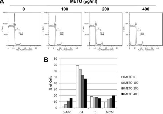
Fig. 3. Induction of G2/M arrest and accumulation of subG1 phase in METO-treated HT29 cells.
Cells were treated with indicated concentrations of METO for 24 h, stained with propidium iodide for 10 min and analyzed by flow cytometry.
DNA-fluorescence histogram (A) and quantitative data of cell distribution (B) are shown.
일어난다[12]. 최근에는이러한암세포의세포주기조절에 의한암세포사멸을유도하는항암제의개발이활발하게이
루어지고있다[32]. 따라서 본 연구에서는 METO에의한
HT29 세포의증식억제현상이세포주기조절에의한것인
지확인하기위하여적정농도의 METO를처리한 HT29에 PI 염색을한후 flow cytometry 분석을통해세포주기의변화 를확인하였다. 그결과, Fig. 3A의 histogram 결과및 Fig.
3B의그래프에서보여지듯이 METO의처리농도가높아질 수록 G2/M기및 SubG1기세포군의분포(%)가점차증가하 였으며반면, G1기및 S기의세포군의분포는 METO 농도 의존적으로감소하였다. 이러한 METO 처리에따른각주 기별세포분포수는정량화하여 Table 1에나타내었다. Table 1에서알수있듯이대조군에서 9.22%였던 G2/M기세포분 포는 METO 농도의존적으로증가하여최고농도인 400 µg/
ml에서는 20%의세포가 G2/M기에정체되어있음을확인하 였다. 또한 apoptosis 유발군으로추정할수있는 SubG1기 의세포분포는대조군에서 2.66%였으며 METO 400 µg/ml 에서는 16%로증가되었다. 반면, G1기와 S기의세포분포는 각각 68.7%에서 47.3%, 18.4%에서 15.3%로감소하였다. 따 라서이와같은결과는 METO에의해 HT29 세포의 G2/M arrest 및 apoptosis가유도됨을시사한다.
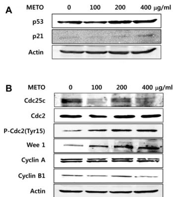
G2/M기 조절 관련 단백질의 발현 변화
세포주기분석결과 G2/M arrest 유발이관찰되었으므
로, 그분자적기전분석을위하여 G2/M기조절관련단백
질들의발현에미치는 METO의영향에관하여조사하였다.
종양억제유전자로널리알려진 p53은정상세포가손상을 입었을때발현증가또는전사후변형(post-transcriptional modification) 등에의해활성화되고, 활성화된 p53은세포 주기억제 단백질인 p21을비롯하여 FAS, Bax 등다양한 apoptosis 관련단백질의발현을촉진하고 caspase의활성 화를유도하여세포주기의진행을억제시키거나 apoptosis 를 유발한다[29, 33]. 따라서 METO를 농도별로 처리한 HT29의 p53 및 p21의발현변화를분석한결과, Fig. 4A와 같이 p53과 p21의발현이 METO 농도의존적으로증가하는
것을확인하였다. 다음으로 G2/M기조절에관여하는 cyclin 의발현을확인한결과, cyclin A의발현은큰변화가관찰 되지않았으나 cyclin B1은 METO 농도가증가할수록그발 현량이감소하였다(Fig. 4B). Cdk의경우, Cdc2 (Cdk1)의발 현에는 큰 변화가 없었으나 Cdc2의 inactive form인 phospho-Cdc2 (Tyr15)의발현량이 METO 농도가증가함에 따라유의적으로증가함을알수있었다. 또한 Cdc2의인산 화를유도하여 Cdc2를 inactivation 시키는 Wee1 kinase의
발현은 METO 농도가 높아질수록 증가하였으며, 반면
phospho-Cdc2의탈인산화를일으켜 Cdc2를 activation시키 는 phosphatase인 Cdc25C의발현량은감소하였다. 이상의
결과는 METO에 의해 Wee1 kinase의 발현이 증가되고
Cdc25C phosphatase의발현이감소되어 Cdc2의인산화가 유도되고, 따라서 Cdc2의 inactive form인 phospho-Cdc2가
증가하게 됨을 시사한다[4, 18, 19]. 결과적으로 증가된
inactive form의 Cdc2는 M 기로의진행을불가능하게하여 G2/M arrest가유도된다고사료된다[7].
Table 1. Cell cycle distribution of Typha orientalis extract- treated HT29 cells.
METO (µg/ml)
% of cell
SubG1 G1 S G2/M
0 02.66 68.7 18.4 09.22
100 05.69 62.7 16.4 14.2
200 11.4 53.3 17.4 15.9
400 16 47.3 15.3 20
Fig. 4. Modulation of the expression of tumor suppressor p53 and G2/M checkpoint proteins in METO-treated HT29 cells.
Cells were exposed to various concentrations of METO for 24 h.
The cells were lysed and proteins were then separated by SDS- PAGE, followed by Western blot analysis. Actin was used as an internal control. (A) Induction of tumor suppressor p53 and CDK inhibitor p21 by METO treatment. (B) Down-regulation of Cdc25C phosphatase and Cyclin B1, and up-regulation of p-Cdc2 and Wee1 kinase.
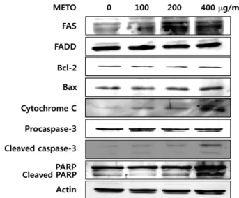
세포사멸 관련 단백질의 발현 변화
이상의결과에서 HT29에 METO를처리했을때농도의존
적으로 apoptosis 유발군인 SubG1기세포분포가증가되었 으며(Fig. 3), METO 처리에의해 p53 단백질의발현이증가 되는것을확인하였으므로(Fig. 4A), 다음으로 apoptosis에 관련된단백질의발현변화를 Western blot analysis로확인 하였다. 세포내에서 apoptosis 관련단백질은 death receptor family, Bcl-2 family, caspase, PARP등이있으며특히 Bcl- 2 family는 anti-apoptotic (Bcl-2, Bcl-xL) 단백질과 pro- apoptotic (Bax, Bak, Bid) 단백질로구성되어있어세포내 균형을이루고있다. 그러나세포내자극에의해균형이깨 어지고 pro-apoptotic 단백질의발현이증가될경우, 미토콘 드리아막전위에변화를유도하여미토콘드리아내막에서 cytochrome C가방출된다[10, 11]. 이러한 apoptosis 유발
인자들에 의해 caspase cascade가 활성화 되고 활성화된
effector caspase-3는여러종류의기질단백질들을분해하 여 apoptosis를일으키게된다. 여러기질단백질중 PARP
는세포의핵내에존재하는효소로서 DNA 회복, 유전자전
사, 세포주기진행등에작용하며활성화된 caspase-3에의 해분해되어 apoptosis 유발에관여하는것으로알려져있다 [30]. METO를처리한 HT29에서이러한 apoptosis 관련단 백질들의발현변화를확인한결과, Fig. 5에서와같이 death receptor인 FAS와 pro-apoptotic 단백질인 Bax의 발현이 METO 농도의존적으로증가하였다. 반면 death receptor인
Fas에결합하는 Fas associated death domain (FADD) 및 anti-apoptotic 단백질인 Bcl-2의발현량은큰변화가관찰되 지않았다. 또한 cytosolic cytochrome C의양은 METO 농 도의존적으로증가하였으며, cleaved caspase-3 및 cleaved
PARP 또한 METO의농도가높아질수록증가하였다. 이와
같은 결과를 통해 METO가 HT29 세포의 p53 발현 및 death receptor인 FAS 발현을 유도시키고 Bcl-2 family의 변화를유도하여 caspase-3 경로를통하여 apoptosis를유도 하는것을알수있었다. 따라서이상의결과들로부터 METO 가인체대장암세포 HT29의 G2/M arrest 유도에의해세포 주기를조절하여세포증식을억제하고, apoptosis에의한세 포사멸을유도한다는것을확인하였다.
최근까지의연구결과들로부터항암활성을보유하고있다 고알려진다양한약재및천연소재들의항암활성기전이밝 혀지고있으며, 특히세포주기조절및 apoptosis 유도에의 한항암활성을보유하는소재들에관한연구는매우활발히
진행되고있다[4, 23]. 최근에보고된연구결과에따르면계
혈등(Spatholobus suberectus)의열수추출물에의해인체유 방암세포주 MCF-7과대장암세포주 HT29의 G2/M arrest 및 apoptosis가유도되고경구투여에의해마우스 xenograft 모델의종양부피가감소되었으며, 땅꽈리(Physalis angulata) 의메탄올추출물또한유방암세포주에서 G2/M arrest를유 도하였다[16, 34]. 이들 추출물들은 Cdc2의 인산화 증가,
Cdc25C 발현감소및 Wee1 발현증가등본연구와유사한
결과를보였으며, 특히계혈등추출물의경우 Bcl-2의발현
감소와 Bax 및 cytosolic cytochrome C의발현증가등에따 른 apoptosis를유도하여본연구결과와유사하였으나, p53 의발현증가는관찰되지않았다[16, 34]. 이러한 CDK/cyclin complex의활성을조절하는 Cdc25 분자및 Wee1에대한연 구결과는많이보고되고있으며, 이들분자는항암제의 target 분자로작용할수있어신약개발을위한중요한분자중하 나이다[7, 19]. 특히이러한세포주기조절분자를 target으 로하는항암제는기존의다른암치료제, 즉 alkylating agent, pyrimidine-purine antimetabolites, DNA topoisomerase 등에 비해정상세포에대한부작용을줄일수있다는점에서관
심이 집중되고 있다[19]. 따라서포황 추출물이 암세포의
Cdc25C 및 Wee1의발현을조절하여 G2/M arrest를유도하
고나아가 apoptosis를유발함을확인한본연구결과는포황
의또다른생리활성인항암작용의기전을해석하였을뿐 아니라, 나아가새로운천연물유래항암제후보물질을연 구하는데에있어귀중한자료로사용될것이라사료된다. 또한이러한기전연구를바탕으로실질적인대장암치료제
로서의사용가능성을확인하기위해서는마우스 xenograft
모델을사용한 in vivo 실험등, 더욱다양한추가실험이수 행되어야할것이다.
Fig. 5. Effects of METO on the levels of apoptosis-related pro- tein expression in HT29 cells.
Cells were harvested and lysed after incubation with the indicated doses of METO for 24 h. Proteins were subjected to Western blot analysis using antibodies against FAS, FADD, Bcl-2, Bax, Cyto- chrome C, Caspase-3 and PARP. Actin was used as an internal control.
요 약
본연구에서는인체대장암세포인 HT29를사용하여포황 메탄올추출물(Methanol extract of Typha orientalis, METO) 의항암활성및그분자적기전에관하여분석하였다. 먼저
METO가다양한암세포의증식에미치는영향을분석한결
과, 인체대장암세포, 폐암세포, 간암세포등의세포증식 을억제하였으며그중에서도대장암세포인 HT29에대해 강한세포증식억제효과를나타내었다. METO에의한세
포 증식 억제 기전을 분석하기 위하여 Flow cytometry
analysis를수행한결과, METO 농도의존적으로 HT29 세포 의 G2/M기세포분포가증가하고아울러 apoptosis 유발군
인 SubG1기세포분포가증가하는것을확인할수있었다.
METO에의한 HT29 세포의 G2/M arrest는 Cdc2의 inactive form인 phospho-Cdc2의증가에의한 G2/M checkpoint 관 련 단백질의 활성억제에 의한 것이라 사료된다. 이러한 phospho-Cdc2의증가는 METO에의해발현이증가된 Wee1 kinase와발현이감소된 Cdc25C phosphatase에의해야기 된 것임을 확인하였다. 또한 METO에 의한 HT29의 apoptosis 유도에 관한분자적 기전분석을위해 Western blot analysis를수행한결과, METO 농도가증가할수록종 양억제유전자인 p53, death receptor인 FAS, Bcl-2 family 중 pro-apoptotic 단백질인 Bax 및 cytosolic cytochrome C
의 발현이 증가되고, Caspase-3가 활성화되어 단편화된
Caspase-3의증가가관찰되었다. 또한활성화된 Caspase-3 의기질단백질인 PARP의단편화가일어나 apoptosis가유 도되는것을알수있었다. 이상의결과들로부터 METO는 인체대장암세포 HT29의 G2/M arrest 및 apoptosis 유도에 의한항암활성을보유함을확인하였다.
Acknowledgments
This research was supported by the Dong-Eui University Grant (2013AA082).
References
1. Ashkenazi A, Dixit VM. 1999. Apoptosis control by death and decoy receptors. Curr. Opin. Cell Biol. 11: 255-260.
2. Bishayee K, Ghosh S, Mukherjee A, Sadhukhan R, Mondal J, Khuda-Bukhsh AR. 2013. Quercetin induces cytochrome-c release and ROS accumulation to promote apoptosis and arrest the cell cycle in G2/M in cervical carcinoma: signal cas- cade and drug-DNA interaction. Cell Prolif. 46: 153-163.
3. Booher RN, Holman PS, Fattaey A. 1997. Human Myt1 is a cell cycle regulated kinase that inhibits Cdc2 but not Cdk2 activity. J. Biol. Chem. 272: 22300-22306.
4. Chang KL, Kung ML, Chow NH, Su SJ. 2004. Genistein arrests hepatoma cells at G2/M phase: involvement of ATM activation and upregulation of p21waf1/cip1 and Wee1. Biochem.
Pharmacol. 67: 717-726.
5. Cory S, Adams JM. 2002. The Bcl2 family: regulators of the cellular life or death switch. Nat. Rev. Cancer 2: 647-656.
6. Cui JC, Cui Y, Huang MH, Kim HJ, Jin CY, Park C, 2010.
Apoptosis induction of human leukemia U937 cells by water extract of wild Ganoderma lucidum spores. Caner Prev. Res.
15: 347-357.
7. Donzelli M, Draetta GF. 2003. Regulating mammalian check- points through Cdc25 inactivation. EMBO Reports 4: 671-677.
8. Dorai T, Aggarwal BB. 2004. Role of chemopreventive agents in cancer therapy. Cancer Lett. 215: 129-140.
9. Elledge SJ, Harper JW. 1994. Cdk inhibitors: on the threshold of checkpoints and development. Curr. Opin. Cell Biol. 6: 847- 852.
10. Fulda S, Debatin KM. 2006. Extrinsic versus intrinsic apopto- sis pathways in anticancer chemotherapy. Oncogene 25:
4798-4811.
11. Gupta S. 2003. Molecular signaling in death receptor and mitochondrial pathways of apoptosis. Int. J. Oncol. 22: 15-20.
12. Hartwell LH, Kastan MB. 1994. Cell cycle control and cancer.
Science 266: 1821-1828.
13. Hartwell LH, Weinert TA. 1989. Checkpoints: controls that ensure the order of cell cycle events. Science 246: 629-633.
14. Hengartner MO. 2000. The biochemistry of apoptosis. Nature 407: 770-776.
15. Hong SH, Choi YH. 2012. Bufalin induces apoptosis through activation of both the intrinsic and extrinsic pathways in human bladder cancer cells. Oncol. Rep. 27: 114-120.
16. Hsieh WT, Huang KY, Lin HY, Chung JG. 2006. Physalis angu- late induced G2/M phase arrest in human breast cancer cells.
Food Chem. Toxicol. 44: 974-983.
17. Jin KS, Oh YN, Lee JY, Son BY, Choi WB, Lee EW, et al.
2013. Anti-oxidative and anti-inflammatory activities of seven medicinal herbs including Tetrapanax papyriferus and Piper longum Linne. Korean J. Microbiol. Biotechnol. 41: 253-262.
18. Jin P, Gu Y, Morgan DO. 1996. Role of inhibitory CDC2 phos- phorylation in radiation-induced G2 arrest in human cells. J.
Cell Biol. 134: 963-970.
19. Kawabe T. 2004. G2 checkpoint abrogators as an anticancer drugs. Mol. Cancer Ther. 3: 513-519.
20. Khan N, Adhami VM, Mukhtar H. 2008. Apoptosis by dietary agents for prevention and treatment of cancer. Biochem.
Pharmacol. 76: 1333-1339.
21. Lee BC, Park HM, Sim HS, Kim GS, Gu JH, Oh MJ. 2009.
Biological activity and chemical analysis of Cattail Pollens. J.
Agri. Sci. 36: 185-197.
22. Lee H, Kang R, Yoon Y. 2010. SH21B, an anti-obesity herbal composition, inhibits fat accumulation in 3T3-L1 adipocytes and high fat diet-induced obese mice through the modulation of the adipogenesis pathway. J. Ethnopharmacol. 127: 709-
717.
23. Lee SM, Kwon JI, Choi YH, Eom HS, Chi GY. 2008. Induction of G2/M arrest and apoptosis by water extract of Stychni semen in human gastric carcinoma AGS cells. Phytother.
Res. 22: 752-758.
24. McGowan CH, Russell P. 1993. Human Wee1 kinase inhibits cell division by phosphorylating p34cdc2 exclusively on Tyr 15. EMBO J. 12: 75-85.
25. Meiler J, Schuler M. 2006. Therapeutic targeting of apoptotic pathways in cancer. Curr. Drug. Targets 7: 1361-1369.
26. Neergheen VS, Bahorun T, Taylor EW, Jen LS, Aruoma OI.
2010. Targeting specific cell signaling transduction pathways by dietary and medicinal phytochemicals in cancer chemopre- vention. Toxicology 278: 229-241.
27. Nicholson DW, Thornberry NA. 1997. Caspase: killer pro- teases. Trends Biochem. Sci. 22: 299-306.
28. O’Connor PM. 1997. Mammalian G1 and G2 phase check- points. Cancer Surv. 29: 151-182.
29. Ryan KM, Phillips AC, Vousden KH. 2001. Regulation and function of the p53 tumor suppressor protein. Curr. Opin. Cell Biol. 13: 332-337.
30. Schreiber V, Dantzer F, Ame JC, de Murcia G. 2006. Poly
(ADP-ribose): novel functions for an old molecule. Nat. Rev.
Mol. Cell Biol. 7: 517-528.
31. Taylor WR, Stark GR. 2001. Regulation of the G2/M transition by p53. Oncogene 20: 1803-1815.
32. Vermeulen K, Van Bockstaele DR, Berneman ZN. 2003. The cell cycle: a review of regulation, deregulation and therapeutic targets in cancer. Cell Prolif. 36: 131-149.
33. Vogelstein B, Lane D, Levine AJ. 2000. Surfing the p53 net- work. Nature 408: 307-310.
34. Wang ZY, Wang DM, Loo TY, Cheng Y, Chen LL, Shen JG, et al. 2011. Spatholobus suberectus inhibits cancer cell growth by inducing apoptosis and arresting cell cycle at G2/M check- point. J. Ethnopharmacol. 133: 751-758.
35. Xin H, Kong Y, Wang Y, Zhou Y, Hu Y, Li D, et al. 2013. Lign- ans extracted from Vitex negundo possess cytotoxic activity by G2/M phase cell cycle arrest and apoptosis induction. Phy- tomedicine 20: 640-647.
36. Xiong Y, Hannon G, Zhang H, Casso D, Kobayashi R, Beach D. 1993. p21 is a universal inhibitor of cyclin kinases. Nature 366: 701-704.
37. Zhou BB, Elledge SJ. 2000. The DNA damage response: put- ting checkpoints in perspective. Nature 408: 433-439.