Induction of G2/M Cell Cycle Arrest by Glutamine Deprivation in Human Prostate Carcinoma PC3 Cells
Dong Yeok Shin1, Sung Hyun Choi 2, Dong Il Park3 and Yung Hyun Choi4,5*
1Dongnam Institute of Radiological & Medicine Sciences, Busan 619-953, Korea
2Department of System Management, Korea Lift College, Geochang 670-802, Korea
Departments of 3Internal Medicine and4Biochemistry, Dongeui University College of Oriental Medicine, Busan 614-052, and 5Anti-Aging Research Center & Blue-Bio Industry RIC, Dongeui University, Busan 614-714, Korea
Received April 28, 2013 /Revised June 24, 2013 /Accepted June 26, 2013
In this study, it was investigated the possible mechanisms by which glutamine deprivation exerts its anti-proliferative action in cultured human prostate carcinoma PC3 cells. Glutamine deprivation re- sulted in inhibition of growth and G2/M arrest of the cell cycle in a time-dependent manner without apoptosis induction, as determined by MTT assay, DAPI staining and flow cytometry analyses. The induction of G2/M arrest by glutamine deprivation was associated with the inhibition of expression of Cdc2, cyclin A and cyclin B1, and up-regulation of the expression of cyclin-dependent kinase (Cdk) inhibitor p21(WAF1/CIP1) in both transcriptional and translational levels. Moreover, glutamine depri- vation increased the phosphorylation of checkpoint kinase (Chk)1 and Chk2; however, the levels of Cdc25C phosphorylation were decreased in response to glutamine deprivation in a time-dependent manner. Our data provide a first biochemical evidence that glutamine deprivation suppresses cell via- bility through G2/M phase arrest without induction of apoptosis in PC3 cells.
Key words : Glutamine deprivation, PC3 cells, cell cycle arrest
*Corresponding author
*Tel:+82-51-850-7413, Fax:+82-51-853-4036
*E-mail : [email protected]
This is an Open-Access article distributed under the terms of the Creative Commons Attribution Non-Commercial License (http://creativecommons.org/licenses/by-nc/3.0) which permits unrestricted non-commercial use, distribution, and reproduction in any medium, provided the original work is properly cited.
Journal of Life Science 2013 Vol. 23. No. 6. 832~837 DOI : http://dx.doi.org/10.5352/JLS.2013.23.6.832
서 론
체내의 비필수아미노산으로 가장 풍부하게 존재하는 글루 타민(L-glutamine)은 질소의 교환과 pH 항상성 유지에 중요 한 역할을 하며, 그들의 생체 내 함량은 단백질이나 글리코겐 의 turnover와는 별개로 독립적으로 조절되는 것으로 알려져 있다[5, 18, 22]. 최근 많은 암환자의 혈액 내에서 글루타민의 수준이 급격히 저하되어 있음이 확인되면서 글루타민 함량 조절이 암환자의 치료 보조요법으로 사용할 수 있는 가능성이 제시되었다. 이는 글루타민이 미토콘드리아 tricarboxylic acid 회로의 기능 회복에 결정적인 영향을 미칠 것으로 생각되어지 기 때문인데[2, 6, 30], 예를 들어, 최근 암화학요법이나 방사선 에 의해 상처를 받은 위장관의 치료에 글루타민 결핍을 적용 하기도 하며 글루타민의 결핍에 의하여 다양한 고형암의 증식 억제효과가 확인된 바도 있다[29]. 또한 글루타민 결핍에 노출 된 암세포는 apoptosis 유발을 통하여 증식이 억제되었으며 [20], 반대로 글루타민의 보충은 산화적 스트레스와 열충격에
의한 위장세포의 apoptosis를 방지하였다[3, 31]. 비록 최근 연 구에 의하면 혈청 내 글루타민 수준의 조절로 암세포의 이동 성과 침윤의 억제[11, 21] 및 글루타민 결핍에 따른 암세포의 apoptosis 유발[9, 13, 14, 23, 27, 32]에 대한 보고가 있어 왔으 나, 세포주기 조절에 관한 연구는 거의 이루어지고 있지 않고 있는 실정이다. 따라서 본 연구에서는 글루타민의 결핍과 과 잉 공급에 따른 암세포의 반응에 관한 정확하고 체계적인 연 구의 진행을 위하여, 인체 전립선암세포를 대상으로 글루타민 결핍 조건에서 나타나는 암세포의 증식 억제에서 세포주기 진행의 변화 여부를 조사하였다.
재료 및 방법 세포배양 및 MTT assay
본 연구에 사용된 PC3 전립선암세포는 American Type Culture Collection (Rockville, MD, USA)에서 분주 받았으며, 10% fetal bovine serum (FBS)에 1%의 penicillin 및 strepto- mycin이 포함된 RPMI-1640 배지(Gibco BRL, Grand Island, NY, USA)를 이용하여 37℃, 5% CO2조건에서 배양하였다.
글루타민 결핍에 따른 증식 억제 여부를 조사하기 위하여 세 포 배양용 6 well plate에 PC3 세포를 1×10⁵개/ml로 분주하고 24시간 동안 안정화시킨 다음 배지를 제거한 후, 글루타민이 결핍된 90%의 RPMI-1640 (Gibco BRL) 배지에 10% FBS, 1%의 penicillin 및 streptomycin이 포함된 성장배지를 사용하여 각 - Note -
well에 분주하였으며 배양하였다. 적정 시간 후 배지를 제거하 고 tetrazolium bromide salt (MTT, Amresco, Solon, Ohio, USA)를 0.5 mg/ml 농도가 되게 배지로 희석하여 3시간 동안 CO2incubator에서 배양시킨 다음 MTT 시약을 깨끗하게 제거 하고 dimethylsulfoxide (DMSO, Amresco)를 이용하여 생성 된 formazin을 모두 녹인 후 96 well plate에 옮겨서 ELISA reader (Molecular Devices, Sunnyvale, CA, USA)로 540 nm 에서 흡광도를 측정하였다.
DAPI staining 및 flow cytometry 분석
글루타민 결핍에 의한 PC3 세포의 증식억제가 apoptosis 유도와 연관성이 있는지의 여부를 조사하기 위하여 4',6-dia- midino-2-phenylindole (DAPI) 염색을 이용한 핵의 형태적 변 화를 관찰하였다. 이를 위하여 동일 조건에서 배양된 PC3 세 포를 37% formaldehyde 용액과 PBS를 1 : 9의 비율로 섞은 fixing solution을 이용하여 상온에서 10분 동안 고정하였다.
이들 세포를 PBS로 2~3회 washing하고 0.2%의 triton X-100 (Amresco)을 첨가하여 상온에서 10분간 고정한 후, DAPI (Sigma-Aldrich Chemical Co., St. Louis, MO, USA) 용액으로 염색시켰다. 15분 정도 염색시킨 후, mounting solution을 처 리한 후 형광 현미경(Carl Zeiss, Germany)을 이용하여 400배 의 배율로 핵의 형태 변화를 관찰하였다. 아울러 세포주기 진 행에 미치는 영향의 분석을 위해서는 글루타민이 결핍된 배지 에서 적정시간 동안 배양시킨 PC3 세포들을 CycleTEST PLUS DNA REAGENT Kit (Becton Dickinson, San Jose, CA, USA) 를 이용하여 고정 및 염색을 하여 4℃, 암실에서 30분 동안 반응을 시킨 다음 DNA flow cytometry (Becton Dickinson)에 적용시켜 형광반응에 따른 histogram을 분석하였다.
RT-PCR 및 Western blot analysis
Reverse transcription-polymerase chain reaction (RT- PCR)을 위하여 준비된 PC3 세포들의 RNA를 TRIzol reagent (Invitrogen Co., Carlsbad, CA)를 이용하여 분리하였다. 분리 된 RNA를 정량한 후, 각각의 primer, DEPC water 그리고 ONE-STEP RT-PCR PreMix Kit (Intron, Korea)를 넣고 Mastercycler gradient (Eppendorf, Hamburg, Germany)를 이용하여 증폭하였다. 각 PCR 산물들의 양적 차이를 확인하 기 위하여 1× TAE buffer로 1% agarose gel을 만들고 well 당 각각의 primer에 해당하는 PCR 산물에 DNA gel loading solution을 섞어서 loading 한 후 50 V에서 전기영동을 행하였 다. 전기영동으로 DNA 분리가 끝난 gel을 ethidium bromide (EtBr)을 이용하여 염색한 후 UV 하에서 발현의 차이를 확인하 였으며, glyceraldehyde-3-phosphate dehydrogenase (GAPDH) 를 internal control로 사용하였다. 또한 Western blot analysis 를 위하여 동일 조건에서 배양된 PC3 세포에 적당량의 lysis buffer [25 mM Tris-Cl (pH 7.5), 250 mM NaCl, 5 mM EDTA,
1% NP-40, 1 mM phenymethylsulfonyl fluoride (PMSF), 5 mM dithiothreitol (DTT)]를 첨가하여 4℃에서 30분간 반응시 킨 후, 14,000 rpm으로 30분간 원심분리하여 그 상층액을 취하 였다. 상층액의 단백질 농도는 Bio-Rad 단백질 정량 시약 (Bio-Rad, Hercules, CA, USA)을 이용하여 정량 한 다음 Laemmli sample buffer (Bio-Rad)를 섞어서 sample을 만들었 다. 이렇게 만든 동량의 단백질을 sodium dodecyl sulphate (SDS)-polyacrylamide gel을 이용하여 전기영동으로 분리하 였다. 분리된 단백질을 함유한 acrylamide gel을 nitro- cellulose membrane (Schleicher and Schuell, Keene, NH, USA)으로 전이시킨 후, 해당 항체를 처리하여 한 후 Enhanced Chemiluminoesence (ECL) 용액(Amersham Life Science Corp., Arlington Heights, IL, USA)을 적용시킨 다음 암실에 서 X-ray film에 감광시켜 특정단백질의 양을 분석하였다.
결과 및 고찰
Fig. 1A 및 B의 결과에서 알 수 있듯이 글루타민이 결핍된 배지에서 배양된 PC3 세포는 배양된 시간이 증가할수록 세포 의 밀도 감소와 함께 생존율의 감소가 관찰되었는데, 48시간 및 72시간 후에는 각각 30% 및 47% 정도의 생존율 억제를 보였다. 이러한 글루타민 결핍에 의한 PC3 세포의 증식억제 가 apoptosis 유도와 연관성이 있는지의 여부를 DAPI 염색을 통하여 관찰해 본 결과는 Fig. 1C에 나타낸 바와 같이, 정상조 건에서 배양된 PC3 세포에서 뿐만 아니라 글루타민이 결핍된 조건에서 배양된 PC3 세포에서도 apoptosis가 일어났을 경우 관찰되는 염색질의 응축에 의한 apoptotic body의 형성은 관 찰되지 않았다. 이는 A549 폐암세포[7]나, U937 또는 HL-60 혈구암세포[9, 26]에서와는 다소 상반된 결과이며, 특히 글루 타민 결핍에 의한 apoptosis가 c-myc 의존적으로 일어난다는 점과 비교할 때[12, 27, 32], PC3 세포는 c-myc의 발현이 정상 적으로 이루어지는 세포[8]인 점을 고려하여 apoptosis와의 연계성에 관해서는 추가적인 연구가 필요한 부분이라고 생각 된다.
비록 글루타민 결핍 조건에서 배양된 PC3 세포에서 apop- tosis 유발이 관찰되지는 않았지만, PC3 세포의 증식이 억제되 었기에 이는 아마도 세포주기 진행에 어떤 영향을 미쳤을 것 으로 기대되어 동일 조건에서 배양된 세포들을 대상으로 세포 주기 분포도 변화를 조사한 결과는 Fig. 1D에 나타낸 바와 같 다. 결과에서 알 수 있듯이 글루타민이 결핍된 조건에서 배양 된 시간이 증가 할수록 G2/M기에 해당되는 세포의 빈도가 매우 증가하였음을 알 수 있었는데, 정상 조건에서 배양된 PC3 세포의 G2/M기에 해당되는 세포의 빈도가 약 19.56%
정도임에 비하여 48시간 및 72시간 후에는 각각 29.2% 및 32.2% 이상으로 증가되었으며, G1기 및 S기에 해당되는 세포 의 빈도는 상대적으로 감소되었다. 이는 글루타민 결핍에 의
A B
C
D
Fig. 1. Glutamine deprivation induces the growth inhibition and G2M arrest in human prostate carcinoma PC3 cells. PC3 cells were seeded into 6-well plate at 1×10⁵cells/ml and incubated in the normal media for 24 hr. (A) Media was then changed to glutamine-free media and the cells were collected at the indicated time to measure cell viability using MTT assay. Results are expressed as the means±S.D. of three independent experiments. The significance was determined by a Student’st-test (*p<0.05, compared with control). (B) Cells were visualized by an inverted light microscope. Magnification, ×200. (C) Stained nuclei with DAPI solution were photographed with a fluorescent microscope using a blue filter. Magnification, ×400. (D) The cells were collected, fixed, and stained with PI for flow cytometry analysis. The percentages of cells in the each phase are presented. The data represent the average of two independent experiments.
한 PC3 세포의 증식 억제가 G2/M 세포주기 억제를 통해 이루 어짐을 의미하는 결과이다. Gaglio et al. [15]에 의하면 글루타 민 결핍에 따른 증식억제가 S기 arrest 유발과 연관성이 있다 고 하였으나, 그들이 사용한 세포는 종양조직에서 유래한 암 세포가 아니라 K-ras transformed fibroblast였으며, Fu et al.
[10]의 결과에서는 글루타민을 포함한 특별한 종류의 아미노 산들이 결핍될 경우 G1 arrest의 유발 가능성을 보여주었으나 글루타민 단독 결핍에 의한 본 연구의 결과와는 직접적인 비 교가 어려웠다.
이상의 결과에서 글루타민 결핍에 의한 PC3 세포의 증식억 제는 G2/M arrest와 연관성이 있을 가능성을 관찰하였기에, 이에 관한 기전 연구의 실시를 위하여 세포주기 G2기에서 M 기로의 전이에 중요한 조절인자들의 발현 변화를 전사 및 번 역 수준에서 조사하였다. PC3 세포에서 글루타민 결핍에 의한 세포주기 교란의 시기인 G2/M기는 G1기와 함께 세포주기 진행에 중요한 check point로 세포분열이 시작되기 전 DNA 손상에 대한 복구를 시행하여 세포증식과 분열에 대한 문제를
해결하는 시기이다[4]. 그리고 세포주기 각각의 단계는 cyclin, Cdk 및 Cdk 억제자의 상호 작용에 의하여 조절되는데, 특히 S기에서 M기로의 진입과 G2/M기 전이에는 cyclin A 및 cyclin B의 발현 증가와 cyclin-dependent kinase (Cdk)2 및 Cdc2의 인산화가 요구되는데, Cdc2의 인산화는 Cdc25c의 탈 인산화 과정과 Wee1의 인산화를 통하여 조절된다[16, 28]. 또 한 check-point kinases (Chks)는 G2/M 조절에 중요한 ataxia telangiectasia-mutated (ATM) 및 Rad3-related (ATR)에 의하 여 인산화가 조절되는 serine/threonine kinases로서 그들의 인산화로 인하여 Cdc25 phosphatases의 기능을 억제하는 것 으로 알려져 있다. Cdc25의 인산화 억제는 Cdk2 및 Cdc2의 활성을 유발한다[1, 24]. 또한 Chks와 함께 G2/M checkpoint 의 중요한 조절인자인 Cdc2의 인산화는 G2 후반기 동안 Wee1 kinase에 의하여 이루어지며, Cdc25 tyrosine phosphatase에 의하여 탈인산화되면서 M기로의 진입이 이루어진다[17, 25].
본 연구의 결과에 의하면 글루타민 결핍 시간의 증가에 따라 세포주기 양성조절 인자인 cyclin A 및 cyclin B1의 발현이
A
B
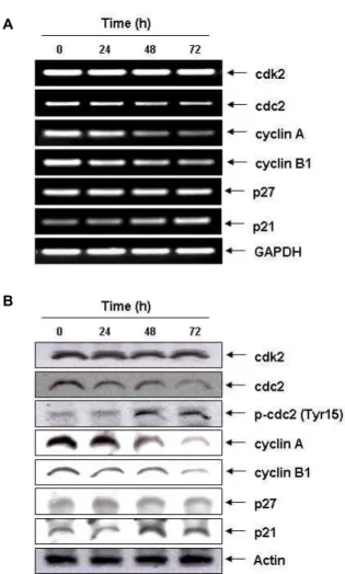
Fig. 2. Modulation of cell cycle-related factors by glutamine dep- rivation in PC3 cells. Cells were incubated in gluta- mine-free media for the indicated times. (A) Total RNAs were isolated and reverse-transcribed. The resulting cDNAs were subjected to PCR with indicated primers and the reaction products were subjected to electro- phoresis in 1% agarose gel and visualized by EtBr staining. GAPDH was used as an internal control. (B) The cells were lysed and then cellular proteins were sep- arated by SDS-polyacrylamide gels and transferred onto nitrocellulose membranes. The membranes were probed with the indicated antibodies. Proteins were visualized using an ECL detection system. Actin was used as an internal control.
전사 및 번역 수준에서 현저히 감소되었으나, Cdk2의 발현은 큰 변화가 없었다. 그러나 흥미롭게도 글루타민 결핍에 따라 Cdc2의 발현 자체는 전사 및 번역수준에서 억제되었으나 Cdc2 단백질의 인산화는 오히려 시간의 경과에 따라 다소 증 가되었다. 뿐만 아니라 Cdk 저해제이면서 광범위한 세포주기 진행의 음성조절인자에 해당되는 p21(WAF1/CIP1)의 발현이 또한 증가되었는데, 특히 PC3 세포는 정상적인 종양 억제인자 p53이 존재하지 않는 세포[19]이기 때문에 글루타민 결핍에
Fig. 3. Effects ofglutamine deprivation on the levels of Chks,g Cdc25C and Wee1 in PC3 cells. Cells were incubated in glutamine-free media for the indicated times. The cells were lysed and the cellular proteins were then separated by electrophoresis on SDS-polyacrylamide gels and transferred onto nitrocellulose membranes. Next, the membranes were probed with the indicated antibodies and the proteins were visualized using an ECL detection system. Actin was used as an internal control.
의한 p21의 발현 증가는 p53 비의존적임을 알 수 있었다(Fig.
2). 아울러 PC3 세포에서 글루타민 결핍 시간의 증가에 따라 Chk1 및 Chk2의 인산화가 증가되었으며, 이와 동반되어 Cdc25C의 인산화는 억제되었으며, Cdc25C의 인산화 억제는 그들 단백질들의 발현 수준 억제와 유사한 경향을 보여주었으 며, 글루타민 결핍 초기시기에 Wee1의 발현은 증가되었으며, 글루타민 결핍 시간의 증가에 따라 점차 감소되는 경향을 보 여주었다(Fig. 3).
비록 현재까지 본 연구와 비교할 수 있는 선행연구가 없다 는 점에서 글루타민 결핍에 의한 G2/M arrest에 관한 구체적 기전 제시가 어렵지만, 본 연구의 결과에서 의하면 글루타민 결핍에 따른 PC3 세포의 G2/M기 arrest에는 아마도 p21의 발현 증가와 Chks의 인산화 증가 및 cyclins의 발현 감소가 중요한 요소로서 작용하는 것 같다.
감사의 글
이 논문은 2010년도 정부(교육과학기술부)의 재원으로 한 국연구재단의 지원을 받아 수행된 기초연구사업임(2010- 0004415).
References
1. Boutros, R., Dozier, C. and Ducommun, B. 2006. The when and wheres of CDC25 phosphatases.Curr Opin Cell Biol18, 185-191.
2. Brasse-Lagnel, C., Lavoinne, A. and Husson, A. 2009.
Control of mammalinan gene expression by amino acids, especially glutamine. FEBS J 276, 1826-1844.
3. Chow, A. and Zhang, R. 1998. Glutamine reduces heat shock-induced cell death in rat intestinal epithelial cells.J Nutr 128, 1296-1301.
4. Cuddihy, A, 2003. O'Connell M. Cell-cycle responses to DNA damage in G2. Int Rev Cytol 222, 99-140.
5. Curthoys, N. P. and Watford, M. 1995. Regulation of gluta- minase activity and glutamine metabolism.Annu Rev Nutr 15, 133-159.
6. DeBerardinis, R. J., Lum, J. J., Hatzivassiliou, G. and Thompson, C. B. 2008. The biology of cancer: metabolic re- programming fuels cell growth and proliferation.Cell Metab 7, 11-20.
7. Drogat, B., Bouchecareilh, M., North, S., Petibois, C., Déléris, G., Chevet, E., Bikfalvi, A. and Moenner, M. 2007. Acute L-glutamine deprivation compromises VEGF-a upregula- tion in A549/8 human carcinoma cells. J Cell Physiol212, 463-472.
8. Eaton, C. L., Davies, P. and Phillips, M. E. 1998. Growth factor involvement and oncogene expression in prostatic tumours. J Steroid Biochem30, 341-345.
9. Exner, R., Weingartmann, G., Eliasen, M. M., Gerner, C., Spittler, A., Roth, E. and Oehler, R. 2002. Glutamine defi- ciency renders human monocytic cells more susceptible to specific apoptosis triggers. Surgery 131, 75-80.
10. Fu, Y. M., Yu, Z. X., Li, Y. Q., Ge, X., Sanchez, P. J., Fu, X. and Meadows, G. G. 2003. Specific amino acid depend- ency regulates invasiveness and viability of androgen-in- dependent prostate cancer cells. Nutr Cancer 45, 60-73.
11. Fu, Y. M., Yu, Z. X., Lin, H., Fu, X. and Meadows, G. G.
2008. Selective amino acid restriction differentially affects the motility and directionality of DU145 and PC3 prostate cancer cells. J Cell Physiol 217, 184-193.
12. Fu, Y. M., Yu, Z. X., Pelayo, B. A., Ferrans, V. J. and Meadows, G. G. 1999. Focal adhesion kinase-dependent apoptosis of melanoma induced by tyrosine and phenyl- alanine deficiency. Cancer Res 59, 758-765.
13. Fuchs, B. C. and Bode, B. P. 2006. Stressing out over survival:
glutamine as an apoptotic modulator.J Surg Res131, 26-40.
14. Fumarola, C., Zerbini, A. and Guidotti, G. G. 2001.
Glutamine deprivation-mediated cell shrinkage induces li- gand-independent CD95 receptor signaling and apoptosis.
Cell Death Differ 8, 1004-1013.
15. Gaglio, D., Soldati, C., Vanoni, M., Alberghina, L. and Chiaradonna, F. 2009. Glutamine deprivation induces abor- tive s-phase rescued by deoxyribonucleotides in k-ras trans- formed fibroblasts. PLoS One4, e4715.
16. Gould, K. L. and Nurse, P. 1989. Tyrosine phosphorylation of the fission yeast cdc2+ protein kinase regulates entry into mitosis. Nature342, 39-45.
17. Han, S. J. and Conti, M. 2006. New pathways from PKA to the Cdc2/cyclin B complex in oocytes: Wee1B as a poten- tial PKA substrate. Cell Cycle5, 227-231.
18. Haussinger, D. 1998. Hepatic glutamine transport and
metabolism. Adv Enzymol Relat Areas Mol Biol72, 43-86.
19. Isaacs, W. B., Carter, B. S. and Ewing, C. M. 1991. Wild-type p53 suppresses growth of human prostate cancer cells con- taining mutant p53 alleles. Cancer Res 51, 4716-4720.
20. Ko, Y. G., Kim, E. K., Kim, T., Park, H., Park, H. S., Choi, E. J. and Kim, S. 2001. Glutamine-dependent antiapoptotic interaction of human glutaminyl-tRNA synthetase with apoptosis signal-regulating kinase 1. J Biol Chem 276, 6030-6036.
21. Koochekpour, S., Majumdar, S., Azabdaftari, G., Attwood, K., Scioneaux, R., Subramani, D., Manhardt, C., Lorusso, G.
D., Willard, S. S., Thompson, H., Shourideh, M., Rezaei, K., Sartor, O., Mohler, J. L. and Vessella, R. L. 2012. Serum glu- tamate levels correlate with Gleason score and glutamate blockade decreases proliferation, migration, and invasion and induces apoptosis in prostate cancer cells.Clin Cancer Res 18, 5888-5901.
22. Lee, W. J., Hawkins, R. A., Wina, J. R. and Peterson, D.
R. 1998. Glutamine transport by the blood-brain barrier: a possible mechanism for nitrogen removal.Am J Physiol Cell Physiol 274, C1101-C1107.
23. Matés, J. M., Segura, J. A., Alonso, F. J. and Márquez, J.
2006. Pathways from glutamine to apoptosis. Front Biosci 11, 3164-3180.
24. Morgan, M. A., Parsels, L. A., Parsels, J. D., Mesiwala, A.
K., Maybaum, J. and Lawrence, T. S. 2005. Role of check- point kinase 1 in preventing premature mitosis in response to gemcitabine. Cancer Res 65, 6835-6842.
25. Niida, H. 2006. Nakanishi M. DNA damage checkpoints in mammals. Mutagenesis21, 3-9.
26. Petronini, P. G., Urbani, S., Alfieri, R., Borghetti, A. F. and Guidotti, G. G. 1996. Cell susceptibility to apoptosis by glu- tamine deprivation and rescue: survival and apoptotic death in cultured lymphoma-leukemia cell lines.J Cell Physiol169, 175-185.
27. Qing, G., Li, B., Vu, A., Skuli, N., Walton, Z. E., Liu, X., Mayes, P. A., Wise, D. R., Thompson, C. B., Maris, J. M., Hogarty, M. D. and Simon, M. C. 2012. ATF4 regulates MYC-mediated neuroblastoma cell death upon glutamine deprivation. Cancer Cell 22, 631-644.
28. Raleigh, J. M. and O’Connell, M. J. 2000. The G(2) DNA damage checkpoint targets both Wee1 and Cdc25.J Cell Sci 113, 1727-1736.
29. Rosenthal, D. I. and Trotti, A. 2009. Strategies for managing radiation-induced mucositis in head and neck cancer.Semin Radiat Oncol 19, 29-34.
30. Weinberg, F. and Chandel, N. S. 2009. Mitochondrial metab- olism and cancer. Ann NY Acad Sci 1177, 66-73.
31. Wischmeyer, P. E., Musch, M. W., Madonna, M. B., Thisted, R. and Chang, E. B. 1997. Glutamine protects intestinal epi- thelial cells: role of inducible HSP70. Am J Physiol 272, G879-G884.
32. Yuneva, M., Zamboni, N., Oefner, P., Sachidanandam, R.
and Lazebnik, Y. 2007. Deficiency in glutamine but not glu- cose induces MYC-dependent apoptosis in human cells.J Cell Biol 178, 93-105.
초록:글루타민 결핍에 의한 PC3 인체 전립선 암세포의 G2/M 세포주기 억제 유발 신동역1․최성현2․박동일3․최영현4,5*
(1동남권원자력의과학원,2한국승강기대학교 승강기시스템관리과, 동의대학교 한의과대학3내과학교실, 4생 화학교실, 5항노화연구소 및 블루바이오소재개발센터)
본 연구에서는 생체 내 구성요소 및 에너지원으로서 중요한 역할을 하는 글루타민 결핍에 의한 인체 전립선 PC3 암세포의 증식에 관한 기전 연구를 실시하였다. 글루타민 결핍에 의한 PC3 세포의 증식억제는 세포주기 G2/M arrest와 연관성이 있었으나, apoptosis 유발 현상은 관찰되지 않았다. 글루타민 결핍에 의한 G2/M arrest 는 전사 및 번역 수준에서 Cdc2, cyclin A 및 cyclin B1의 발현 억제 및 p53 비의존적인 p21(WAF1/CIP1)의 발현 증가와 연관성이 있었다. 아울러 글루타민 결핍은 Chk1 및 Chk2의 인산화를 증가시켰으나, Cdc25C의 인산화는 감소시켰다. 본 연구의 결과는 글루타민 결핍에 의한 PC3 세포의 증식억제가 apoptosis 유발과는 상관없이 G2/M arrest를 유발시킨다는 첫 번째 증거이다.