서 론
Cisplatin (cis-diamminedichloroplatinum[Ⅱ])은 세포에서 성장정지와 apoptosis를 강력하게 유도하는 물질로서 암치료를 위한 화학요법 치료제로 광범위하게 사용되고 있지만1), 신부전 증을 유발하는 신장독성 때문에 사용이 제한되어 있다2,3). Cisplatin의 세포독성은 1차적으로 DNA의 퓨린계열 염기의 친핵성 N7부위와 상호작용에 기인하며, DNA-protein 또는 DNA-DNA interstrand 또는 intrastrand crosslink를 형성하기 때문이다4). DNA와 cisplatin의 상호작용에 의해 형성된 DNA 부
가물은 암세포의 apoptosis를 유도하는 mitogen-activated protein kinases (MAPKs)와 같은 신호전달경로들을 활성화시킨 다5). MAPKs는 세포증식, 분화, apoptosis와 같은 다양한 세포반 응들의 조절에 관여하는 serine/threonin kinases이다6,7). 독성물질, 국소빈혈, 방사선에 노출된 세포는 DNA damage 에 대해 조사하고 복구하기 위해 G1/S와 G2/M에서 세포주기 정지를 유도하는 것으로 알려져 있다8-11). Cyclin-dependent kinase (CDK) inhibitor들인 p21과 p27은 cisplatin에 의해 유도 된 DNA damage후에 유도되고12,13), G1/S와 G2/M기 검사점 (checkpoint)에서 세포주기를 정지시키는 역할을 수행한다12,14,15). 암 억제단백질인 p53은 DNA 손상제에 대한 세포의 반응에 관여하고 있다16). 정상세포에서 손상된 DNA로부터 발생한 신호 는 p53 활성경로를 이끌고 세포주기 정지 및 DNA 복구 또는
保正防癌湯加減方이 cisplatin으로 유도된 인간 근위세뇨관 HK-2세포의 G2/M phase arrest에 미치는 영향
박성철․이수경․염승룡․권영달*․송용선
원광대학교 한의과대학 한방재활의학과교실
Inhibitory Effect of Bojungbangam-tang Kakambang on Cisplatin-Induced G2/M Phase Arrest in Human Renal Proximal Tubular HK-2 Cells
Sung Cheul Park, Su Kyung Lee, Seung Ryong Yeom, Young Dal Kwon*, Yung Sun Song
Department of Oriental Rehabilitation Medicine, College of Oriental Medicine, Wonkwang University
To idenifty effect of Bojungbangam-tang kakambang on Cisplatin-Induced G2/M Phase Arrest in Human Renal Proximal Tubular HK-2 Cells. Cytotoxicity of cisplatin was detected in HK-2 cells and the value of IC50 is about 25 μM. The treatment of cisplatin to HK-2 showed the G2/M phase cell cycle arrest. The ethanol extract of Bojungbangam-tang kakambang (EBTKB), a new herbal prescription composed of ten crude herbs, inhibited cisplatin-induced G2/M phase arrest in HK-2 cells. EBTKB increased G0/G1 peak in cisplatin-treated HK-2 cells. p53, p21 and p27 expression were increased in cisplatin-treated HK-2 cells. Inhibitory effect of EBTKB on cisplatin-induced G2/M phase arrest was accomplished through inhibition of p53, p21 and p27 expression. Also, reduced CDK2 and cyclin A expression by cisplatin were increased by EBTKB, but cyclin E was not changed. Reduction of ERK activation and increment of p38 activation by cisplatin were increased ERK activation and decreased p38 activation by EBTKB.
Cisplatin had no effect on JNK activation, but EBTKB increased JNK activation. These results can suggest that EBTKB inhibits cisplatin-induced G2/M phase arrest in HK-2 cell through reduction of p53-dependent p21 and p27 protein, ERK activation and p38 inactivation.
Key words : HK-2, Cisplatin, Nephrotoxicity, Cell cycle, G2/M phae arrest, p53, p21, p27, ERK, p38, JNK,
Bojungbangam-tang kakambang (EBTKB)
* 교신저자 : 권영달, 광주시 남구 주월동 543-8 원광대학교 광주한방병원
․E-mail : [email protected], ․Tel : 062-670-6452
․접수 : 2007/09/28 ․채택 : 2007/10/31
apoptosis를 유도한다17). 또한 p53 단백질은 p21의 전사적 활성 에 의해 G1/S arrest를 유도한다. 그러나 p21은 G2/M arrest에 기여하기 때문에 이것은 논란의 여지가 있고. 몇몇 연구에서는 p53이 G2/M 제한점 (Restriction point)에 관여하는 것으로 보고
되었다18,19).
Extracellular signal-regulated kinase (ERK), c-Jun N-terminal kinase (JNK/SAPK), p38 kinase 등은 세가지 주요한 MAPKs subfamily들이다. 각각의 MAPKs는 특정한 인산화 케스 케이드 경로를 통하여 활성화된다. ERK 경로는 주로 mitogen과 성장인자들에 의해 활성화 되며, 세포성장 및 생존과 분화를 조 절하는데 중요한 역할을 수행한다20,21). 대조적으로 JNK와 p38 경로는 화학적 또는 환경적 스트레스에 대한 반응에 의해 활성 화 되고 주로 apoptosis 유도에 관여한다21,22).
이전 연구에서 保正防癌湯은 간으로 암전이에 대한 항전이 효과와 면역기능증강효과가 있다는 것을 보고하였고23), rat mesangial cells에서는 cisplatin에 의해 유도된 apoptosis에 대한 보호효과를 나타내었고24), 혈관내피세포에서 cisplatin에 의한 세 포고사의 방어효과25) 및 혈관형성 저해작용이 있는 것으로 나타 났다26). 그러나 保正防癌湯에 in vivo에 대한 연구가 이루어지지 는 못하였다.
본 연구에서는 cisplatin에 의해 유도된 HK-2세포의 G2/M arrest에 대한 保正防癌湯加減方의 억제효과를 관찰하고, 이에 관여하는 세포주기조절단백질의 발현을 조사하고, MAPKs 신호 기전의 역할을 규명하고자 하였다.
재료 및 방법
1. 시약 및 항체
Cisplatin, 3-(4,5-dimethylthiazol-2-yl)-2,5-diphenyl tetrazolium bromid (MTT), propidium iodine (PI), Ribonuclease A (RNase A), protease inhibitor cocktail은 Sigma-Aldrich Chemical (St.
Louis, MO, USA)로부터 구입하였다. Anti-p53, anti-p21, anti-p27, anti-CDK2, anti-cyclin A, anti-cyclin E 항체는 Santa Cruz Biotechnology, INC. (Santa Cruz, CA, USA)로부터 구입하 였다. anti-ERK, anti-phospho-ERK, anti-p38, anti-phospho-p38, anti-JNK, anti-phospho-JNK 항체들은 Cell Signaling Technology INC. (Beverly, MA, USA)로부터 구입하였다.
2. 保正防癌湯加減方 제조
保正防癌湯加減方은 Table 1과 같이 10가지 한약재로 구성 되어 있다. 保正防癌湯加減方 에탄올추출물 (Ethanol extract of Bojeongbang-tang kakambang; EBTKB)는 건조된 10가지 한약재 를 분말로 만들어 혼합한 후 1첩분량(54 g)을 에탄올에 3×부피로 3회 씩 3 일 동안 상온에서 추출하였다. 추출물은 감압하여 동결 건조하였으며, 수율 9.21%인 약 5.16 g의 분말을 획득하였다. 동 결건조된 추출물은 dimethylsulfoxide (DMSO)로 200 ㎎/mL의 농도로 용해시켜 -20℃에 보관하였고 실험에 사용하기 전에 희 석하여 사용하였다.
Table 1. The botanical origins and ration of crude herbs of Bojungbangam-tangⅢ
Crude herbs Botanical origin Weight (g)
동충하초(冬蟲夏草) Cordyceps Sinensis 10
황기(黃芪) Astragal Radix 8
황정(黃精) Polygonati Rhizoma 8
인삼(人蔘) Ginseng Radix 4
보골지(補骨脂) Psoraleae Semen 4
백출(白朮) Atractylodis Macrocephalae Rhizoma 4
맥문동(麥門冬) Liriopes Radix 4
당귀(當歸) Angelicae Gigantis Radix 4
백복령(白茯苓) White Poria Cocas 4
진피(陳皮) Citri Pericarpium 4
3. 세포배양
Human proximal tubular cell인 HK-2세포는 American Type Culture Collection (ATCC, Rockville, MD, USA)로부터 분 양받았고, 10% fetal calf serum (FCS, Invitrogen, Burlington, ON, Canada), 100 U/mL penicillin, 100 μg/mL streptomycin이 포함된 Dulbecco's Modified Eagle's medium (Invitrogen, Burlington, ON, Canada)를 사용하여 37℃와 5% CO2 상태에서 배양하였다. 세포는 0.05% trypsin-0.53 mM EDTA용액을 사용하 여 계대 배양하였으며, 세포는 cisplatin과 EBTKB를 처리하기 24 시간 전에 배양접시에 seeding하였다.
4. 세포생존률측정
세포생존률은 MTT 방법을 사용하여 측정하였다. MTT 검사 는 미토콘드리아의 숙신산 탈수소효소에 의해 MTT가 formazan 으로 환원되는 원리를 기초로 하고 있다. 세포는 5×10⁴ cells/mL 로 12-well 플레이트에 24시간동안 배양하였다. 세포는 cisplatin 과 EBTKB를 일정한 간격으로 농도를 증가시켜 24 시간 또는 48 시간 동안 처리하였다. 배양 후, 배지를 제거하고 500 μL의 MTT (0.5 ㎎/mL in PBS) 용액을 각 well에 첨가하여 37℃에서 4시간 동안 배양하였다. 그 후 상층액을 제거하고 500 μL의 DMSO를 각 well에 첨가하여 비수용성의 보라색 formazan을 용해시켰다.
Formazan의 양은 SpectraMAX 250 microplate spectrophotometer (Molecular Devices, Sunnyvale, CA)를 이용하여 570 ㎚에서 측 정하였다. 세포생존률의 확률은 다음 방정식으로 계산하였다.
(mean OD of treated cells/mean OD of control cells) × 100=viability(%)
5. 세포주기분석
배양된 세포는 cisplatin과 EBTKB를 단독 또는 함께 48 시간 동안 처리한 후, 부착되어 있는 또는 부유되어 있는 세포 모두를 수확하여 PBS로 세척하였다. 세척한 세포는 냉각된 70% 에탄올 로 4℃에서 1 시간 동안 고정하였다. 고정된 세포는 PBS로 다시 세척하고 1mL 의 PI (10 μg/mL)/RNase A(100 μg/mL)용액으 로 재부유시켜 37℃에서 암실조건으로 1 시간 동안 배양하였다.
세포주기분석은 fluorescence-activated cell sorting (FACS) Calibur (BD Biosciences, CA, USA)를 사용하여 측정하였다. G0/G1, S, G2/M population 비율은 CellQuest Pro (BD Biosciences, CA,
USA) software를 사용하여 측정하였다.
6. Western blot analysis
시료처리 후 배양이 끝난 세포는 ice-cold PBS로 세척하였고 1% protease inhbitor cocktail이 포함된 ice-cold lysis buffer (50 mM Tris-HCl, pH 7.4, 150 mM NaCl, 1% NP-40, 0.5% sodium deoxycholate, 0.1% SDS, 1 mM sodium vanadate)로 얼음 위에서 30 분 동안 용해하였다. 세포용해액은 저온원심분리기로 14,000 ×g 상태에서 15 분 동안 원심분리 하였고, 단백질 농도는 Bradford assay를 이용하여 측정하였다. 총 30 μg의 단백질 샘플을 10% 또 는 12% SDS-PAGE 젤로 분리하였고, 분리된 SDS-PAGE 젤 상의 단백질을 trans blot kit를 사용하여 40 V에서 3 시간 동안 nitrocellulose membrane으로 이동시켰다. Membrane은 5% milk 가 포함된 Tris-buffered saline+Tween-20 (20 mM Tris-HCl, pH 7.6, 150 mM NaCl, 0.05% Tween-20)으로 차단한 후 1차 항체를 결 합시켰다. 반응활성도는 퍼옥시아제가 붙어있는 anti-rabbit 또는 anti-mouse 2차 항체를 사용하여 SuperSignal West Pico Chemiluminescent (Pierce, Rockford, IL, USA)에 의해 탐지하였다.
7. 통계처리
모든 데이터의 결과는 마이크로소프트 오피스 엑셀 (EXCEL) 2003 프로그램을 통하여 통계처리 하여 mean±standard deviation (SD)로 기록하였다.
실험결과
1. HK-2세포에 대한 Cisplatin 및 保正防癌湯加減方의 세포독성효과 A
0 2 0 4 0 6 0 8 0 1 0 0 1 2 0
0 5 1 0 1 5 2 0 2 5 3 0 3 5
C o n c e n t r a t i o n o f C i s p l a t i n ( u M ) Cell viability (% of control)
2 4 h t r e a t m e n t 4 8 h t r e a t m e n t
B
0 2 0 4 0 6 0 8 0 1 0 0 1 2 0
0 5 0 1 0 0 1 5 0 2 0 0 2 5 0
C o n c e n t r a t i o n o f E B T K B ( u g / m L ) Cell viability (% of control)
2 4 h t r e a t m e n t 4 8 h t r e a t m e n t
Fig. 1. Effect of cisplatin or ethanol extract of bojungbangam-tang kakambang (EBTKB) on cytotoxicity in HK-2 cells. The cells were treated with various concentrations of cisplatin (A) and EBTKB (B) for 24 h or 48 h and the cells were tested for viability by MTT assay. Value are means ± SD, N = 3.
HK-2세포의 cisplatin에 대한 세포독성의 효과를 조사하기 위해 cisplatin을 일정한 간격으로 농도를 증가시켜 5~30 μM로 24 시간 또는 48시간동안 처리하였다. 세포생존률은 MTT검사에 의하여 측정되었다. Cisplatin은 농도의존적으로 HK-2세포의 생 존률을 감소시켰다(Fig. 1A).
48시간 처리 후, Cisplatin의 IC50값은 약 25 μM이었다. 保 正防癌湯加減方 (EBTKB)의 HK-2세포에 대한 세포독성의 효과 를 조사하기위해 50, 100, 150, 200 μg/mL 농도로 24시간 또는 48시간 동안 처리하였다. 保正防癌湯加減方은 HK-2세포의 생존 률을 24시간 또는 48시간 처리 후에도 거의 감소시키지 못하였 다(Fig. 1B).
2. Cisplatin에 의해 유도된 G2/M phase arrest에 대한 保正防癌 湯加減方의 억제효과
Cisplatin에 의해 유도된 G2/M phase arrest에 대한 保正防 癌湯加減方의 효과를 조사하기 위하여 flow cytometry를 통하여 세포주기를 분석하였다.
A B
Control 5 μM Cisplatin
Cell cycle % Sub-G1 5.49±1.28
G0/G1 63.00±5.29
S 12.51±3.79
G2/M 19.40±2.48
Cell cycle % Sub-G1 9.61±1.91
G0/G1 10.98±5.14
S 25.64±3.60
G2/M 54.27±2.50
C D
5 μM Cisplatin + 50 μg/mL EBTKB 5 μM Cisplatin + 100 μg/mL EBTKB Cell cycle %
Sub-G1 10.51±1.61 G0/G1 17.06±2.53
S 22.19±5.04
G2/M 50.90±3.65
Cell cycle % Sub-G1 13.00±5.00
G0/G1 21.68±1.70
S 26.02±3.46
G2/M 39.71±2.54 Fig. 2. Effect of ethanol extract of bojungbangam-tang kakambang (EBTKB) on cisplatin-induced G2/M phase arrest in HK-2 cells. The cells were preincubated with 50, 100 μg/mL of EBTKB for 3 hr, and then treated with 5 μM cisplatin for 48 hr. The cells were fixed and stained with PI and the DNA content was analyzed by flow cytometry.
대조군과 cisplatin 단독실험군에서 G0/G1 phase는 63.00±
5.29%에서 10.98±5.14%로 크게 감소하였고, G2/M phase는 19.40±2.48%에서 54.27±2.50%로 크게 증가하였다. 또한 Sub-G1
에서는 큰 차이를 보이지 않아 5 μM의 cisplatin농도에서는 세포 고사가 거의 일어나지 못했다. 保正防癌湯加減方와 cisplatin을 같이 처리한 실험군에서 G0/G1 phase는 cisplatin 단독처리군보 다 17.06±2.53% (50 μg/mL EBTKB)와 21.68±1.70% (100 μg/mL EBTKB)로 증가하였고, G2/M phase는 50.90±3.65% (50 μg/mL EBTKB)와 39.71±2.54% (100 μg/mL EBTKB)로 감소하였다(Fig. 2).
3. Cisplatin에 의해 유도된 p53, p21, p27 단백질발현에 대한 保 正防癌湯加減方의 억제효과
Cisplatin에 의해 유도된 HK-2세포의 G2/M phase arrest상 에 세포주기조절 단백질인 p21과 p27의 단백질 발현수준과 保正 防癌湯加減方가 p21과 p27의 단백질 발현에 미치는 효과를 조사 하였다. Cisplatin은 HK-2세포에서 p21과 p27의 단백질 발현을 각각 1.37배와 1.55배 모두 증가시켰다. 保正防癌湯加減方를 같 이 처리한 실험군에서는 p21 단백질은 1.20 (50 μg/mL EBTKB), 1.15 (100 μg/mL EBTKB), 1.09 (200 μg/mL EBTKB)배 감소시켰 고, p27 단백질은 1.47 (50 μg/mL EBTKB), 1.29 (100 μg/mL EBTKB), 1.08배 (200 μg/mL EBTKB) 감소하였다. 암억제 단백질 p53의 발현에서도 cisplatin처리군은 1.45배 증가시켰고, 保正防 癌湯加減方를 같이 처리한 실험군에서는 1.36 (50 μg/mL EBTKB), 1.33 (100 μg/mL EBTKB), 1.30배 (200 μg/mL EBTKB) 감소시켰다(Fig. 3).
A
B
Fig. 3. Effect of ethanol extract of bojungbangam-tang kakambang (EBTKB) on expression of p53, p21 and p27 proteins in cisplatin-treated HK-2 cells. The cells were preincubated with 50, 100 and 200 μ g/mL of EBTKB for 3 hr, and then treated with 5 μM cisplatin for 48 hr. (A) Cells were the lysed, and the supernatants were subjected to Western blot analysis using anti-p53, anti-p21, anti-p27. (B) Densitometric analysis of the specific bands was done using GS-800 Calibrated Densitometer (Bio-Rad Laboratories, Inc. Hercules, CA)
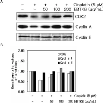
4. Cisplatin에 의해 억제된 세포주기조절 단백질 CDK2, Cyclin A, Cyclin E 단백질발현에 대한 保正防癌湯加減方의 효과 Cisplatin에 의해 유도된 HK-2세포의 G2/M phase arrest상
에 세포주기조절 단백질인 CDK2, cyclin A, cyclin E의 단백질 발현수준과 保正防癌湯加減方가 CDK2, cyclin A, cyclin E의 단 백질 발현에 미치는 효과를 조사하였다. Cisplatin은 HK-2세포에 서 CDK2와 cyclin A의 단백질 발현은 각각 0.56배와 0.55배 감소 시켰지만, cyclin E의 단백질 발현에는 아무런 차이가 없었다. 保 正防癌湯加減方를 같이 처리한 실험군에서는 CDK2 단백질은 0.71 (50 μg/mL EBTKB), 0.95 (100μg/mL EBTKB), 0.89 (200 μ g/mL EBTKB)배 증가시켰고, cyclin A 단백질은 0.85 (50 μ g/mL EBTKB), 1.14 (100 μg/mL EBTKB), 0.99배 (200 μg/mL EBTKB) 증가시켰다(Fig. 4). 그러나 cyclin E 단백질 발현에는 아 무런 영향을 미치지 못했다.
A
B
Fig. 4. Effect of ethanol extract of bojungbangam-tang kakambang (EBTKB) on expression of the cell cycle-related proteins in cisplatin-treated HK-2 cells. The cells were preincubated with 50, 100 and 200 μ g/mL of EBTKB for 3 hr, and then treated with 5 μM cisplatin for 48 hr. (A) Cells were the lysed, and the supernatants were subjected to Western blot analysis using anti-CDK2, anti-cyclin A, anti-cyclin E. (B) Densitometric analysis of the specific bands was done using GS-800 Calibrated Densitometer (Bio-Rad Laboratories, Inc. Hercules, CA)
5. Cisplatin에 의한 MAPK의 활성에서 保正防癌湯加減方의 효과 Cisplatin에 의한 HK-2세포의 MAPK활성에 대한 保正防癌 湯加減方의 효과를 조사하였다. Cisplatin은 HK-2세포에서 ERK활성을 0.58배 감소시켰고 p38활성은 1.3배 증가시켰다. 그 러나 JNK활성에는 아무런 영향을 미치지 못하였다. 保正防癌湯 加減方는 cisplatin에 의해 감소된 ERK활성을 를 같이 처리한 실험군에서는 CDK2 단백질은 0.71 (50 μg/mL EBTKB), 1.07 (100 μg/mL EBTKB), 1.20 (200 μg/mL EBTKB)배 증가시켰고, 증가된 p38활성을 1.16 (50 μg/mL EBTKB), 1.10 (100 μg/mL EBTKB), 0.91배 (200 μg/mL EBTKB) 감소시켰다. 그러나 cisplatin활성에 아무런 영향을 받지 않은 JNK활성은 保正防癌 湯加減方에 의해 1.33 (100 μg/mL EBTKB), 1.73배 (200 μg/mL EBTKB) 증가되었다(Fig. 5).
A
B
Fig. 5. Effect of ethanol extract of bojungbangam-tang kakambang (EBTKB) on activation of MAPKs in cisplatin-treated HK-2 cells. The cells were preincubated with 50, 100 and 200 μg/mL of EBTKB for 3 hr, and then treated with 5 μM cisplatin for 12 hr. (A) Cells were the lysed, and the supernatants were subjected to Western blot analysis using anti-ERK, anti-p-ERK, anti-p38, anti-p-p38, anti-JNK and anti-p-JNK antibodies. (B) Densitometric analysis of the specific bands was done using GS-800 Calibrated Densitometer (Bio-Rad Laboratories, Inc. Hercules, CA)
고 찰
대부분의 항암제들과 DNA 손상제는 G1, S 또는 G2/M기에 서 cell cycle을 arrest시키고 그 후에 apoptosis를 유도한다27-29). 세포주기검사점은 DNA를 복구하기위한 시간을 확보하는 기능 을 하는 반면에 apoptosis는 복구할 수 없는 세포나 손상된 세포 를 제거하는 기능을 한다30,31). 포유동물세포에서 세포주기진행은 cyclin과 cyclin-dependent kinase (CDK)에 의해 조절이 된다32). cyclin/CDK 복합체의 활성은 CDK inhibitor (CDKI)에 의해 음 성적으로 조절된다33). 이 CDKI는 세포주기 전반에 걸쳐서 CDK 의 활성을 억제하여 세포주기진행을 조절하는데 중요한 역할을
수행한다33-36). CDKI는 현재 2가지 종류로 분류가 된다. 한 그룹
은 p21, p27, p57을 포함하는 CIP/KIP 패밀리로 모든 CDK활성
에 광범위하게 작용하고33,36), 다른 하나는 p15, p16 p18, p19을 포함하는 INK4 패밀리로 CDK4와 CDK6를 표적으로 한다33).
HK-2세포는 5 μM의 cisplatin 농도에서 G2/M phase arrest 를 유도하였다(Fig. 2). Cisplatin에 의한 G2/M phase arrest는 CDK inhibitor인 p21과 p27 단백질 발현을 통하여 이루어졌다 (Fig. 3). p21과 p27 단백질은 G1기 에서 S기 또는 G2기에서 M 기로의 전이를 조절한다12,14,15,30)
. 특히 p27 단백질은 cyclin D/CDK4/6 와 cyclin E/CDK2 복합체와 결합하여 활성을 억제 하여 그로인해 G1/S 또는 G2/M기에서 cell cycle arrest를 유도 하는 것으로 알려져 있다14,15,37). p21단백질의 발현은 wild-type p53핵단백질에 의해 유도된 수 있다38). 몇몇 연구에서 다양한 자 극에 의해 유도된 cell cycle arrest가 p53의존적 p21활성을 통하 여 이루어졌다고 보고되었다39-41). 본 연구에서도 cisplatin은 HK-2세포에서 p53의 발현을 증가시켰고, 또한 p21단백질이 발 현이 p53의존적임을 확인 할 수 있었다(Fig. 3). 최근 연구에서 cisplatin에 의해 유도된 tubular cell의 apoptosis에서 p53의 역할 에 대해서 보고되고 있다. p53은 cisplatin을 처리하는 동안 활성
화된다42,43). 특히 Cislpatin에 의해 유도된 tubular cell의
apoptosis는 p53의 pharmacological inhibitor인 pifithrin-α에 의 해 감소된다42,43). Cisplatin의 신장독성은 p53활성과 밀접한 관련 이 있지만 그 신호전달기전에 대해선 알려져 있지 않다.
활성산소종(Reactive oxygen species, ROS)은 다양한 병리학 적 상태의 신장손상에서 증가하고 특히 p53조절에 관여하고 있
다44,45). 활성산소종은 DNA손상을 유도하고, p53활성을 강하게
촉진한다. 게다가 활성산소종은 p53을 인산화시키고 활성시키는 다양한 protein kinase를 수반하는 신호전달기전을 활성화 시킨 다46). 이런 활성산소종은 세포주기에서 양성적으로 또는 음성적 으로 생성이 되는데47), 특히 세포주기진행을 방해는 것으로 알려 져 있다48). 현재 cisplatin의 신장독성과 관련하여 p53과 활성산 소종에 대한 연구가 활발하게 이루어지고 있다.
본 연구에 사용된 保正防癌湯加減方은 동충하초, 황기, 황 정, 인삼 보골지, 백출, 맥문동, 당귀, 백복령, 진피로 구성되었다.
이중 君藥인 冬蟲夏草는 麥角菌科에 속한 식물인 冬蟲夏草菌의 포자 및 거기에 기생하는 곤충인 綠蝙蝠蛾의 유충사체등의 복합 체이고, 性味는 甘 溫 無毒하고 肺와 腎으로 歸經하며, 滋肺補腎 하고 止咳化痰한다. 冬蟲夏草는 滋肺陰작용과 補腎陽의 작용을 구비하고 있기 때문에 君藥으로 쓰였다. 黃芪는 豆科에 속한 다 년생 초본인 단너삼의 根으로 性味는 甘 微溫 無毒하고, 脾肺로 歸經하고, 補氣升陽 固表止汗 托毒排膿 利水退腫한다. 補氣扶正 의 효력으로 癰疽로 正氣가 부족할 때 쓰이므로 臣藥으로 쓰였 다. 黃精은 백합과에 속한 다년생초본인 죽대둥글레의 根莖이고, 性味는 甘 平無毒하고, 歸經은 脾肺腎이고, 그 효능은 補裨 潤肺 生津한다. 補中益氣하기 때문에 臣藥으로 쓰였다. 人蔘은 오갈피 나무과에 속한 다년생 초본인 人蔘의 根이다. 그 性味는 甘, 微 苦, 溫 無毒하고, 脾,肺로 歸經한다. 그 효능은 大補元氣, 補脾益 (肺)氣, 生津, 寧神益智이다. 補骨脂는 콩과에 속한 일년생초본 인 파고지의 성숙한 種子이다. 그 性味는 辛, 溫 無毒하며, 그 歸 經은 腎經이다. 효능은 補腎壯陽이다. 白朮은 菊花科에 속한 다
년생초본인 흰삽주의 塊根이다. 그 性味는 甘, 苦, 微溫, 無毒이 며, 歸經은 脾胃이다. 그 효능은 補脾益氣, 燥濕利水, 固表止汗, 安胎이다. 麥門冬은 백합과에 속한 다년생초본인 소엽맥문동의 塊根인데, 그 性味는 甘, 微苦, 寒 無毒이며, 그 歸經은 心肺胃이 다. 그 효능은 滋陰淸熱 潤肺生津 淸心利尿이다. 當歸는 미나리 과에 속한 다년생초본인 참당귀 및 일당귀의 根이다. 그 성미는 甘,辛 溫, 無毒이며, 그 歸經은 心肝脾이다. 그 효능은 補血和血, 調經止痛, 潤燥滑腸이다. 茯苓은 구멍쟁이버섯과에 속하며, 松樹 根에 기생하는 不定形의 균핵인 茯苓이다. 그 性味는 甘,淡,平 無 毒하며, 歸經은 心脾肺이다. 그 효능은 利尿滲濕 健脾補中 寧心 安神이다. 陳皮는 산초과에 속한 상록소교목인 귤나무의 성숙한 果皮이다. 그 性味는 辛,苦,溫 無毒이며, 歸經은 脾肺이다. 그 효 능은 理氣健脾, 燥濕化痰이다. 保正防癌湯加減方은 이상의 약재 들의 효능으로 볼때, 세포들이 항암제로 인하여 괴사되어가고 말 라들어가는 것을 진액을 보충하고, 補氣함으로써 생명력을 증강 시키는 쪽으로 방향이 설정된 처방이다. 保正防癌湯加減方는 HK-2세포에서 cisplatin에 의해 유도된 G2/M arrest에 대한 억 제효과를 보여 주었다. 保正防癌湯加減方는 HK-2세포에 대해서 세포독성을 거의 보여주지 못하였다(Fig. 1B). 또한 Cisplatin 단 독처리군 보다 G2/M기의 비율을 크게 감소시켰고 (Fig. 2), 이러 한 保正防癌湯加減方의 G2/M arrest의 억제효과는 cisplatin에 의해 유도된 p21과 p27단백질의 발현을 감소시켜서 이루어졌다 (Fig. 3). p53단백질 발현 또한 保正防癌湯加減方에 의해 감소되 었고, p21단백질 발현이 감소가 p53단백질 발현의 감소에 의해 이루어졌음을 확인 할 수 있었다. 또한 cisplatin에 의해 감소된 세포주기조절 단백질인 CDK2와 cyclin A가 保正防癌湯加減方 에 의해 증가하는 것을 확인 할 수 있었다 (Fig. 4). 이러한 결과 들을 통하여 保正防癌湯加減方가 cisplatin에 의해 유도된 G2/M arrest를 억제하는데 효과적임을 확인할 수 있었다.
MAPKs는 세포증식과 분화를 조절하는데 중요한 신호기전 이다. 최근연구에서 MAPK는 세포주기와 apoptosis를 조절한다 고 보고되었다50,51). 일반적으로 ERK cascade는 성장인자에 의해 활성화되고 증식과 생존에 대해 중요하다52). 그러나 JNK와 p38 은 성장인자에 의해 약하게 활성화되지만 tumor necrosis factor, hyperosmotic stress, ultraviolet irradiation과 같은 다양한 스트 레스자극에 대해 크게 활성이 되고, 이 활성들은 대부분 apoptosis를 유도와 관련되어 있다53). ERK와 p38은 세포주기에 서 G2/M 전이를 조절하는데 관여한다. ERK는 정상적인 세포주 기의 G2/M 진행과 DNA 손상에 의해 유도된 G2/M 정지에 필
요하다54,55). p38 또한 G2/M의 DNA 손상과 관련된 세포주기 검
사점에 작용하는 것으로 알려졌다56,57). 특히 p38은 최근 연구에 서 세포주기진행의 조절에서 중요한 역할을 수행하는 것으로 보 고되었다. p38은 ultraviolet irradiation과 ionizing irradiation에 의한 G2/M arrest를 촉발시키고58,59), p38의 억제는 ultraviolet raddiation후에 인간과 마우스세포에서 G2/M 검사점의 개시를 막았다58). 또한 UV 또는 특정한 항암제와 함께 처리된 세포에서 p38은 p53을 인산화시켜 활성화 시켰다60-62). 본 연구에서 cisplatin을 처리한 HK-2세포는 ERK활성은 감소하였지만, p38의
활성은 증가하였다(Fig. 5). 또한 cisplatin에 의해 증가된 p38활 성이 증가된 p53을 활성화 시켜서 HK-2세포의 G2/M arrest를 유도한 것으로 생각된다. JNK활성은 cisplatin에 의해 아무런 변 화가 없었다. Cisplatin에 의해 감소된 ERK활성은 세포증식과 생 존이 감소시키는 것으로 생각된다. Cisplatin은 DNA에 치명적인 손상을 일으켜 cell cycle arret를 유도 후, 손상된 DNA 복구할 수 없을 경우 apoptosis를 유도한다. 최근 연구에서 난소암세포 에서 cisplatin에 의해 유도된 ERK의 활성이 apoptosis가 아닌 세포생존을 증가시켰다는 연구결과가 보고되었다63-65).
保正防癌湯加減方는 cisplatin에 의해 억제된 ERK활성과 JNK활성을 증가시킨 반면, 증가된 p38활성은 감소시켰다(Fig.
5). 이 결과들은 保正防癌湯加減方의 G2/M arrest의 억제효과가 ERK활성증가와 p38활성감소를 통하여 이루어지는 것으로 생각 된다. 保正防癌湯加減方의한 JNK활성증가는 cisplatin에 의한 HK-2세포의 DNA손상에 따른 G2/M arrest통한 apoptosis유도가 保正防癌湯加減方에 의해 G2/M arrest 억제되어 apoptosis유도 하지 못하여 또 다른 apoptosis 신호기전을 활성화 시키는 것으로 생각된다. 이에 대한 부분에 대해서 앞으로 연구가 더 필요하다.
결론적으로 保正防癌湯加減方는 cisplatin에 의해 유도되는 G2/M arrest를 억제효과는 p38활성억제를 통하여 p53활성 및 발현을 감소시켜 p53의존적인 p21단백질 발현 감소와 p27단백 질 발현을 감소를 통하여 이루어지는 것으로 생각된다. 그러나 保正防癌湯加減方의 cisplatin에 대한 HK-2세포의 보호효과는 완전하지 못한 것으로 생각된다. 그 이유는 保正防癌湯加減方는 cisplatin의 G2/M arrest통한 apoptosis를 억제하지만, JNK활성 을 통한 apoptosis활성 기전에는 아무런영향을 미치지 못했기 때 문이다. 保正防癌湯加減方에 여러 약재를 가감하고 보완하여 保 正防癌湯을 항암치료 부작용을 억제하는 처방으로서 기능을 할 수 있을 것으로 생각되며 이에 대한 연구를 지속적으로 할 필요 가 있을 것으로 생각된다.
결 론
保正防癌湯加減方가 항암제인 cisplatin으로 유도된 G2/M arrest에 대한 억제효과 및 기전을 분자생물학적 방법을 통하여 다음과 같은 결과를 얻을 수 있었다.
Cisplatin에 의해 유도된 HK-2 세포의 G2/M arrest는 p53의 존적인 p21단백질과 p27단백질의 발현증가와 관련되어 있음을 확인하였다. Cisplatin에 의해 유도된 HK-2 세포의 G2/M arrest 에서 保正防癌湯加減方는 p53 감소를 통한 p21단백질와 p27단 백질 발현억제를 통하여 이루어졌음을 확인하였다. Cisplatin에 의해 유도된 HK-2세포의 ERK활성감소와 p38활성증가를 확인하 였다. Cisplatin에 의해 감소된 ERK활성과 증가된 p38활성은 保 正防癌湯加減方에 의해 ERK 활성이 증가하고 p38 활성이 감소 하는 것을 확인하였다.
이상의 결과는 保正防癌湯加減方이 HK-2세포에서 cisplatin에 의해 유도된 G2/M phase arrest에 대한 억제효과를 확인 할 수 있 었다. 또한 保正防癌湯加減方이 항암제 부작용을 완화할 수 있는
가능성에 대한 in vivo 연구가 앞으로 필요할 것으로 생각된다.
감사의 글
본 연구는 2006년도 원광대학교 교비 연구비 지원에 의해 수행되었습니다.
참고문헌
1. Timmer-Bosscha, H., Mulder, N.H., de Vries, E.G.
Modulation of cis-diamminedichloroplatinum(II) resistance:
a review. Br J Cancer 66: 227-238, 1992.
2. Goldstein, R.S., Mayor, G.H. Minireview. The nephrotoxicity of cisplatin. Life Sci 32: 685-690, 1983.
3. Safirstein, R., Winston, J., Goldstein, M., Moel, D., Dikman, S., Guttenplan, J. Cisplatin nephrotoxicity. Am J Kidney Dis 8: 356-367, 1986.
4. Eastman, A. The formation, isolation and characterization of DNA adducts produced by anticancer platinum complexes. Pharmacol Ther 34: 155-166, 1987.
5. Siddik, Z.H. Cisplatin: mode of cytotoxic action and molecular basis of resistance. Oncogene 22: 7265-7279, 2003.
6. Cross, T.G., Scheel-Toellner, D., Henriquez, N.V., Deacon, E., Salmon, M., Lord, J.M. Serine/threonine protein kinases and apoptosis. Exp Cell Res 256: 34-41, 2000.
7. Pearson, G., Robinson, F., Beers Gibson, T., Xu, B.E., Karandikar, M., Berman, K., Cobb, M.H. Mitogen-activated protein (MAP) kinase pathways: regulation and physiological functions. Endocr Rev 22: 153-183, 2001.
8. Bedi, A., Barber, J.P., Bedi, G.C., el-Deiry, W.S., Sidransky, D., Vala, M.S., Akhtar, A.J., Hilton, J., Jones, R.J.
BCR-ABL-mediated inhibition of apoptosis with delay of G2/M transition after DNA damage: a mechanism of resistance to multiple anticancer agents. Blood 86:
1148-1158, 1995.
9. Latif, C., Harvey, S.H., O'Connell, M.J. Ensuring the stability of the genome: DNA damage checkpoints.
ScientificWorldJournal 1: 684-702, 2001.
10. Schwartz, D., Almog, N., Peled, A., Goldfinger, N., Rotter, V. Role of wild type p53 in the G2 phase: regulation of the gamma-irradiation-induced delay and DNA repair.
Oncogene 15: 2597-2607, 1997.
11. Tomasevic, G., Kamme, F., Stubberod, P., Wieloch, M., Wieloch, T. The tumor suppressor p53 and its response gene p21WAF1/Cip1 are not markers of neuronal death following transient global cerebral ischemia. Neuroscience 90: 781-792, 1999.
12. Lin, Z., Lim, S., Viani, M.A., Sapp, M., Lim, M.S.
Down-regulation of telomerase activity in malignant lymphomas by radiation and chemotherapeutic agents. Am J Pathol 159: 711-719, 2001.
13. Miyaji, T., Kato, A., Yasuda, H., Fujigaki, Y., Hishida, A.
Role of the increase in p21 in cisplatin-induced acute renal failure in rats. J Am Soc Nephrol 12: 900-908, 2001.
14. Shankland, S.J., Wolf, G. Cell cycle regulatory proteins in renal disease: role in hypertrophy, proliferation, and apoptosis. Am J Physiol Renal Physiol 278: F515-529, 2000.
15. Sherr, C.J., Roberts, J.M. CDK inhibitors: positive and negative regulators of G1-phase progression. Genes Dev 13: 1501-1512, 1999.
16. Kastan, M.B., Onyekwere, O., Sidransky, D., Vogelstein, B., Craig, R.W. Participation of p53 protein in the cellular response to DNA damage. Cancer Res 56: 892-898, 1996.
17. Harris, C.C. Structure and function of the p53 tumor suppressor gene: clues for rational cancer therapeutic strategies. J Natl Cancer Inst 88: 1442-1455, 1996.
18. Stewart, N., Hicks, G.G., Paraskevas, F., Mowat, M.
Evidence for a second cell cycle block at G2/M by p53.
Oncogene 10: 109-115, 1995.
19. Agarwal, M.L., Agarwal, A., Taylor, W.R., Stark, G.R. p53 controls both the G2/M and the G1 cell cycle checkpoints and mediates reversible growth arrest in human fibroblasts. Proc Natl Acad Sci USA 92: 8493-8497, 1995.
20. Cobb, M.H. MAP kinase pathways. Prog Biophys Mol Biol 71: 479-500, 1999.
21. Xia, Z., Dickens, M., Raingeaud, J., Davis, R.J., Greenberg, M.E. Opposing effects of ERK and JNK-p38 MAP kinases on apoptosis. Science 270: 1326-1331, 1995.
22. Davis RJ. Signal transduction by the JNK group of MAP kinases. Cell 103: 239-252, 2000.
23. Lee, S.J., Saiki, I., Hayakawa, Y., Nunome, S., Yamada, H., Kim, S.H. Antimetastatic and immunomodulating properties of a new herbal prescription, Bojung-bangam- tang. Int Immunopharmacol 3: 147-157, 2000.
24. Kim, N.S., Ju, S.M., Kwon, Y.D., Shin, B.C., Ahn, K.S., Kim, S.H., Song, Y.S., Jeon, B.H. Anti-apoptotic effect of bojungbangam-tang ethanol extract on cisplatin-induced apoptosis in rat mesnagial cells. J Orient Physiol Pathol 20: 1664-1671, 2006.
25. Kwon, K.B., Kim, E.K., Lee, Y.R., Ju, S.M., Ryu, D.G., Kim, S.H., Jeon, B.H. Protective effects of sanyakbojungbangam-tang ethanol extracts on cisplatin-induced apoptosis in ECV304 cells. J Orient Physiol Pathol 20: 20-24, 2006.
26. Lee, E.O., Shim, B.S., Surh, Y.J., Jeon, B..H, Ahn, K.S., Kim, S.H. Study on the anti-angiogenic activity of ethanol extract
of bojungbangam-tang. J Orient Physiol Pathol 20: 15-19, 2006.
27. Orren, D.K., Petersen, L.N., Bohr, V.A. Persistent DNA damage inhibits S-phase and G2 progression, and results in apoptosis. Mol Biol Cell 8: 1129-1142, 1997.
28. Fujimoto, K., Hosotani, R., Doi, R., Wada, M., Lee, J.U., Koshiba, T., Miyamoto, Y., Tsuji, S., Nakajima, S., Imamura, M. Induction of cell-cycle arrest and apoptosis by a novel retinobenzoic-acid derivative, TAC-101, in human pancreatic-cancer cells. Int J Cancer 81: 637-644, 1999.
29. Gamet-Payrastre, L., Li, P., Lumeau, S., Cassar, G., Dupont, M.A., Chevolleau, S., Gasc, N., Tulliez, J., Terce, F.
Sulforaphane, a naturally occurring isothiocyanate, induces cell cycle arrest and apoptosis in HT29 human colon cancer cells. Cancer Res 60: 1426-1433, 2000.
30. O'Connor, P.M., Ferris, D.K., Pagano, M., Draetta, G., Pines, J., Hunter, T., Longo, D.L., Kohn, K.W. G2 delay induced by nitrogen mustard in human cells affects cyclin A/cdk2 and cyclin B1/cdc2-kinase complexes differently. J Biol Chem 268: 8298-8308, 1993.
31. Murray, A.W. Recycling the cell cycle: cyclins revisited.
Cell 116: 221-234, 2004.
32. Covacci, V., Bruzzese, N., Sgambato, A., Di Francesco, A., Russo, M.A., Wolf, F.I., Cittadini, A. Magnesium restriction induces granulocytic differentiation and expression of p27Kip1 in human leukemic HL-60 cells. J Cell Biochem 70: 313-322, 1998.
33. Sherr, C.J. Roberts, J.M. Inhibitors of mammalian G1 cyclin-dependent kinases. Genes Dev 9: 1149-1163, 1995.
34. Peter, M. Herskowitz I. Joining the complex: cyclin-dependent kinase inhibitory proteins and the cell cycle. Cell 79:
233-244, 1994.
35. Elledge, S.J. Harper, J.W. Cdk inhibitors: on the threshold of checkpoints and development. Curr Opin Cell Biol 6:
847-852, 1994.
36. Brugarolas, J., Chandrasekaran, C., Gordon, J.I., Beach, D., Jacks, T., Hannon, G.J. Radiation-induced cell cycle arrest compromised by p21 deficiency. Nature 377: 552-557, 1995.
37. Owa, T., Yoshino, H., Yoshimatsu, K., Nagasu, T. Cell cycle regulation in the G1 phase: a promising target for the development of new chemotherapeutic anticancer agents.
Curr Med Chem 8: 1487-1503, 2001.
38. el-Deiry, W.S., Tokino, T., Velculescu, V.E., Levy, D.B., Parsons, R., Trent, J.M., Lin, D., Mercer, W.E., Kinzler, K.W., Vogelstein, B. WAF1, a potential mediator of p53 tumor suppression. Cell 75: 817-825, 1993.
39. He, G., Siddik, Z.H., Huang, Z., Wang, R., Koomen, J., Kobayashi, R., Khokhar, A.R., Kuang, J. Induction of p21 by p53 following DNA damage inhibits both Cdk4 and
Cdk2 activities. Oncogene 24: 2929-2943, 2005.
40. Igata, M., Motoshima, H., Tsuruzoe, K., Kojima, K., Matsumura, T., Kondo, T., Taguchi, T., Nakamaru, K., Yano, M., Kukidome, D., Matsumoto, K., Toyonaga, T., Asano, T., Nishikawa, T., Araki, E. Adenosine monophosphate-activated protein kinase suppresses vascular smooth muscle cell proliferation through the inhibition of cell cycle progression. Circ Res 97: 837-844, 2005.
41. Wright, J.W., Stouffer, R.L., Rodland, K.D. High-dose estrogen and clinical selective estrogen receptor modulators induce growth arrest, p21, and p53 in primate ovarian surface epithelial cells. J Clin Endocrinol Metab 90: 3688-3695, 2005.
42. Cummings, B.S., Schnellmann, R.G. Cisplatin-induced renal cell apoptosis: caspase 3-dependent and -independent pathways. J Pharmacol Exp Ther 302: 8-17, 2002.
43. Jiang, M., Yi, X., Hsu, S., Wang, C.Y., Dong, Z. Role of p53 in cisplatin-induced tubular cell apoptosis: dependence on p53 transcriptional activity. Am J Physiol Renal Physiol 287: F1140-1147, 2004.
44. Baliga, R., Ueda, N., Walker, P.D., Shah, S.V. Oxidant mechanisms in toxic acute renal failure. Drug Metab Rev 31: 971-997, 1999.
45. Nath, K.A., Norby, S.M. Reactive oxygen species and acute renal failure. Am J Med 109: 665-678, 2000.
46. Martindale, J.L., Holbrook, N.J. Cellular response to oxidative stress: signaling for suicide and survival. J Cell Physiol 192: 1-15, 2002.
47. Takahashi, Y., Ogra, Y., Suzuki, K.T. Synchronized generation of reactive oxygen species with the cell cycle. Life Sci 75:
301-311, 2004.
48. Helt, C.E., Rancourt, R.C., Staversky, R.J., O'Reilly, M.A.
p53-dependent induction of p21(Cip1/WAF1/Sdi1) protects against oxygen-induced toxicity. Toxicol Sci 63: 214-222, 2001.
49. 권승봉. 원색임상본초학. 영림사, 1992.
50. Xia, Z., Dickens, M., Raingeaud, J., Davis, R.J., Greenberg, M.E. Opposing effects of ERK and JNK-p38 MAP kinases on apoptosis. Science 270: 1326-1331, 1995.
51. Klekotka, P.A., Santoro, S.A., Wang, H., Zutter, M.M. Specific residues within the alpha 2 integrin subunit cytoplasmic domain regulate migration and cell cycle progression via distinct MAPK pathways. J Biol Chem 276: 32353-32361, 2001.
52. Roulston, A., Reinhard, C., Amiri, P., Williams, L.T. Early activation of c-Jun N-terminal kinase and p38 kinase regulate cell survival in response to tumor necrosis factor alpha. J Biol Chem 273: 10232-10239, 1998.
53. Nagata, Y., Todokoro, K. Requirement of activation of JNK and p38 for environmental stress-induced erythroid differentiation and apoptosis and of inhibition of ERK for
apoptosis. Blood 94: 853-863, 1999.
54. Guadagno, T.M., Ferrell, J.E. Jr. Requirement for MAPK activation for normal mitotic progression in Xenopus egg extracts. Science 282: 1312-1315, 1998.
55. Wright, J.H., Munar, E., Jameson, D.R., Andreassen, P.R., Margolis, R.L., Seger, R., Krebs, E.G. Mitogen-activated protein kinase kinase activity is required for the G(2)/M transition of the cell cycle in mammalian fibroblasts. Proc Natl Acad Sci USA 96: 11335-11340, 1999.
56. Bulavin, D.V., Higashimoto, Y., Popoff, I.J., Gaarde, W.A., Basrur, V., Potapova, O., Appella, E., Fornace, A.J. Jr.
Initiation of a G2/M checkpoint after ultraviolet radiation requires p38 kinase. Nature 411: 102-107, 2001.
57. Wang, X., McGowan, C.H., Zhao, M., He, L., Downey, J.S., Fearns, C., Wang, Y., Huang, S., Han, J. Involvement of the MKK6-p38gamma cascade in gamma-radiation-induced cell cycle arrest. Mol Cell Biol 20: 4543-4552, 2000.
58. Bulavin, D.V., Higashimoto, Y., Popoff, I.J., Gaarde, W.A., Basrur, V., Potapova, O., Appella, E., Fornace, A.J. Jr.
Initiation of a G2/M checkpoin after ultraviolet radiation requires p38 kinase. Nature 411: 102-107, 2001.
59. Wang, X., McGowan, C.H., Zhao, M., He, L., Downey, J.S., Fearns, C., Wang, Y., Huang, S., Han, J. Involvement of the MKK6-p38gamma cascade in gamma-radiation-induced cell cycle arrest. Mol Cell Biol 20: 4543-4552, 2000.
60. Huang, C., Ma, W.Y., Maxiner, A., Sun, Y., Dong, Z. p38 kinase mediates UV-induced phosphorylation of p53 protein at serine 389. J Biol Chem 274: 12229-12235, 1999.
61. Takekawa, M., Adachi, M., Nakahata, A., Nakayama, I., Itoh, F., Tsukuda, H., Taya, Y., Imai, K. p53-inducible Wip1 phosphatase mediates a negative feedback regulation of p38 MAPK-p53 signaling in response to UV radiation.
EMBO J 19: 6517-6526, 2000.
62. Sanchez-Prieto, R., Rojas, J.M., Taya, Y., Gutkind, J.S. A role for the p38 mitogen-activated protein kinase pathway in the transcriptional activation of p53 on genotoxic stress by chemotherapeutic agents. Cancer Res 60: 2464-2472, 2000.
63. DeHaan, R.D., Yazlovitskaya, E.M., Persons, D.L. Regulation of p53 target gene expression by cisplatin-induced extracellular signal-regulated kinase. Cancer Chemother.
Pharmacol 48: 383-388, 2001.
64. Hayakawa, J., Ohmichi, M., Kurachi, H., Ikegami, H., Kimura, A., Matsuoka, T., Jikihara, H., Mercola, D., Murata, Y. Inhibition of extracellular signal-regulated protein kinase or c-Jun N-terminal protein kinase cascade, differentially activated by cisplatin, sensitizes human ovarian cancer cell line. J Biol Chem 274: 31648-31654, 1999.
65. Persons, D.L., Yazlovitskaya, E.M., Pelling, J.C. Effect of extracellular signal-regulated kinase on p53 accumulation in response to cisplatin. J Biol Chem 275: 35778-357785, 2000.