- 33 - 대한두경부종양학회지, 제34권 제1호, 2018. pp.33-36
Korean Journal of Head & Neck Oncology, Vol.34, No.1
http://doi.org/10.21593/kjhno/2018.34.1.33 ISSN 1229-5183(Print) / ISSN 2586-2553(Online)
흡인 세포검사에서 유두상 암종으로 오인된 갑상선 유리질 소주형 종양 1예
홍석정1⋅김은주2⋅김승우1+
중앙보훈병원 이비인후과1, 병리과2
A Case of Thyroid Hyalinizing Trabecular Tumor Mistaken for Papillary Carcinoma in Aspiration Cytology
Seok Jung Hong, MD1, Eun Ju Kim, MD2, Seung Woo Kim, MD1+
Department of Otolaryngology-Head and Neck Surgery1 and Pathology,2 Veterans Health Service Medical Center, Seoul, Korea
= Abstract =
Hyalinizing trabecular tumor (HTT) of the thyroid gland is a rare neoplasm and only less than 100 cases have been reported so far. It is characterized by hyalinizing stroma with trabecular growth pattern and has an indolent clinical course. Because of its histologic features, it is frequently misdiagnosed as papillary or medullary carcinoma in fine needle aspiration cytologic findings. The tumor is benign or low malignant potential and thyroid lobectomy is recommended for adequate treatment. We recently experienced a case of thyroidal HTT in a 57-year-old man, who presented with a right thyroid nodule that was suspicious of papillary carcinoma in aspiration cytology. We report the unique and rare disease entity with brief literature review.
Key W ords : Hyalinizing trabecular tumor, Papillary thyroid carcinoma, Aspiration cytology
R eceived R e v i s e d A ccepted
: February 24, 2018 : March 26, 2018 : March 27, 2018
+Corresponding author: Seung Woo Kim, MD
Department of Otolaryngology-Head and Neck Surgery, Veterans Health Service Medical Center, 53, Jinhwangdo-ro 61-gil, Gangdong-gu, Seoul, Korea. 05368
Tel: +82-2-2225-1384 Fax: +82-2-2225-1385 E-mail: [email protected]
서 론
갑상선 유리질 소주형 종양(hyalinizing trabecular tu- mor, HTT)은 완만한 임상 경과를 보이는 매우 드문 갑상 선 종양으로, 종양세포들의 뚜렷한 소주형 배열과 주변 의 현저한 유리질화 간질 양상이 병리학적 특징이다.1) 그러나 이 종양은 사종체(psammoma body)로 의심되는 석 회화와 핵구(nuclear groove), 핵 내 세포질 봉입(intranuclear inclusion) 등도 관찰될 수 있어서, 유두상 암종으로 오진
될 수 있고, 심한 유리질의 섬유화는 아밀로이드와 유사 한 모양을 보여 갑상선 수질암으로 진단될 수 있다.1,2) 이 종양은 수술로 완치되므로 정확한 감별진단이 중요하 다.3)저자들은 흡인 세포검사에서 갑상선 유두상 암종이 의심되어 수술을 위해 내원한 57세 남자 환자에서 갑상 선 유리질 소주형 종양으로 최종 진단된 매우 드문 증례 를 문헌고찰과 함께 보고한다.
증 례
57세 남자 환자가 우측 갑상선결절을 주소로 내원하였 다. 환자는 건강 검진을 위해 시행한 갑상선 초음파에서 우측 갑상선 하부에 1.4 × 0.9 × 1.4cm 크기의 앞뒤가 긴 모양(taller than wide)의 비균질한 등에코성 결절이 관찰 되어 세침흡인검사를 시행하였다(Fig. 1A and B). 그 결 과 핵 내 가성 세포질 봉입체와 유두상 구조를 보이는
- 34 - A
B
C
Fig. 1. Radiologic findings. Transverse scan of neck ultra- sonography shows 1.4 × 0.9 X 1.4cm sized heterogeneously iso- echoic nodule with mild lobulated margin in the right thyroid gland (arrow)(A). Longitudinal scan shows the nodule appears to be localized within the thyroid capsule (arrow)(B). The en- hanced axial image of neck computed tomography scan shows that the thyroid nodule was not clearly defined (C).
Fig. 2. Fine needle aspiration cytologic findings. Elongated cells have nuclear pseudoinclusions and show papillary-like structure (arrows)(Pap stain, X400)
세포들이 관찰되어 Bethesda분류 V로 보고되었다(Fig.
2). 신체 검사에서 경부에 비정상적으로 촉지되는 종물 은 없었고, 양측 후두 움직임도 정상이었다. 갑상선 기능 검사 및 항체 검사에서는 정상 범위였다. 갑상선 종물과
주변 구조물과의 관계를 평가하기 위해 시행한 경부 전 산화 단층 촬영에서 우측 갑상선 종물은 명확하게 관찰 되지 않았다(Fig. 1C).
이상의 소견을 종합하여 우측 갑상선 유두상 암종을 의심하고, 우측 갑상선 엽 절제술과 동측의 중심구역 림 프절 절제술을 계획하였다. 수술 소견은 결절은 우측 갑 상선 하부에 위치하였으며, 주변 조직과 다소 유착 소견 이 있었다. 우측 기관주변 림프절과 전기관 림프절 제거 술을 같이 시행하였고, 부갑상선 및 되돌이 후두 신경은 보존하였다. 술 중의 동결절편 검사는 종양의 크기가 4cm 미만이고, 초음파 소견에서 피막 외 침범 및 림프절 전이의 소견이 없어서 시행하지 않았다.
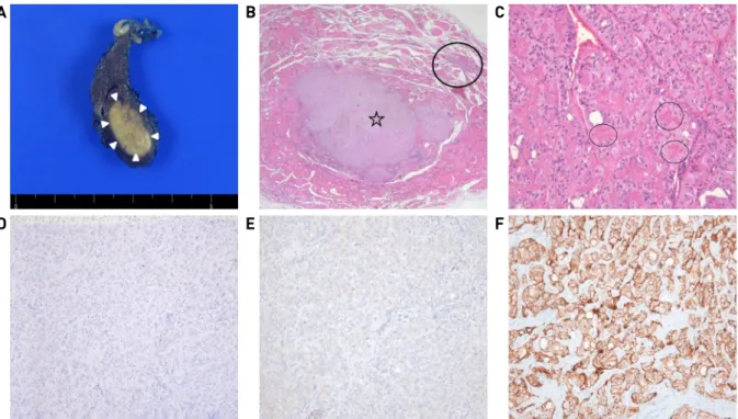
병리 소견에서는 절제된 종물의 절단면은 1.5 × 0.8cm 크기의 경계가 분명하고 단단한 양상의 결절이 갑상선의 하부에 존재하고 있었다(Fig. 3A). 현미경 소견에서는 종 양 세포는 소주형 배열과 함께 주변의 풍부한 유리질성 간질이 관찰되었고, 1 × 2mm 크기의 미세유두상 암종도 공존하였다(Fig. 3B and C). 면역 조직화학 염색에서는 cytokeratin 19, galactin-3 등에는 음성이었고(Fig. 3D and E), MIB(mindbomb E3 ubiquitin protein ligase)-1에는 종양 세포의 핵은 음성 반응을 보이나 세포막을 따라 강한 양성 반응을 보였다(Fig. 3F). 이상의 병리 소견으로 미세 유두상 암종과 공존한 갑상선 유리질 소주형 종양으로 최종 진단되었다. 환자는 수술 후 합병증 없이 퇴원하였 고, 9개월이 지난 현재까지 재발 없이 추적 관찰 중이다.
고 찰
갑상선 유리질 소주형 종양은 1987년 처음 기술된 이 후로 전세계적으로는 약 100례 정도 보고되었고, 현재까
- 35 -
A B C
D E F
Fig. 3. Pathological findings. The surgical specimen shows that 1.5 X 0.8cm sized well-defined ovoid solid mass is present in the lower pole of right thyroid (arrowheads)(A). There are two nodular lesions. The larger one is a hyalinizing trabecular tumor (HTT) (star) and the other is papillary microcarcinoma (circle)(Hematoxylin & Eosin, X10)(B). Tumor cells are arranged in trabecular and alveolar growth patterns with a prominent hyaline stroma (circle)(Hematoxylin & Eosin, X200)(C). HTT cells react negatively for cytokeratin 19 (D) and galactin-3 immunostaining (X100)(E). It reveals strong positive for mindbomb E3 ubiquitin protein ligase-1 along cytoplasmic membranes without nuclear staining (X200)(F).
지 국내에는 26예 보고된 매우 드문 질환이다.1-6)그러나 본 증례와 같이 미세유두상 암종이 공존하는 경우는 이 제까지 보고된 바가 없다. 주로 40대 여성에서 더 흔하며 병인은 불명확하지만, 다결절성 갑상선종, 만성 림프구 성 갑상선염, 갑상선 유두상 암종 등과 연관성이 있다고 알려져 있다.2,6)
종양은 육안 소견에서는 대부분 피막형성이 잘 되어 있어 좋은 경계를 가지며, 현미경 소견에서는 다각형 또 는 방추형 모양의 세포들이 소주형으로 무리 지어서 배 열되어 있으며, 주변에 풍부한 유리질의 간질이 분포한 다.3-5)술 전 진단은 수술 범위를 결정하는데 중요한 요소 이므로 다른 갑상선 악성 종양과 감별이 중요하지만, 진 단이 용이하지 않다. 술 전 세침흡인검사는 진단에 도움 을 줄 수 있으나, 이 종양의 빈도가 매우 드물고 과세포성, 사종체, 핵구 및 핵내 가성 세포질 봉입 등이 관찰되기 때 문에 종종 유두상 암종으로 오인된다.3,4,6)또한, Bethesda 분류 V나 VI 등인 경우, 술 후 병리 검사에서 갑상선 유두 상 암종으로 보고되지 않는 빈도는 2~10% 정도로 알려져 있고, 이런 경우에 최종 조직검사에서 75~85%가 결절성 과증식으로 진단되며, 그 외에 여포성 선종 및 유리질 소주형 종양 등이 원인이 될 수 있다.7,8) 임상적으로 술 전 세포 흡인검사에서 유두상 암종인 경우 초음파 검사 소견에서 석회화가 없으면서 뚜렷한 저에코를 보이면 유
리질 소주형 종양을 의심할 수 있다는 보고가 있다.9)이 외에도 초음파 소견에서 결절이 평행한 방향성(parallel orientation)을 보이거나, 혈관성(presence of vascularity) 등 의 비 특이적인 소견을 보일 수도 있으며,9) 본 증례와 같이 비균질한 등에코성 결절인 경우도 있다. 결론적으 로 본 종양만의 특징적인 초음파 소견은 정립되지 않았 고, 조직학적 확진으로만 최종 진단이 가능하다. 술 중 동결절편 검사에서는 유두상 암종과 정확한 감별은 어렵 지만, 종양 세포의 소주형 배열이 확인되면 더욱 의심할 수 있다.9,10)
병리학적 확진을 위하여 면역조직화학 염색이 필수적 이다. 갑상선 유리질 소주형 종양은 cytokeratin 19와 ga- lactin-3에서 음성이거나 약한 양성을 보이는 반면, 유두 상 암종에서는 강한 양성으로 나타난다.11)또한, 갑상선 글로불린에 양성이고, 칼시토닌에 음성을 보이는 것으 로 수질암과 감별할 수 있다.6)특히, MIB-1 항체는 대부 분 음성 반응을 보이는 반면, 종양의 세포막을 따라서는 특징적으로 강한 양성을 보여, 감별 진단에 유용하다.2,11)
매우 소수의 증례에서 갑상선 유리질 소주형 종양의 피막 침윤과 림프절 전이를 보고하면서 독립된 양성 질 환보다는 갑상선 유두상 암종의 변형된 형태로 분류해야 한다는 의견이 있었다.12)그러나 유두상 암종에서 보이 는 BRAF변이와 N-ras변이 등이 관찰되지 않는 점에서
- 36 - 유두상 암종의 변형으로 보기에는 무리가 있다는 의견이 우세하며, 장기간 추적관찰 결과 재발이나 전이 소견 없 는 양성 종물의 경과를 보인다.13)따라서 일측 엽에 국한 된 갑상선 유리질 소주형 종양의 치료는 엽 절제술로 충분하고, 악성화 가능성이 완전히 배제된 것은 아니므 로 지속적인 추적 관찰은 필요하다.3,5)
갑상선 유리질 소주형 종양은 악성 종양과 비슷한 세 포 모양 및 비특이적 영상학적 소견으로 악성으로 오진 되는 경우가 많고 빈도가 드물어 술 전에 진단하는 것에 는 한계가 있다. 저자들은 이번 증례를 통해 흡인 세포검 사에서 악성이 의심되는 경우라도 드물지만, 최종 조직 검사 결과에서 양성 종양으로 보고될 수 있을 가능성을 염두에 두고, 이에 대해서 환자 및 보호자에게 충분한 설명을 해야 한다는 필요성을 느꼈다. 향후 유사한 증례 를 많이 채집하여, 유리질 소주형 종양만의 임상적 특징 을 특정하여, 술 전 감별할 수 있는 임상 진단 기준에 대한 추가 연구가 필요하다고 생각된다.
References
1) Jang KY, Kim JH, Chung MJ, Moon WS, Kang MJ. Hyalinizing trabecular carcinoma of the thyroid gland: A report of two cases.
Korean J Pathol. 2000;34:318-322.
2) Bârsu C, Bârsu M. Medico-historical overview and histopatho- logical comments about a hyalinizing trabecular tumor case of thyroid gland. Rom J Morphol Embryol. 2014;55:989-992.
3) Howard BE, Gnagi SH, Ocal IT, Hinni ML. Hyalinizing tra- becular tumor masquerading as papillary thyroid carcinoma on fine-needle aspiration. ORL J Otorhinolaryngol Relat Spec.
2013;75:309-313.
4) Lee HK, Kim HS, Hur MH, Kang SS, Lee JH, Lee SK.
Hyalinizing trabecular adenoma of thyroid gland. J Korean Surg Soc. 2002;62:87-90.
5) Park KS, Kim SW, Min HS, Han WS, Noh DY, Park SH, et al.
Hyalinizing trabecular adenoma of thyroid. J Korean Surg Soc.
2003;65:572-575.
6) Seo JH, Kim JP, Woo SH. A case of hyalinizing trabecular ad- enoma of the thyroid gland. Int J Thyroidol. 2017;10:46-49.
7) Yi KI, Ann S, Park DY, Lee JC, Lee BJ, Wang SG, et al.
False-positive cytopathology results for papillary thyroid carci- noma: A trap for thyroid surgeons. Clin Otolaryngol. 2017;42:
1153-1160.
8) Jang EK, Song DE, Gong G, Baek JH, Choi YM, Jeon MJ, et al.
Positive cytology findings and a negative histological diagnosis of papillary thyroid carcinoma in the thyroid: Is it a false-positive cytology or a disappearing tumor? Eur Thyroid J. 2013;2:
203-210.
9) Jang H, Park CK, Son EJ, Kim EK, Kwak JY, Moon HJ, et al.
Hyalinizing trabecular tumor of the thyroid: Diagnosis of a rare tumor using ultrasonography, cytology, and intraoperative fro- zen sections. Ultrasonography. 2016;35:131-139.
10) Sung SY, Shen HY, Hsieh CB, Duh QY, Su TF, Chan DC, et al.
Hyalinizing trabecular tumor of thyroid: Does frozen section prevent unnecessarily aggressive operation? six new cases and a literature review. J Chin Med Assoc. 2014;77:573-577.
11) Lee S, Hong S, Koo JS. Immunohistochemical subclassification of thyroid tumors with a prominent hyalinizing trabecular pattern. APMIS. 2011;119:529-536.
12) Papotti M, Volante M, Giuliano A, Fassina A, Fusco A, Bussolati G, et al. RET/PTC activation in hyalinzing trabecular tumors of the thyroid. Am J Surg Pathol. 2000;24:1615-1621.
13) Carney JA, Hirokawa M, Lloyd RV, Papottie M, Sebo TJ.
Hyalinzing trabecular tumors of the thyroid gland are almost all benign. Am J Surg Pathol. 2008;32:1877-1889.