Vol.21 No.2 p224-230, Dec. 2004
서 론
1)
갑상선 결절은 전체 인구의 40∼50%가 가지
책임저자:이재교, 대구시 남구 대명동 317-1, 영남대학교 의과대학 진단방사선과학교실, Tel: (053) 620-3047, Fax: (053) 653-5484, E-mail: [email protected]
고 있는 흔한 질환이나
1)촉지되는 결절은 5%
미만이고 대부분의 무증상 비촉지 결절이다.
2)최근 고해상 초음파 검사의 발달로 갑상선
갑상선 유두암의 초음파 소견
이 재 교
영남대학교 의과대학 진단방사선과학교실
Sonographic Findings of Thyroid Papillary Carcinoma Jaekyo Lee
Department of Radiology,
College of Medicine, Yeungnam University, Daegu, Korea
-Abstract-
Background: To determine the various sonographic findings in a papillary carcinoma of the thyroid.
Materials and Methods: 48 patients with a proven papillary carcinoma of the thyroid were involved. The sonographic features analyzed were the size, shape, content, margin, internal echo, and calcification pattern.
Results: Common sonographic features of a papillary carcinoma include the hypoechoic texture (94%), an ill defined margin (81%), a solid nodule (100%), irregular shape (48%), and microcalcifications (35%), or no calcifications (42%). The uncommon features included a hyperechoic or mixed echo texture, cystic elements, a well defined margin, and a coarse or peripheral calcifications.
Conclusion: Ill-defined hypoechoic solid nodule with microcalcification is a characteristic ultrasonographic finding of a thyroid papillary carcinoma.
Key Words: Thyroid neoplasm, Ultrasonography
결절의 발견률은 높아졌지만 영상소견 만으로 는 양성 혹은 악성질환의 감별에 제한점이 많 다.
3)유두암은 갑상선 암 중 가장 흔한 형태이 나 초음파 소견에 대한 보고는 드물다. 따라서 본 저자는 유방 초음파 시행 시 함께 시행한 갑상선 초음파 선별검사에서 발견된 갑상선 유 두암의 특징적인 초음파 소견을 알아보고자 하 였다.
대상 및 방법
2001년 12월부터 2003년 12월까지 영남대학 교 의료원에서 유방초음파 검사와 갑상선 초음 파 선별검사를 함께 시행한 7,026명의 여자 환 자 중 갑상선 유두암으로 확진된 45명 48개의 결절을 대상으로 하였고, 수질암으로 확진된 1 예는 대상에서 제외하였다. 모든 예에서 수술 혹 은 흡입 생검에서 유두암으로 확진되었다. 사용
한 초음파 기기는 HDI-5000(Phillips Medical System, Bothell, USA) 기종의 5-10 MHz 선 형 탐촉자를 사용하였다.
영상 분석은 종괴의 크기, 모양, 성상, 경계 면의 양상, 내부에코 및 석회화 유무로 세분화 하였다. 종괴의 모양은 원형, 엽상형, 불규칙형 으로 구분하였고, 성상은 충실성, 낭성 및 복합 성으로, 경계면은 분명한 것과 불분명한 것으 로, 내부에코는 저에코, 등에코, 고에코 등으로 구분하였다.
결 과
유방 초음파와 함께 갑상선 선별 초음파 검 사를 시행한 환자는 모두 7,026명이고 1,364명 에서(19%) 갑상선 결절을 발견할 수 있었다.
조직학적으로 악성 결절로 확진된 증례는 46명 (3.4%)에서 49개 결절이었고, 1명은 수질암이
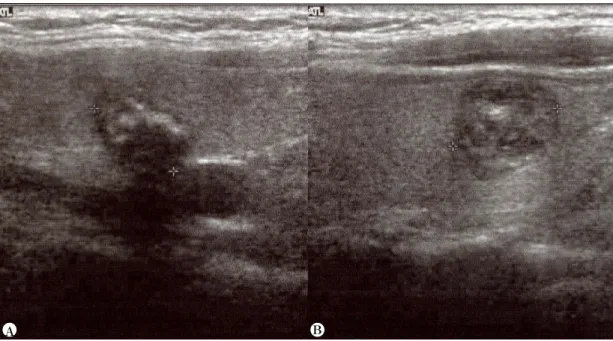
Fig. 1. Two nodules at both thyroid glands in 56 year old female. Left side nodule (A) shows irregular shape, but right one (B) shows round.
A B
었다. 갑상선 유두암으로 확진된 증례는 45명 에서 48개 결절이었고 3명에서 2개의 결절이 각각 유두암으로 확진되었다. 환자들의 나이는 평균 53.6(38-82)세였고, 결절의 평균 크기는 12.1(3-26) mm 였다.
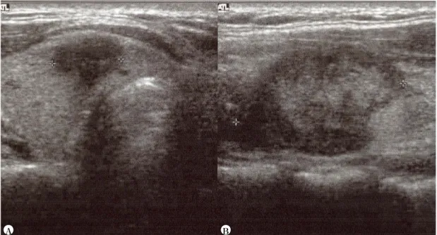
갑상성 유두암의 초음파 소견에서 결절의 모양은 불규칙한 것이 23예(48%)(Fig. 1a), 둥 근형이 19예(40%)(Fig. 1B), 엽상형이 6예(12%) 였고, 경계는 불분명한 것이 39예(81%)(Fig. 2a), 경계가 좋은 것이 9예(19%)(Fig. 2B)였다. 내 부 성상은 전 예에서 충실성 결절로(Fig. 2B) 관찰되었고 낭성이나 혹은 복합성은 없었으며, 종괴의 내부에코는 저에코로 보인 것이 45예 (94%)(Fig. 3A), 고에코로 보인 것이 2예(4%) (Fig. 3B), 등에코로 보인 것이 1예(2%)였다.
종괴의 석회화는 없는 것이 20예(42%), 미세 석회화가 17예(35%)(Fig. 4A), 큰 석회화가 7 예(15%)(Fig. 4B), 주변부 석회화가 4예(8%)였 다(Table 1).
Table. 1. Summary of songraphic findings of thyroid papillary carcinima
Sonographic findings
Number of patients (%) Shape
Irregular Round Lobulating Margin Ill defined Well defined Internal echo Hypoechoic Isoechoic Hyperechoic Calcification None
Microcalcification Dense calcification Wall calcification
23 (48) 19 (40) 6 (12)
39 (81) 9 (19)
45 (94) 1 ( 2) 2 ( 4)
20 (42) 17 (35) 7 (15) 4 ( 8)
Fig. 2. Two patients with ill-defined (A) and well-defined (B) papillary carcinoma.
A B
Fig. 4. Two patients with nodules with calcification. Microcalcification (A) and dense calcification (B) are well demonstrated.
Fig. 3. Two nodules at both thyroid gland in 52 year old female. Right side nodule (A) shows hypoechoic and ill-defined nodule, but left (B) one shows hyperechoic nodule.
A B
A B
고 찰
고해상 초음파 기기를 이용한 갑상선 질환 의 진단은 촉지되지 않는 갑상선종의 양상, 단 발성 혹은 다발성의 유무, 주위조직으로 침범 등을 비침습적이며 간단하고 효과적인 방법으 로 알 수 있어 널리 사용되고 있다.
4-7)일반인 에서 갑상선 결절은 흔한 질환이고, 최근 고해 상 초음파 검사을 발달로 발견율이 높아지고 있으나 발견되는 대부분의 결절은 양성이고, 비 촉지 결절의 조직검사가 증가함에도 악성 결절의 빈도는 5∼6.5%에 불과하다.
8)갑상성 암에서 가장 흔한 유두암의 초음파 소견에 대한 보고는 흔하지만 아직 악성과 양 성의 구별점은 뚜렸하지 않다.
3)최근 Papini 등
8)의 연구에서 8∼15mm 이하 크기의 비 촉 지 유두암의 초음파 영상 분석은, 87%에서 저 에코, 77%에서 불규칙한 경계, 74%에서 내부 혈관음영, 29%에서 미세석회화가 보였고, 양성 결절에서는 저에코 57%, 불규칙한 경계 15%, 내부 혈관음영 19%, 미세석회화 4% 등으로 보 고하며 그 감별점은 불분명하다, 또한 Frates 등
9)이 209 결절의 color doppler 검사에서 고 형의 고혈관 결절은 낭성 저혈관 결절에 비해 악성도가 높다고 발표하였으나, 이 또한 결절 내 혈류 정도에 대한 중복이 심해 감별에 믿을 만한 지표는 아니다.
이상의 다양한 연구와 비교하면 갑상선암의 가장 흔한 형태인 유두암의 특징적인 초음파 소견은 저에코이다. 본 연구에서 48 결절중 45 예(94%)에서 저에코로 보여 다른 연구에서 나 타난 결과 보다 높게 나타났고,
10-11)3 증례에 서만 등에코 혹은 고에코를 보였다. 이전의 연 구에서도 고에코 유두암에 대한 보고는 드물
고, 본 연구에서도 2예(4%)에서 주변 갑상선 조직에 비해 증가된 에코를 보였다.
결절 전체가 낭성 변화를 보인 유두암의 보 고는 드물고, 본 연구에서도 모든 예에서 고형 결절의 형태를 보였다. 이는 타 보고자들에 낭 포성 결절에서 악성의 빈도가 낮은 결과와 일 치하였다.
12)기존의 발표에서 유두암은 경계는 60∼79%
에서 불규칙하다고 보고하였고,
8,11,13)본 연구에 서도 39예(81%)에서 경계가 불규칙하게 보여 유두암의 특징적인 소견으로 볼 수 있다.
종괴의 모양은 불규칙한 것이 23예(48%)로 가장 많았으며 둥근형이 19예(40%), 엽상이 6 예(12%)로 나타났으며 특징적인 양상은 없었다.
결절 내부에 혈류 증가는 갑상선 유두암의 특징적인 소견으로 보고되고 있다.
9)Chan 등
14)은 결절 주변부 혈류증가에 비해 내부 혈류 증 가가 악성 결절의 특징으로 설명하였고, 특히 혈류증가를 보이지 않는 결절에서는 악성 증례 가 하나도 없는 것으로 보고하였다. 본 연구에 서는 유방 초음파와 병행하여 갑상선 선별검사 를 시행한 이유로 혈류 검사를 시행하지 못하 였다.
종괴 내부의 석회화는 24예(50%), 주변부 석 회화가 4예(8%)에서 보였고 석회화를 보이지 않은 경우가 20예(42%)였다. 내부 석회화는 미 세 석회화가 17예(35%), 거대 석회화가 7예(15%) 에서 보여 이는 기존의 연구와 다르지 않다.
종괴 내부의 석회화 그 자체는 악성 종양의 특
이한 소견은 아니라고 알려져 있다. 그러나 보
고자에 따라 유두암의 경우는 1 mm 이하의
psammomatous calcification이 특징적이며, 불
균질한 저에코의 충실성 종괴에서 내부 석회화
를 동반한 경우, 특히, 그 양상이 다발성의 작
은 점상이며 불규칙하게 나타날 때는 감상선암 의 소견으로서 의의가 있다고 하였다.
7,12,15)또 한 석회화가 뚜렷이 보이지 않은 경우에도 술 후 조직 검사상 미세석회화가 포함된 경우가 많아 갑상선 초음파 검사에서 내부 석회화의 유무를 발견하는 것이 고형의 저음영 결절에서 는 중요하다 하겠다.
즉, 기존의 연구에서 갑상선 유두암의 다양 한 악성 소견을 기술하였으나, 본 연구에서는 경계가 불규칙한 저에코의 고형 결절과 동반된 미세석회화가 악성을 시사하는 소견으로 나타 났다.
요 약
유방암 검사와 함께 시행한 갑상선 선별 초 음파 검사에서 나타난 전형적인 갑상선 유두암 의 소견은 경계가 불분명한 고형의 저에코 결 절로 나타나고 점상 혹은 미세석회화를 보일 수 있어 이러한 결절에서는 조직 검사를 통한 확진이 필요하다.
참 고 문 헌
1. Mortensen JD, Woolner LB, Bennett WA.
Gross and microscopic findings in clinically normal thyroid glands. J Clin Endocrinol Metab 1955 Oct;15(10):1270-80.
2. Ross DS. Nonpalpable thyroid nodules: managing an epidemic. J Clin Endocrinol Metab 2002 May;87(5): 1938-40.
3. Clark OH, Duh QY. Thyroid cancer. Med Clin North Am 1991 Jan;75(1):211-34.
4. Muller N, Cooperberg PL, Suen KC. Needle aspiration biopsy in cystic papillary carcinoma
of thyroid. AJR 1985 Feb;144(2):251-3.
5. Simenone JF, Daniels GH, Hall DA, McCarthy K, Kopans DB, Butch RJ, et al. Sonography in the follow up of 100 patients with thyroid carcinoma AJR 1987 Jan;148(1):45-9.
6. Hajek PC, Salomonuitz E, Turk R, Tscholakoff D, Kumpan W, Czembirek H. LN of neck evaluation with US. Radiology 1986 Mar;158(3) :739-42.
7. Solbiati L, Volterrani L, Rizzatto G, Bazzocchi M, Busilacci P, Candiani F, et al. The thyroid gland with low uptake lesion evaluation by ultrasound. Radiology 1985 Apr;155(1):187-91.
8. Papini E, Guglielmi R, Bianchini A, Crescenzi A, Taccogna S, Nardi F, et al. Risk of malignancy in nonpalpable thyroid nodules:
predictive value of ultrasound and color-Doppler features. J Clin Endocrinol Metab 2002 May;
87(5): 1941-6.
9. Frates MC, Benson CB, Doubilet PM, Cibas ES, Marqusee E. Can color doppler sonography aid in th prediction of malignancy of thyroid nodules? J Ultrasound Med 2003 Fed;22(2) :127-31.
10. Watters DA, Ahuja AT, Evans RM, Chick W, King WW, Metreweli C, et al. Role of ultrasound in the management of thyroid nodules Am J Surg 1992 Dec;164(6):654-7.
11. Lu C, Chang TC, Hsiao YL Kuo MS.
Ultrasonographic findings of papillary thyroid carcinoma and their relation to pathologic changes. J Formos Med Assoc 1994 Nov-Dec;
93(11-12):933-8.
12. 박용현, 이종태, 유형식. 결절성 갑상선종의 초 음파 검사의 의의. 대한초음파의학회지 1987;
6(2):106-113.
13. Takashima S, Matsuzuka F, Nagareda T, Tomiyama N, Kozuka T. Thyroid nodules associated with Hashimoto thyroiditis:
assessment with US. Radiology 1992 Oct;185
(1):125-30.
14. Chan BK, Desser TS, McDougall IR, Weigel RJ, Jeffrey RB Jr. Common and uncommon sonographic features of papillary thyroid carcinoma. J Ultrasound Med 2003 Oct;22(10)
:1083-90.
15. 박해원, 박성학, 박인규, 김용주, 강덕식. 갑상선 종괴의 초음파 소견. 대한방사선의학회지 1984;
20(4):789-94.