갑상선 유두상 암종의 한 종류인 미만성 경화성 갑상선 유 두상 암종(Diffuse Sclerosing Variant of papillary thyroid carcinoma, 이하 DSPC)은 다른 종류의 갑상선 유두상 암종 (papillary thyroid carcinoma, 이하 PTC)보다 임상 경과가 빠 르고 좋지 않은 예후를 보인다(1). 대부분의 PTC는 영상진단 에서 갑상선의 결절성 병소로 보이지만, DSPC는 갑상선에 결 절성 병변없이 한쪽 엽이나 양 엽의 미만성 증대로 나타난다 (1-3). PTC의 미만성 경화형에 대한 방사선학적 보고는 알려 진 바가 없다. 저자들은 최근 경험한 DSPC 1예의 초음파 (ultrasonography, US) 및 컴퓨터 단층 촬영 (computed tomography, 이하 CT) 소견을 보고하고자 한다.
증례 보고
평소 건강하게 지내던 38세 여자가 경부 전하방에 4개월 전 부터 만져지는 종괴를 주소로 내원하였으며, 내원 1주 전부터 목이 쉬고 기침도 동반되었다. 과거력상 특이소견은 없었다. 초 음파에서 갑상선이 전체적으로 상당히 커져 있었으며, 갑상선 실질의 에코가 거칠게 감소하고, 미세석회화가 미만성으로 관 찰되고, 색 도플러 상에서 경한 갑상선 실질의 혈관성 증가도 동반되었으나, 갑상선 내 결절성 병변은 관찰되지 않았다. 상 당히 커진 경부 림프절들이 양측성으로 관찰되었고, 림프절의 에코가 거칠게 감소하고, 갑상선 초음파에서와 마찬가지로 미 세석회화 소견을 관찰할 수 있었으며, 림프절의 정상적인 고에 코의 림프절 문부의 소실도 보였다. CT 상에서, 갑상선은 결 절성 병변이 없이 미만성 증대를 보이며 실질 감쇠 소견을 보 이고 커진 림프절들이 양측성으로 관찰되었다. 종격동에서도
경부보다는 작지만 커진 림프절들이 보이고, 종격동의 지방음 영이 증가되고, 1.5 cm의 폐 결절이 좌하엽에서 관찰되었다.
폐 음영 영상에서 몇 개의 작은 전이성 폐 결절들과 림프관성 폐 전이암을 시사하는 소견이 보였다(Fig. 1).
양쪽 갑상선 엽과 오른쪽 경부 림프절에 대한 초음파 유도 하 미세침흡인생검술(fine needle aspiration biopsy, FNAB) 을 실시하였으며, 세포병리학적 검사상에서 전형적인 PTC의 소견이 관찰되었다(Fig. 2). 하지만, 갑상선과 경부 림프절 등 에 대한 외과적 수술이나 자동총생검, 좌하엽 폐 결절에 대한 생검이나 기관지 폐포 세척(bronchoalveolar lavage, BAL) 등 은 시행하지 않았다.
미세흡인생검의 병리결과가 나오자마자 서둘러 환자가 다른 병원으로의 전원을 원해 더 이상의 환자 경과를 알 수 없었으 나, 3개월 후 사망한 사실이 확인되었다.
토 론
PTC는 세포학적으로 다양한 종류의 아형들이 있다. 1953년 Crile 과 Fisher는 처음으로 갑상선 유두상 암종의 미만성 경 화형 변이를 보고하였다(4). 1985년에는 Vickery 등(5)이 조 직학적으로 DSPC를 정립하였다. 갑상선의 악성종양 중 약 90%가 PTC이며, PTC의 1.3-3.4%가 DSPC이라고 한다(2).
DSPC는 전형적인 PTC와 마찬가지로 여자에서 더 흔하나 평 균 발생 연령은 다른 PTC 형보다 어리다(3). 임상적으로 DSPC 는 전형적인 PTC보다 질병의 진행 속도가 빠르며 불량한 예 후를 가진다(1). 초음파 유도하 미세침 흡인 생검술은 PTC의 진단에는 높은 예민도와 특이도를 보이지만, 미세침 흡인 생검 술만으로는 DSPC를 진단하기는 힘들다. 즉, DSPC의 세포학 적 특징은 세포핵 색소 감소증(nuclear hypochromatism), 핵
─ 49 ─ 대한영상의학회지 2006;55:49-52
갑상선 유두상 암종의 미만성 경화형 변종: 증례 보고1
이 승 찬・김 동 욱
미만성 경화형 갑상선 유두상 암종(DSPC)은 갑상선 유두상 암종의 변종이다. 그러나 갑상 선 유두상 암종에 비해 임상경과가 빠르고 나쁜 예후를 보인다. 대부분의 갑상선 유두상 암종 은 결절성 병변으로 보이나 DSPC 는 미만성으로 침습적인 형태를 보이며 결절성 병변은 보이 지 않는다. 저자들은 저자들이 관찰한 1예의 DSPC에서 관찰된 초음파 소견과 컴퓨터 단층 촬 영소견을 보고하고자 한다.
1성균관대학교 의과대학 마산삼성병원 영상의학과
이 논문은 2006년 1월 5일 접수하여 2006년 4월 6일에 채택되었음.
─ 50 ─
이승찬 외: 갑상선 유두상 암종의 미만성 경화형 변종
A B
C D
E F
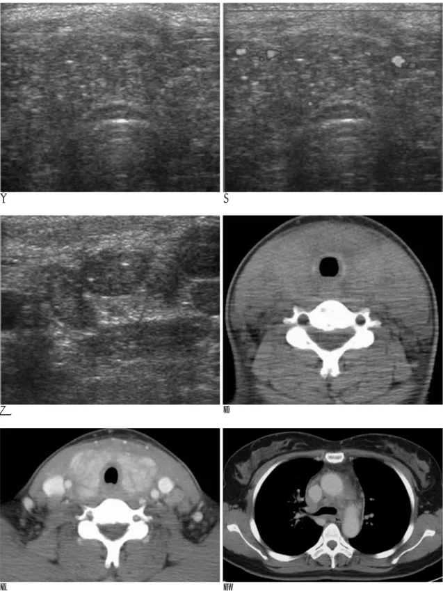
Fig. 1. Radiologic findings on sonography and CT scan
A, B. Ultrasonography of the thyroid shows diffuse glandular enlargement, decreased parenchymal echogenecity, scattered micro- calcification and slightly increased parenchymal vascularity.
C. Ultrasonography of cervical lymph nodes shows moderate enlargement, decreased parenchymal echogenecity and loss of echogenic hila.
D. On non-contrast enhanced CT scan, thyroid shows diffuse enlargement and decreased parenchymal attenuation.
E. On axial view of contrast enhanced CT scan, thyroid shows inhomogeneous parenchymal enhancement and without focal nodu- lar (or mass-like) lesion, and enlarged cervical lymph nodes show inhomogeneous enhancement.
F. On soft tissue setting view of a mediastinal CT scan, mildly enlarged, several mediastinal lymph nodes and increased fat attenua- tion of mediastinum are shown.
내 가성 봉입체(nuclear pseudoin-clusions), 핵 구(nuclear grooves), 유두상 구조(papillary structure) 및 다수의 사종체 (psammoma bodies)등으로 전형적인 PTC와 같다. PTC와 구 별되는 DSPC만의 조직학적 소견은 현저한 경화증과 수많은 사종체들, 심한 림프구의 침습, 그리고 편평 상피화생 등이다 (2, 3, 6). 이 중 DSPC의 가장 특징적인 조직학적 소견은 갑
상선과 그 외 부위에 나타나는 악성 세포의 점진적인 림프관 성 침습이며, 이는 갑상선에 결절성 병변이 없이 한쪽 혹은 양 쪽 갑상선의 미만성 증대를 보이는 병리기전이다. 경부 림프절 전이가 DSPC의 중요한 특징이며, 폐 전이도 흔히 동반된다.
비록 하나의 증례에 불과하지만, 다른 갑상선 질환과 구분 되는 DSPC의 특징직인 소견을 볼 수 있다. 본 증례에서는 좌
─ 51 ─ 대한영상의학회지 2006;55:49-52
G H
I
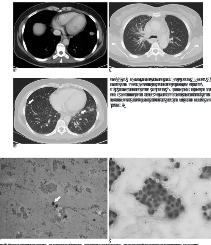
Fig. 1. G. On soft tissue setting view of a chest CT scan, 1.5 cm sized pulmonary nodule is observed at left lower lobe.
H, I. Lung setting view of chest CT scan shows the smooth or some nodular thickening of peribronchial interstitia, thickened interlobular septa and fissures, and small pulmonary nodules (arrow).
A B
Fig. 2. Cytologic findings from the thyroid (random aspiration biopsy from both lobes) and a right cervical lymph node.
A. Several papillary structures and psammoma bodies (arrow) are revealed (×40).
B. The nuclear hypochromasia, intranuclear inclusions, nuclear groove and mitosis are observed (×400).
하엽에 1.5 cm의 폐결절이 일차성 악성 폐 종양 인지 전이성 폐 결절 인지를 알 수 있는 조직검사를 시행하지 않아서 림프 관성 폐 전이암의 소견이 좌하엽의 폐 종양에서 기인한 것인 지 DSPC에서 병발되었는지를 감별할 수 없지만, 갑상선 자체 의 병변과 경부 림프절 전이는 DSPC에 의한 병변으로 생각 할 수 있다. 요약하면, PTC의 세포학적 소견이 갑상선 양 엽 과 경부 림프절에서 관찰되며 다음과 같은 DSPC의 특징적인 방사선학적 소견 즉, 결절성 병변이 없이, 갑상선의 미만성 증 대가 보이며, 미세석회화 소견이 갑상선과 경부 림프절에서 관 찰될 때 DSPC를 진단할 수 있을 것으로 생각되며, DSPC에 서 비롯된 종격동의 림프절 전이나 폐 전이도 진단에 도움이 될 수 있다.
DSPC에 대한 감별 진단에는, 경부의 림프절 전이를 동반하 는 갑상선 림프종이나 갑상선 혹은 갑상선외 경부 편평 상피 세포암이 갑상선염과 경부 림프절 전이를 동반하는 경우 등을 포함할 수 있다.
참 고 문 헌
1. Kebapci N, Efe B, Kabukcuoglu S, Akalin A, Kebapci M. Diffuse sclerosing variant of papillary thyroid carcinoma with primary squamous cell carcinoma. J Endocrinol Invest 2002;25:730-734 2. Soares J, Limbert E, Sobrinho-Simoes M. Diffuse sclerosing variant
of papillary thyroid carcinoma. A clinicopathologic study of 10 cas- es. Pathol Res Pract 1989;185:200-206
3. Carcangiu ML, Bianchi S. Diffuse sclerosing variant of papillary thyroid carcinoma. Clinicopathologic study of 15 cases. Am J Surg Pathol 1989;13:1041-1049
4. Crile G Jr, Fisher ER. Simultaneous occurrence of thyroiditis and papillary carcinoma: report of two cases. Cancer 1953;6:57-62 5. Vickery AL Jr, Carcangiu ML, Johannessen JV, Sobrinho-Simoes
M. Papillary carcinoma. Semin Diagn Pathol 1985;2:90-100 6. Hedinger C, Williams ED, Sobin LH. Histological typing of thyroid
tumours. 2nd ed. Berlin: Springer-Verlag, 1988
─ 52 ─
이승찬 외: 갑상선 유두상 암종의 미만성 경화형 변종
J Korean Radiol Soc 2006;55:49-52
Address reprint requests to : Seung-chan Lee, M.D., Department of Radiology, Masan Samsung Hospital, Sungkyunkwan University, School of Medicine, 50 , Hapseoung-dong, Masan 630-522, Korea.
Tel. 82-55-290-6092 Fax. 82-55-290-6087 E-mail: [email protected]
Diffuse Sclerosing Variant of Papillary Thyroid Carcinoma:
Case Report
1Seung-chan Lee, M.D., Dong-Wook Kim, M.D.
1Department of Radiology, Masan Samsung Hospital, Sungkyunkwan University, School of Medicine
Diffuse sclerosing papillary carcinoma (DSPC) is a variant of papillary thyroid carcinoma (PTC), but it shows more aggressive clinical course and a poorer prognosis than the other types of PTC. Most PTCs show a focal nodular pattern in the thyroid on the imaging modalities, but DSPC reveals a diffuse infiltrating configuration in the thyroid without any focal nodular lesion. To our knowledge, there are scant radiological reports of dif- fuse sclerosing variant of papillary thyroid carcinoma. In this report, we present the case of a patient with DSPC who showed the characteristic findings on sonography and computed tomography.
Index words :Thyroid, neoplasms Ultrasound (US)
Computed tomography (CT)