Clinical Study
Better Understanding in the Differentiation of Thyroid
Follicular Adenoma, Follicular Carcinoma, and Follicular
Variant of Papillary Carcinoma: A Retrospective Study
Jung Hyun Yoon, Eun-Kyung Kim, Ji Hyun Youk, Hee Jung Moon, and Jin Young Kwak
Department of Radiology, Severance Hospital, Research Institute of Radiological Science, Yonsei University College of Medicine, 50 Yonsei-ro, Seodaemun-gu, Seoul 120-752, Republic of KoreaCorrespondence should be addressed to Jin Young Kwak; [email protected] Received 9 January 2014; Accepted 8 September 2014; Published 18 September 2014 Academic Editor: Constantinos Pantos
Copyright © 2014 Jung Hyun Yoon et al. This is an open access article distributed under the Creative Commons Attribution License, which permits unrestricted use, distribution, and reproduction in any medium, provided the original work is properly cited.
Background. To evaluate the role of ultrasonography (US), US-guided fine-needle aspiration (USFNA) and intraoperative frozen section (FS) in follicular neoplasm. Methods. US features, USFNA cytology, and FS results were compared based on the pathology results of patients with follicular adenoma (FA), follicular carcinoma (FC), and follicular variant of papillary thyroid carcinoma (FVPTC). Results. FC and FVPTC showed significantly higher rates of suspicious US features (𝑃 < 0.05) and positive findings on either US or cytology, 80.0% and 90.7%, compared to FA, 64.5% (𝑃 = 0.001). Intraoperative FS showed higher malignant rates
in FVPTC and FC (81.8% and 75.0%, resp.), compared to FA (3.8%,𝑃 < 0.001). Conclusion. Suspicious US features were more
significantly seen in FC and FVPTC compared to FA. Intraoperative FS is useful in the differential diagnosis of these lesions and supplements cytology results of USFNA.
1. Introduction
Thyroid nodules showing follicular morphologic features include adenomatous nodule, follicular adenoma (FA), follic-ular carcinoma (FC), and follicfollic-ular variant of papillary
thy-roid carcinoma (FVPTC) [1]. Cytologic features are known
to overlap among these tumors [2,3], and definite diagnosis
of FA, FC, and FVPTC is mostly obtained by pathologic
examination following complete excision of the lesion [1,4,5].
The diagnosis of a solitary, encapsulated nodule with follicular histology features is frequently problematic since a broad range of benign to malignant subtypes of follicular tumors need to be differentiated, such as FA, FC, and
FVPTC [6]. Differential diagnosis of FC from FA is based
on the presence of capsular, vascular, or extrathyroidal tissue
invasion, and nodal or distant metastasis [4,6,7]. Diagnosis
of follicular neoplasm based on cytology alone has always been challenging to both clinicians and cytopathologists,
since it is well known that cytologic features overlap in both
benign follicular adenoma and carcinomas [2,3,8,9]. Studies
have investigated ways in providing additional information that may be helpful in differential diagnosis and surgical
planning of follicular neoplasm [4,6,10,11] but controversy
still remains and clinicians are still skeptical until they see the conclusive reports on permanent section.
Diagnostic criteria for the cytologic diagnosis of FVPTC are in general similar to those of PTC, that is, cells contain-ing fine chromatic, nuclear grooves, intranuclear inclusions,
and overlapping nuclei [12–15], but FVPTC lacks papillary
groups and shows follicular patterns with variable colloid component, which can also be seen in benign and neoplastic
follicular lesions [1,13]. This overlap makes accurate cytologic
diagnosis difficult in FVPTC and results in the low sensitivity (25% to 42%) of fine-needle aspiration (FNA) in the diagnosis of FVPTC, compared to conventional papillary carcinoma
(sensitivity range from 60% to over 90%) [13–15].
Volume 2014, Article ID 321595, 9 pages http://dx.doi.org/10.1155/2014/321595
False-negative cytologic results are also occasionally observed, for example, follicular carcinomas containing ma-crofollicular pattern with abundant background colloid can be easily mistaken as a benign adenomatoid colloid nodule
on cytology [16]. Even with surgery, differential diagnosis
between FA, minimally invasive FC, and FVPTC in a solitary, encapsulated nodule showing follicular histology has been
problematic [6]. While there are several studies focusing
on ways to differentiate these neoplasms [5,7,15,16], little
has been evaluated in association between the ultrasound (US) features or the cytology results of USFNA within these tumors. In this study, we evaluated the differences in US features and the role of US-guided fine-needle aspiration (USFNA) and intraoperative frozen section (FS) in FA, FC, and FVPTC.
2. Materials and Methods
This retrospective study was approved by the institutional review board (IRB) of Severance Hospital, Yonsei University, Seoul, Republic of Korea. Neither patient approval nor informed consent was required for review of medical records or images. Informed consent was signed and obtained from all patients before USFNA or surgery prior to procedures.
2.1. Study Population. From January 2003 to December 2008,
our institutional database was reviewed for patients diag-nosed with FA, FC, and FVPTC after surgical excision. A total of 281 patients with 282 thyroid nodules were included in this study. Among them, 51 patients were excluded because they had either undergone USFNA at an outside clinic or had not undergone preoperative cytologic diagnosis. In total, 230 patients with 231 thyroid nodules were included in this study. Of the 230 patients, 45 (19.6%) were men, and 185 (80.4%) were women. Mean age of the 230 patients included was 44.0 years. Mean size of the 231 thyroid nodules was 27.3 mm. Medical records, US images and radiological reports, and cytopathologic reports of these patients were reviewed, retrospectively.
2.2. US Imaging and Imaging Analyses. US was performed in
all patients using a 7–15 MHz linear array transducer (HDI 3000 or 5000; Philips Medical Systems, Bothell, WA) or a 5–12 MHz linear array transducer (iU22; Philips Medical System). Compound imaging was obtained in all images using HDI5000 or iU22 machines.
Real-time US was performed by 1 of the 5 board-certified radiologists with 1–13 years of experience in thyroid imaging. US features of the thyroid nodules were retrospectively reviewed and analyzed by one dedicated thyroid radiologist (Y.J.H) with 3 years of experience. The radiologist was blinded to the clinical and cytopathological information of the patient during image review. US features of each thyroid nodule were described according to internal components, echogenicity,
margin, calcifications, and shape [5]. Internal components
were divided into solid nodules, mixed solid, and cystic nodules, that is, mainly solid nodules containing more than 50% of solid contents, mainly cystic nodules containing less
than 50% of solid contents, and cysts. Echogenicity was divided into hyper or isoechoic (nodules showing hyperecho-genicity or isoechohyperecho-genicity compared with the adjacent nor-mal thyroid parenchyma), hypoechoic (nodules showing hypoechogenicity compared to the adjacent normal thyroid parenchyma), and markedly hypoechoic (nodules showing hypoechogenicity compared to the adjacent strap mus-cle). Margin was classified as circumscribed or noncircum-scribed (i.e., microlobulated or irregular margins). Calcifi-cations were classified as microcalcifiCalcifi-cations (tiny, punctate,
echogenic foci measuring less than 1 mm) [17] or mixed
microcalcifications with macrocalcifications, macrocalcifica-tions (including eggshell calcificamacrocalcifica-tions), and no calcificamacrocalcifica-tions. Shape was divided into parallel or nonparallel (greater in the anteroposterior dimension than the transverse dimension, or “taller-than-wide”). Malignant US features were defined as marked hypoechogenicity, noncircumscribed margins, microcalcifications or mixed calcifications, and nonparallel
shape, based on previously published criteria [18]. Final
assessments of the thyroid nodules were given as probably benign (when none of the suspicious US features described above was present) or suspicious malignant (when 1 or more suspicious US features above were present).
2.3. USFNA and Cytological Analyses. USFNA was
subse-quently performed by the same radiologist who obtained the real-time US images. USFNA was performed either on the thyroid nodules showing suspicious US features or on the nodule with the largest size without any suspicious US features.
USFNA was performed at least twice from the targeted thyroid nodule using a 23-gauge needle attached to a 20 mL disposable syringe with an aspirator or a 23-gauge needle attached to a 2 mL disposable syringe without an aspirator, depending on the radiologist’s preference. Local anesthesia was not routinely applied. Aspirated material was expelled on to glass slides, smeared, and immediately placed in 95% alcohol for Papanicolaou staining. The remaining material in the syringe was rinsed in normal saline for cell block processing. The cytopathologists were not present during USFNA procedures, and additional staining was performed on a case-by-case basis at the request of the cytopathologist.
One of the 5 cytopathologists specializing in thyroid pathology interpreted the slides obtained from USFNA. During the study period, cytologic reports were divided into the following categories: (1) malignant, (2) suspicious for malignant, (3) indeterminate, (4) benign, and (5) inadequate
[5,19–21]. Malignancy indicated specimen showing abundant
cells with unequivocal cytologic features of malignancy. Suspicious for malignancy was used in specimen showing cytologic atypia, that is, crowded, overlapping, pleomor-phic, and enlarged nuclei, but with insufficient cellularity
for definite diagnosis of malignancy [19, 21]. Indeterminate
cytology included follicular neoplasm and H¨urthle cell neo-plasm, indicating specimen showing monotonous cellular population, scanty colloid, and lacking papillary carcinoma
features [22]. Benign cytology includes colloid nodules,
and postpartum thyroiditis. Inadequate cytology indicates specimen showing less than the required minimum of six groupings of well-preserved thyroid cells, each consisting of
less than 10 cells per group [19,20].
2.4. Surgical Procedures and Intraoperative Frozen Section.
The extent of surgery was performed based on the cytology results and US features. A lobectomy, subtotal thyroidectomy, or total thyroidectomy was performed if cytology findings were diagnosed as malignancy or suspicious for malignancy or if the US features were assessed as suspicious malignant in nodules with benign cytology diagnosis. A lobectomy, or subtotal thyroidectomy, was performed if the cytology results were benign. Of the cytology results was inadequate or indeterminate, the extent of thyroid surgery was based on intraoperative FS during surgery.
Tissue samples including the thyroid nodule and/or the adjacent thyroid parenchyma were obtained and processed for FS analyses. Frozen tissue samples were subsequently cut and stained for diagnosis. After diagnosis was made, results were notified to the operation room. Diagnosis was classified into the following 3 categories in FS: (1) malignant, (2) benign,
and (3) deferred results, including follicular neoplasm [5,21].
2.5. Statistical Analyses. Histopathologic results from surgery
were considered standard reference. In comparison to the mean age and mean size of thyroid nodules on US among the three neoplasms, Analysis of variance (ANOVA) test and
post hoc test was used.𝜒2-test or Fisher’s exact test was used
in comparison to US features among the final pathology of the disease. Diagnostic performances including sensitivity, specificity, positive predictive value (PPV), negative predic-tive value (NPV), and accuracy were calculated for USFNA cytology and intraoperative FS results. In regard to USFNA, inadequate cytology was excluded during calculation of diag-nostic performances, considering benign cytology as negative and indeterminate, suspicious for malignancy, and malignant cytology as positive. For comparison with intraoperative FS, diagnostic performances of USFNA excluding both inade-quate and indeterminate cytology were also calculated. In regard to FS, deferred results were excluded when obtaining
diagnostic performances [5].
𝑃 value of less than 0.05 was considered significant. Sta-tistical analysis was performed by the SAS system (MAGREE SAS Macro program; SAS Institute, Cary, NC).
3. Results
Of the 231 thyroid lesions, 152 (65.8%) were diagnosed as FA, 25 (10.8%) as FVPTC, and 54 (23.4%) as FC on surgical pathology. Mean age and size among the three neoplasms are
summarized inTable 1. Mean age of the nodules diagnosed
as FC was the oldest, 47.2 ± 17.7 years, with statistical
significance (𝑃 = 0.042). When comparing FC to FA, mean age was also significantly older in FC (𝑃 = 0.034). No significant differences were observed in mean age when comparing between FVPTC and FA or between FVPTC and FC (𝑃 = 0.101 and 0.991, resp.). Mean size of the nodules
diagnosed as FVPTC was the smallest,16.3 ± 14.6 mm, with
statistical significance (𝑃 < 0.001). FVPTC was significantly
smaller than FA, 29.7 ± 14.5 mm to 16.3 ± 14.6 mm (𝑃 <
0.001), but tumor size between FC and FA did not show statistical significance (𝑃 = 0.126).
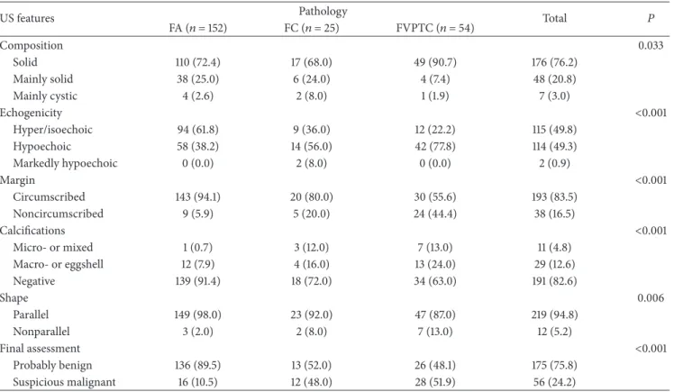
US features of the 231 thyroid nodules are summarized inTable 2. Of the 152 nodules diagnosed as FA, 136 (89.5%) showed no suspicious US features. In contrast, 12 (48.0%) of the 25 nodules diagnosed as FC and 28 (51.9%) of the 54 nodules diagnosed as FVPTC showed one or more suspicious US features. Lesions diagnosed as FC and FVPTC showed significantly higher rates of suspicious US features compared to FA (𝑃 < 0.001). Suspicious US features such as hypoechogenicity or marked hypoechogenicity, noncircum-scribed margins, presence of micro- or macrocalcifications, or nonparallel orientation were significantly associated with FC or FVPTC than FA (𝑃 < 0.05).
Results of USFNA cytology are summarized in Table 3
and Figure 1. Rate of inadequate cytology on USFNA was higher in FA (18.4%) compared to FC (4.0%) and FVPTC (7.4%). Also, rate of benign and indeterminate cytology was relatively higher in FA (23.7% and 36.2%) and FC (24.0% and 52.0%) compared to FVPTC (5.6% and 7.4%, resp.). In con-trast, rate of suspicious for malignancy and malignant cytol-ogy was higher in FVPTC (31.5% and 48.1%) than FA (17.1% and 4.6%) or FC (20.0% and 0.0%, resp.). When considering each type of neoplasm, 88 of 152 (57.9%) nodules diagnosed as FA, 18 of 25 (72.0%) nodules diagnosed as FC, and 47 of 54 (87.0%) nodules diagnosed as FVPTC were diagnosed as indeterminate, suspicious for malignancy or malignancy on cytology, showing statistical significance (𝑃 < 0.001).
Of the 231 thyroid nodules, 156 (67.5%) underwent
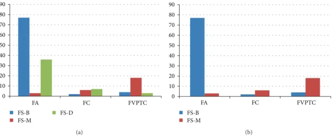
intra-operative FS (Table 3, Figure 2). Among them, 46 (29.5%)
were deferred to final pathology. Malignant results on intra-operative FS significantly correlated to FC or FVPTC on final pathology (𝑃 < 0.001). Two of the 15 nodules diagnosed as FC and 4 of the 25 nodules diagnosed as FVPTC showed false-negative results on intraoperative FS. Five of the 6 (83.3%) nodules showing false-negative FS were diagnosed as suspicious for malignancy or malignancy on USFNA. Also, 3 of the 116 nodules diagnosed as FA showed false-positive results on intraoperative FS.
Diagnostic performances of USFNA and intraoperative
FS are summarized inTable 4. Specificity of USFNA was low,
29.3%, when considering indeterminate cytology as positive. Overall diagnostic performances of intraoperative FS were higher than USFNA.
4. Discussion
Follicular adenomas are well-encapsulated thyroid neo-plasms which do not show the typical invasiveness of follic-ular carcinoma, nor abnormal nuclear features of papillary
carcinomas [7]. FA and FC, along with FVPTC, are
well-encapsulated lesions, sharing many imaging and cytologic
features, and show relatively benign US features [7,23,24]. In
our study, tumor size of FVPTC was significantly smaller than FC or FA, 16.3 mm to 36.4 mm and 29.7 mm, respectively. As
Table 1: Comparison of mean age and size among the 231 thyroid nodules diagnosed as follicular adenoma, follicular carcinoma, and follicular variant of papillary thyroid carcinoma.
Pathology 𝑁 Age (years) 𝑃 Size (mm) 𝑃
Mean± SD Min Max 0.042 Mean± SD Min Max <0.001
FA 152 42.5± 12.8 19.0 72.0 — 29.7± 14.5 6.0 73.0 —
FC 25 47.2± 17.7 15.0 78.0 0.230∗ 36.4± 20.2 13.0 100.0 0.126∗
FVPTC 54 46.9± 9.9 27.0 64.0 0.101∗ 16.3± 14.6 3.0 100.0 <0.001∗
FA: follicular adenoma. FC: follicular carcinoma.
FVPTC: follicular variant papillary thyroid carcinoma. 𝑁: number of cases.
SD: standard deviation.
∗values when compared to follicular adenoma.
Table 2: Comparison of US features among the 231 thyroid nodules diagnosed as follicular adenoma, follicular carcinoma, and follicular variant of papillary thyroid carcinoma.
US features Pathology Total 𝑃
FA (𝑛 = 152) FC (𝑛 = 25) FVPTC (𝑛 = 54) Composition 0.033 Solid 110 (72.4) 17 (68.0) 49 (90.7) 176 (76.2) Mainly solid 38 (25.0) 6 (24.0) 4 (7.4) 48 (20.8) Mainly cystic 4 (2.6) 2 (8.0) 1 (1.9) 7 (3.0) Echogenicity <0.001 Hyper/isoechoic 94 (61.8) 9 (36.0) 12 (22.2) 115 (49.8) Hypoechoic 58 (38.2) 14 (56.0) 42 (77.8) 114 (49.3) Markedly hypoechoic 0 (0.0) 2 (8.0) 0 (0.0) 2 (0.9) Margin <0.001 Circumscribed 143 (94.1) 20 (80.0) 30 (55.6) 193 (83.5) Noncircumscribed 9 (5.9) 5 (20.0) 24 (44.4) 38 (16.5) Calcifications <0.001 Micro- or mixed 1 (0.7) 3 (12.0) 7 (13.0) 11 (4.8) Macro- or eggshell 12 (7.9) 4 (16.0) 13 (24.0) 29 (12.6) Negative 139 (91.4) 18 (72.0) 34 (63.0) 191 (82.6) Shape 0.006 Parallel 149 (98.0) 23 (92.0) 47 (87.0) 219 (94.8) Nonparallel 3 (2.0) 2 (8.0) 7 (13.0) 12 (5.2) Final assessment <0.001 Probably benign 136 (89.5) 13 (52.0) 26 (48.1) 175 (75.8) Suspicious malignant 16 (10.5) 12 (48.0) 28 (51.9) 56 (24.2)
Note: percentages are in parentheses.
mentioned above, thyroid lesions of follicular pattern tend to represent more benign features on US and, therefore, may have not undergone diagnostic procedures such as USFNA unless they have reached sizes over 10 mm or until they have grown to sizes that may have brought about clinical significance such as presence of symptoms.
Common suspicious US features such as microlobulated or irregular margins, marked hypoechogenicity, taller-than-wide shape, and presence of microcalcifications are used in differentiating papillary thyroid carcinoma with high diagnostic accuracy but do not seem to work the same when differentiating between lesions of follicular patterns
[10]. US features reported for follicular neoplasm or FVPTC
are relatively benign appearing, showing well-defined, solid
mass with oval shape, surrounding hypoechoic rim [10, 15,
23, 25], among which findings do not significantly differ
between benign FA or malignant FC or FVPTC. Our results showed that 52.0% of FC and 48.1% of FVPTC had no suspicious US features, consistent with other reports in that malignant form of follicular neoplasm has relatively benign appearance on US. However, several suspicious US features of papillary thyroid carcinoma such as microlobulated or ill-defined margins, microcalcifications, and taller-than-wide shape have been reported to be more significantly seen in the malignancy among nodules showing indeterminate cytology
T a ble 3: C o rr ela ti o n o f US FN A cyt o log y and in tr ao p era ti ve fr o zen se ct io n resul ts to final pa th o log y. C yt o log y 𝑁 (%) P at h o log y FA FC FVPT C To ta l FS-B FS-D FS-M To ta l FS-B FS-D FS-M T o ta l F S-B F S-D F S-M 𝑛 =1 16 ∗ 𝑛 =1 5 † 𝑛 =2 5 ‡ In adeq ua te 33 (1 4.3) 28 (18.4) 18 6 0 1 (4.0) 1 0 0 4 (7 .4) 0 0 3 B enign 4 5 (1 9.5) 36 (2 3.7) 16 2 1 6 (2 4.0) 0 3 0 3 (5.6) 0 0 0 In det er mina te 72 (3 1.2) 55 (3 6.2) 25 22 0 13 (5 2.0) 0 4 5 4 (7 .4) 0 0 3 Su sp icio us fo r m aligna nc y 4 8 (20.8) 26 (1 7. 1) 14 6 2 5 (20.0) 1 0 1 17 (3 1.5) 2 3 6 M aligna n cy 33 (1 4.2) 7 (4.6) 4 0 0 0 (0.0) 0 0 0 26 (4 8.1) 2 6 0 T o ta l 23 1 15 2 77 (6 6.4) 36 (3 1.0) 3 (2.6) 25 2 (13.3) 7 (4 6.7) 6 (4 0.0) 54 4 (16.0) 3 (12.0) 18 (7 2.0) T o ta le xc ludin g d ef er 110 80 77 (9 6.3) — 3 (3.8) 8 2 (2 5.0) — 6 (7 5.0) 22 4 (18.2) — 18 (8 1.8) P erc en ta ge s are in p are n th es es . FS-B: b enign o n fr o zen sec tio n . FS-D: d ef er re d o n fr o zen sec tio n . FS-M: m al igna nc y o n fr o zen sec tio n . ∗36 ca se s w it h o u t F S ex cl u d ed . †10 ca se s w it ho u t FS excl ud ed . ‡29 cas es w it ho u t FS ex cl uded .
Table 4: Diagnostic performances of USFNA and intraoperative frozen section. (%) FNA∗ FNA† FS‡ Sensitivity 87.8 (65/74) 84.2 (48/57) 80.0 (24/30) Specificity 29.0 (36/124) 52.2 (36/69) 96.3 (77/80) PPV 42.5 (65/153) 59.3 (48/81) 88.9 (24/27) NPV 80.0 (36/45) 80.0 (36/45) 92.8 (77/83) Accuracy 51.0 (101/198) 66.7 (84/126) 91.8 (101/110)
FNA: fine needle aspiration; FS: frozen section; PPV: positive predictive value; NPV: negative predictive value.
Note: raw data are in parenthesis.
∗Inadequate cytology results excluded, indeterminate, suspicious for
malig-nancy and malignant cytology results considered positive.
†Inadequate and indeterminate cytology results excluded.
‡46 nodules excluded due to deferred results on FS.
0 10 20 30 40 50 60 FA FC FVPTC In ad eq u at e B enign In det er mina te Su sp icio us f o r ma lig n an cy M aligna nc y
Figure 1: Results of USFNA cytology of the 231 thyroid nodules. Note: numbers in image represent percentages (%).
associated with FC or FVPTC than FA. Although FC or FVPTC do not show the typical suspicious US features as frequently as conventional PTC, the presence of each indi-vidual US features may have a role in leading the radiologists or clinicians in differentiating these lesions from FA.
While USFNA is widely used in discriminating between benign and malignancy in various lesions of the thyroid showing excellent performances (sensitivity 65%–98%,
speci-ficity 72%–100%) [3, 5, 26, 27] this has limited value in
the differential diagnosis of follicular neoplasm, in which
USFNA is considered only as a “screening test” [28]. Nodules
diagnosed as follicular neoplasm or suspicious for follicular neoplasm on cytology mostly undergo surgery for diagnostic purposes, but the true role of USFNA cytology results in pre-dicting diagnosis of follicular neoplasm has not been clarified. Indeed, sensitivity of USFNA in the diagnosis of FVPTC has been reported to be lower than PTC, ranging from 25.0%
to 32.0% [13, 15, 29, 30]. Cytologic diagnosis of follicular
patterned lesions of the thyroid with USFNA is imprecise, although one can predict a diagnosis but cannot reach a final
conclusion based on cytology alone [1]. Results of our study
showed higher rates of benign cytology in FA (23.7%) and FC (24.0%) compared to FVPTC (5.6%). Cytology specimen showing multinodular process with intervening colloid-rich thyroid tissue is often seen not only in follicular neoplasm
but also in benign adenomatoid nodules [1,31], which may
have been a cause for false-negative results. Another cause for benign cytology results in FC may be failure to sample
in FC with cystic areas [32]. Nearly 32.0% of FC included
in our study revealed cystic portions within the tumor on preoperative US, which may have been one of the causes for benign results on USFNA.
The diagnosis rate of FVPTC on USFNA cytology is
low in clinical practice, ranging from 9.0% to 36.0% [13,33,
34]. Unlike conventional papillary carcinoma, the presence
of abundant colloid, subtle nuclear features of papillary carcinoma, and the absence of papillary formations and psammomatous bodies are the known causes that interfere
with the definite diagnosis on cytology [22,32]. But a recent
study suggested that some cytologic features of conventional PTC such as fine chromatic, nuclear grooving, and
intranu-clear inclusions are present at high frequency in FVPTC [13].
Although present with a wide variance, these specific features may help in classifying FVPTC towards indeterminate or suspicious for papillary carcinoma which is enough to lead
towards surgical management [13]. Our study showed higher
rates of suspicious for malignancy or malignant cytology results in FVPTC (31.5% and 48.1%) than FA (17.1% and 4.6%) or FC (20.0% and 0.0%), and the cytology features of FVPTC mentioned above may have contributed to these results. In addition, cystic changes, hemorrhage, and degeneration of
collagen can be found in FA [35–37], and along with the
typical “spoke and wheel” vascularity pattern characteristic for FA may have been the causes for high rates of inadequate cytology (18.4%) compared to FC (4.0%) and FVPTC (7.4%)
[37,38].
Intraoperative FS has been popularly used in the diagno-sis of thyroid nodules, having an important role in deciding
the surgical extent based on its results [39, 40]. Although
it is not useful in the differential diagnosis of benign to
malignant thyroid nodules [21, 41], it is often used as a
supplement to preoperative USFNA. Controversy remains in the role of intraoperative FS in follicular neoplasm. Some proved increased specificity, but lower sensitivity compared
to USFNA, diagnostic accuracy ranging from 50% to 98% [5,
42–44], while others claim that FS does not effectively provide
any additional information in the diagnosis of follicular
neoplasm [45]. In one study on USFNA and FS, both FNA
and FS were highly accurate in predicting final pathology when the diagnosis was papillary carcinoma or benign but
missed 44% of the malignancies in follicular lesions [39].
Diagnostic performances of intraoperative FS when exclud-ing the deferred results in our study showed high sensitivity (80.0%), specificity (96.3%), and accuracy (91.8%), showing
better performances than USFNA as in a recent report [44].
FVPTC and FC showed significantly higher malignant results in intraoperative FS, 81.8% and 75.0%, respectively, compared to FA, 3.8%. These results are similar to a previous study suggesting that with intraoperative FS, nearly 52% to 60% of the malignant subtype of follicular neoplasm do not
0 10 20 30 40 50 60 70 80 90 FA FC FVPTC FS-B FS-M FS-D (a) 0 10 20 30 40 50 60 70 80 90 FA FC FVPTC FS-B FS-M (b)
Figure 2: Results of intraoperative frozen section of the 231 thyroid nodules. Note: numbers in image represent percentages (%).
require secondary procedures [44]. Also, among the nodules
showing false-negative intraoperative FS results, 83.3% (5 of
6 diagnosed as benign on FS, Table 3) were diagnosed as
suspicious for malignancy or malignancy on USFNA, which further supports the complementary relation of USFNA and intraoperative FS in lesions of follicular pattern in thyroid
[39].
There are several limitations to our study. First, this study was in a retrospective design, including patients diagnosed as FA, FC, or FVPTC on surgery. Selection bias may have existed in patient inclusion. Second, 5 cytopathologists were involved in interpretation of cytology, intraoperative FS, and final pathologic diagnosis. Observer variability on the diagnosis of follicular neoplasm may have affected the results of our study
[1]. Third, vascularity of the nodule on Doppler US was not
considered in the analysis of US features among the subtypes of follicular neoplasm included in this study. Controversy remains in the role of vascularity on US in the diagnosis of
thyroid nodules [46,47], and how it would apply to follicular
neoplasm is yet to be explained.
In conclusion, suspicious US features were more signifi-cantly seen in FC and FVPTC compared to FA. Intraoperative FS is useful in the differential diagnosis of these lesions and supplements cytology results of USFNA.
Conflict of Interests
The authors declare that there is no conflict of interests regarding the publication of this paper.
Authors’ Contribution
Jung Hyun Yoon was involved in acquisition of data, analysis and interpretation of data, and paper construction. Eun-Kyung Kim was involved in paper drafting and revision. Ji Hyun Youk participated in study design and paper revision. Hee Jung Moon was involved in paper drafting and revision. Jin Young Kwak mainly contributed to conception and
design, drafting the paper, and final approval of the version to be published.
References
[1] R. Duggal, A. Rajwanshi, N. Gupta, and R. K. Vasishta, “Inter-observer variability amongst cytopathologists and histopathol-ogists in the diagnosis of neoplastic follicular patterned lesions of thyroid,” Diagnostic Cytopathology, vol. 39, no. 4, pp. 235–241, 2011.
[2] T. S. Greaves, M. Olvera, B. D. Florentine et al., “Follicular lesions of thyroid: a 5-year fine-needle aspiration experience,” Cancer, vol. 90, no. 6, pp. 335–341, 2000.
[3] M. J. Kim, E.-K. Kim, B. M. Kim et al., “Thyroglobulin measurement in fine-needle aspirate washouts: the criteria for neck node dissection for patients with thyroid cancer,” Clinical Endocrinology, vol. 70, no. 1, pp. 145–151, 2009.
[4] R. E. Goldstein, J. L. Netterville, B. Burkey, and J. E. Johnson, “Implications of follicular neoplasms, atypia, and lesions sus-picious for malignancy diagnosed by fine-needle aspiration of thyroid nodules,” Annals of Surgery, vol. 235, no. 5, pp. 656–664, 2002.
[5] J. H. Yoon, J. Y. Kwak, E.-K. Kim et al., “How to approach thy-roid nodules with indeterminate cytology,” Annals of Surgical Oncology, vol. 17, no. 8, pp. 2147–2155, 2010.
[6] D. Dosen, M. Turic, J. Smalcelj, R. Janusic, M. P. Grgic, and V. Separovic, “The value of frozen section in intraoperative surgical management of thyroid follicular carcinoma,” Head & Neck, vol. 25, no. 7, pp. 521–528, 2003.
[7] V. V. Vasko, J. Gaudart, C. Allasia et al., “Thyroid follicular adenomas may display features of follicular carcinoma and follicular variant of papillary carcinoma,” European Journal of Endocrinology, vol. 151, no. 6, pp. 779–786, 2004.
[8] B. Miller, S. Burkey, G. Lindberg, W. H. Snyder III, and F. E. Nwariaku, “Prevalence of malignancy within cytologically inde-terminate thyroid nodules,” The American Journal of Surgery, vol. 188, no. 5, pp. 459–462, 2004.
[9] J. Smith, R. E. Cheifetz, N. Schneidereit, K. Berean, T. Thomson, and C. Porter, “Can cytology accurately predict benign follicular nodules?” The American Journal of Surgery, vol. 189, no. 5, pp. 592–595, 2005.
[10] N. Fukunari, M. Nagahama, K. Sugino, T. Mimura, and K. Ito, “Clinical evaluation of color doppler imaging for the differential diagnosis of thyroid follicular lesions,” World Journal of Surgery, vol. 28, no. 12, pp. 1261–1265, 2004.
[11] F. Monzani, N. Caraccio, P. Iacconi et al., “Prevalence of cancer in follicular thyroid nodules: is there still a role for intraoperative frozen section analysis?” Thyroid, vol. 13, no. 4, pp. 389–394, 2003.
[12] D. K. Das, M. K. Mallik, P. Sharma et al., “Papillary thyroid carcinoma and its variants in fine needle aspiration smears: a cytomorphologic study with special reference to tall cell variant,” Acta Cytologica, vol. 48, no. 3, pp. 325–336, 2004. [13] E. M. Kurian, M. Dawlett, J. Wang, Y. Gong, and M. Guo,
“The triage efficacy of fine needle aspiration biopsy for follicular variant of papillary thyroid carcinoma using the Bethesda reporting guidelines,” Diagnostic Cytopathology, vol. 40, no. 1, pp. E69–E73, 2012.
[14] S. R. Shih, C. T. Shun, D. H. Su, Y. L. Hsiao, and T. C. Chang, “Follicular variant of papillary thyroid carcinoma: diagnostic limitations of fine needle aspiration cytology,” Acta Cytologica, vol. 49, no. 4, pp. 383–386, 2005.
[15] H. Y. Jung, E.-K. Kim, W. H. Soon, Y. K. Jin, and J. K. Min, “Sonographic features of the follicular variant of papillary thyroid carcinoma,” Journal of Ultrasound in Medicine, vol. 27, no. 10, pp. 1431–1437, 2008.
[16] G. M. Sclabas, G. A. Staerkel, S. E. Shapiro et al., “Fine-needle aspiration of the thyroid and correlation with histopathology in a contemporary series of 240 patients,” The American Journal of Surgery, vol. 186, no. 6, pp. 702–710, 2003.
[17] J. H. Yoon, E.-K. Kim, E. J. Son, H. J. Moon, and J. Y. Kwak, “Diffuse microcalcifications only of the thyroid gland seen on ultrasound: clinical implication and diagnostic approach,” Annals of Surgical Oncology, vol. 18, no. 10, pp. 2899–2906, 2011. [18] E.-K. Kim, C. S. Park, W. Y. Chung et al., “New sonographic criteria for recommending fine-needle aspiration biopsy of nonpalpable solid nodules of the thyroid,” The American Journal of Roentgenology, vol. 178, no. 3, pp. 687–691, 2002.
[19] J. Y. Kwak, E.-K. Kim, H. J. Kim, M. J. Kim, E. J. Son, and H. J. Moon, “How to combine ultrasound and cytological infor-mation in decision making about thyroid nodules,” European Radiology, vol. 19, no. 8, pp. 1923–1931, 2009.
[20] J. Y. Kwak, E.-K. Kim, M. J. Kim et al., “The role of ultrasound in thyroid nodules with a cytology reading of “suspicious for papillary thyroid carcinoma”,” Thyroid, vol. 18, no. 5, pp. 517–522, 2008.
[21] H. J. Moon, J. Y. Kwak, E.-K. Kim et al., “The combined role of ultrasound and frozen section in surgical management of thyroid nodules read as suspicious for papillary thyroid carcinoma on fine needle aspiration biopsy: a retrospective study,” World Journal of Surgery, vol. 33, no. 5, pp. 950–957, 2009. [22] Z. W. Baloch, M. J. Sack, G. H. Yu, V. A. Livolsi, and P. K. Gupta, “Fine-needle aspiration of thyroid: an institutional experience,” Thyroid, vol. 8, no. 7, pp. 565–569, 1998.
[23] S.-K. Jeh, L. J. So, S. K. Bum, and S. L. Yoen, “Evaluating the degree of conformity of papillary carcinoma and follicular car-cinoma to the reported ultrasonographic findings of malignant thyroid tumor,” Korean Journal of Radiology, vol. 8, no. 3, pp. 192–197, 2007.
[24] S. Jogai, A. O. Adesina, L. Temmim, A. Al-Jassar, T. Amir, and H. G. Amanguno, “Follicular variant of papillary thyroid carcinoma—a cytological study,” Cytopathology, vol. 15, no. 4, pp. 212–216, 2004.
[25] J. C. Sillery, C. C. Reading, J. W. Charboneau, T. L. Henrichsen, I. D. Hay, and J. N. Mandrekar, “Thyroid follicular carcinoma: sonographic features of 50 cases,” American Journal of Roent-genology, vol. 194, no. 1, pp. 44–54, 2010.
[26] N. E. Gulcelik, M. A. Gulcelik, and B. Kuru, “Risk of malignancy in patients with follicular neoplasm: predictive value of clinical and ultrasonographic features,” Archives of Otolaryngology— Head & Neck Surgery, vol. 134, no. 12, pp. 1312–1315, 2008. [27] M. Kitano, R. Rahbari, E. E. Patterson et al., “Expression
profil-ing of difficult-to-diagnose thyroid histologic subtypes shows distinct expression profiles and identify candidate diagnostic MicroRNAs,” Annals of Surgical Oncology, vol. 18, no. 12, pp. 3443–3452, 2011.
[28] E. S. Cibas and S. Z. Ali, “The bethesda system for reporting thyroid cytopathology,” Thyroid, vol. 19, no. 11, pp. 1159–1165, 2009.
[29] L. A. Boyd, R. C. Earnhardt, J. T. Dunn, H. F. Frierson, and J. B. Hanks, “Preoperative evaluation and predictive value of fine-needle aspiration and frozen section of thyroid nodules,” Journal of the American College of Surgeons, vol. 187, no. 5, pp. 494–502, 1998.
[30] D. Ozdemir, R. Ersoy, N. Cuhaci et al., “Classical and follicular variant papillary thyroid carcinoma: comparison of clinical, ultrasonographical, cytological, and histopathological features in 444 patients,” Endocrine Pathology, vol. 22, no. 2, pp. 58–65, 2011.
[31] R. J. de Vos Tot Nederveen Cappel, N. D. Bouvy, H. J. Bonjer, J. M. van Muiswinkel, and S. Chadha, “Fine needle aspiration cytology of thyroid nodules: how accurate is it and what are the causes of discrepant cases?” Cytopathology, vol. 12, no. 6, pp. 399–405, 2001.
[32] G. Sangalli, G. Serio, C. Zampatti, M. Bellotti, and G. Lomuscio, “Fine needle aspiration cytology of the thyroid: a comparison of 5469 cytological and final histological diagnoses,” Cytopathol-ogy, vol. 17, no. 5, pp. 245–250, 2006.
[33] W. M. Goodell, M. H. Saboorian, and R. Ashfaq, “Fine-needle aspiration diagnosis of the follicular variant of papillary carcinoma,” Cancer, vol. 84, no. 6, pp. 349–354, 1998.
[34] S. B. Kesmodel, K. P. Terhune, R. J. Canter et al., “The diagnostic dilemma of follicular variant of papillary thyroid carcinoma,” Surgery, vol. 134, no. 6, pp. 1005–1012, 2003.
[35] W.-J. Moon, H. J. Kwag, and D.-G. Na, “Are there any specific ultrasound findings of nodular hyperplasia (“leave me alone lesion”) to differentiate it from follicular adenoma?” Acta Radiologica, vol. 50, no. 4, pp. 383–388, 2009.
[36] H. W. Muller, S. Schroder, C. Schneider, and G. Seifert, “Sonographic tissue characterisation in thyroid gland diagnosis. A correlation between sonography and histology,” Klinische Wochenschrift, vol. 63, no. 15, pp. 706–710, 1985.
[37] H. S. Seo, D. H. Lee, S. H. Park, H. S. Min, and D. G. Na, “Thy-roid follicular neoplasms: can sonography distinguish between adenomas and carcinomas?” Journal of Clinical Ultrasound, vol. 37, no. 9, pp. 493–500, 2009.
[38] L. Solbiati, V. Osti, L. Cova, and M. Tonolini, “Ultrasound of thyroid, parathyroid glands and neck lymph nodes,” European Radiology, vol. 11, no. 12, pp. 2411–2424, 2001.
[39] S. Akhtar and M. S. Awan, “Role of fine needle aspiration and frozen section in determining the extent of thyroidectomy,” European Archives of Oto-Rhino-Laryngology, vol. 264, no. 9, pp. 1075–1079, 2007.
[40] S. P. Bugis, J. E. M. Young, S. D. Archibald, and V. S. M. Chen, “Diagnostic accuracy of fine-needle aspiration biopsy
versus frozen section in solitary thyroid nodules,” The American Journal of Surgery, vol. 152, no. 4, pp. 411–416, 1986.
[41] H. Gharib, E. Papini, R. Valcavi et al., “American associ-ation of clinical endocrinologists and associazione medici endocrinologi medical guidelines for clinical practice for the diagnosis and management of thyroid nodules,” Endocrine Practice, vol. 12, no. 1, pp. 63–102, 2006.
[42] R. A. Callcut, S. M. Selvaggi, E. MacK, O. Ozgul, T. Warner, and H. Chen, “The utility of frozen section evaluation for follicular thyroid lesions,” Annals of Surgical Oncology, vol. 11, no. 1, pp. 94–98, 2004.
[43] E. Leteurtre, X. Leroy, F. Pattou et al., “Why do frozen sections have limited value in encapsulated or minimally invasive fol-licular carcinoma of the thyroid?” American Journal of Clinical Pathology, vol. 115, no. 3, pp. 370–374, 2001.
[44] J. Liu, B. Singh, G. Tallini et al., “Follicular variant of papillary thyroid carcinoma: a clinicopathologic study of a problematic entity,” Cancer, vol. 107, no. 6, pp. 1255–1264, 2006.
[45] R. Udelsman, W. H. Westra, P. I. Donovan, T. A. Sohn, and J. L. Cameron, “Randomized prospective evaluation of frozen-section analysis for follicular neoplasms of the thyroid,” Annals of Surgery, vol. 233, no. 5, pp. 716–722, 2001.
[46] M. C. Frates, C. B. Benson, P. M. Doubilet, E. S. Cibas, and E. Marqusee, “Can color doppler sonography aid in the prediction of malignancy of thyroid nodules?” Journal of Ultrasound in Medicine, vol. 22, no. 2, pp. 127–131, 2003.
[47] H. J. Moon, J. Y. Kwak, M. J. Kim, E. J. Son, and E.-K. Kim, “Can vascularity at power Doppler US help predict thyroid malignancy?” Radiology, vol. 255, no. 1, pp. 260–269, 2010.
Submit your manuscripts at
http://www.hindawi.com
Stem Cells
International
Hindawi Publishing Corporationhttp://www.hindawi.com Volume 2014
Hindawi Publishing Corporation
http://www.hindawi.com Volume 2014
INFLAMMATION
Hindawi Publishing Corporation
http://www.hindawi.com Volume 2014
Behavioural
Neurology
Endocrinology
International Journal ofHindawi Publishing Corporation
http://www.hindawi.com Volume 2014
Hindawi Publishing Corporation
http://www.hindawi.com Volume 2014
Disease Markers
Hindawi Publishing Corporation
http://www.hindawi.com Volume 2014
BioMed
Research International
Oncology
Journal ofHindawi Publishing Corporation
http://www.hindawi.com Volume 2014
Hindawi Publishing Corporation
http://www.hindawi.com Volume 2014
Oxidative Medicine and Cellular Longevity
Hindawi Publishing Corporation
http://www.hindawi.com Volume 2014
PPAR Research
The Scientific
World Journal
Hindawi Publishing Corporation
http://www.hindawi.com Volume 2014
Immunology Research
Hindawi Publishing Corporation
http://www.hindawi.com Volume 2014
Journal of
Obesity
Journal ofHindawi Publishing Corporation
http://www.hindawi.com Volume 2014
Hindawi Publishing Corporation
http://www.hindawi.com Volume 2014
Computational and Mathematical Methods in Medicine
Ophthalmology
Journal ofHindawi Publishing Corporation
http://www.hindawi.com Volume 2014
Diabetes Research
Journal ofHindawi Publishing Corporation
http://www.hindawi.com Volume 2014
Hindawi Publishing Corporation
http://www.hindawi.com Volume 2014
Research and Treatment
AIDS
Hindawi Publishing Corporation
http://www.hindawi.com Volume 2014
Gastroenterology Research and Practice
Hindawi Publishing Corporation
http://www.hindawi.com Volume 2014