http://www.enm-kes.org Received: 21 January 2011, Accepted: 26 July 2011
Corresponding author: Yeo Joo Kim
Division of Endocrinology, Department of Internal Medicine, Soonchunhyang University Cheonan Hospital, 31 Suncheonhyang 6-gil, Dongnam-gu, Cheonan 330-930, Korea
Tel: +82-41-570-3685, Fax: +82-41-574-5762, E-mail: [email protected]
Papillary Thyroid Carcinoma Manifesting as an Autonomously Functioning Thyroid Nodule
Ji Hyun Kim, Gyeong Jae Na, Ki Won Kim, Hee Ja Ko, Sung Wan Jeon, Yeo Joo Kim, Sang Jin Kim, Hyeun Duk Jo1, Chang Jin Kim1
Division of Endocrinology, Department of Internal Medicine, Department of Pathology1, Soonchunhyang University College of Medicine, Cheonan, Korea
Hyperfunctioning thyroid carcinoma is very rare. Hence, radionuclide imaging of thyroid hot nodules usually suggests a benign tu- mor, and less than 4% of cases have been reported as malignant. We would like to present a case of a hyperfunctioning papillary thy- roid carcinoma that was initially treated with radioactive iodine. A 58-year-old woman was referred to our hospital for palpable thy- roid nodule and a 5-kg weight loss within 6 months. Thyroid function test revealed thyrotoxicosis, and thyroid autoantibodies were absent. 99mTc thyroid scintigraphy showed a 2 × 2 cm-sized hyperactive hot nodule at the left lobe. Despite radioactive iodine treat- ment with a dose of 10 mCi 131I, thyroid function did not improve. Fine needle aspiration revealed papillary thyroid cancer. The pa- tient underwent total thyroidectomy. Although clinical features and thyroid scans suggest a benign nodule, the possibility of malig- nancy should not be ruled out. Malignant thyroid hot nodules are rare; however, its possibility should be taken into account. There- fore, we suggest that ruling out malignancy by existing diagnostic guidelines can misdiagnose even a typical case with benign fea- tures. As thyroid nodule detection is getting sensitive and accurate, we present this case to discuss whether additional diagnostic approaches would be necessary for thyroid nodules. (Endocrinol Metab 27:59-62, 2012)
Key Words: Thyroid nodule, Papillary thyroid cancer Endocrinol Metab 27(1):59-62, March 2012
http://dx.doi.org/10.3803/EnM.2012.27.1.59 CASE REPORT
INTRODUCTION
Thyroid nodule is common disorder which occurs in about 3-7%
of adults by physical examination, and 6-20% of nodules present autonomously functioning thyroid nodule [1]. When thyroid scinti- graphic imaging reveals hot uptake, it is generally accepted as be- nign feature because malignancy is very rare in hot nodule [2,3].
From the existing guideline, fine needle aspiration biopsy is not recommended for hot nodule, and treatment should be started with iodine ablation. In this report, we present a rare case of malignant thyroid hot nodule which had been treated with radioactive iodine without success.
CASE REPORT
A 58-year-old female patient visited to Soonchunhyang Univer-
sity Hospital because of 5 kg of weight loss during last 6 months.
Her weight was 50.1 kg, height 157.5 cm and body mass index 20.2.
She didn’t have any family history of thyroid cancer and radiation exposure, and she didn’t complain any typical symptoms as palpi- tation, sweating or general weakness. On physical examination, a palpable 2 × 2 cm sized mass was found on left lobe of the thyroid, and the laboratory investigation showed: T3 2.77 ng/mL (0.8-2), free T4 2.91 ng/dL (0.93-1.7), thyroid stimulating hormone (TSH) 0.006 μIU/
mL (0.27-4.2), anti-thyroglobulin antibody 10.11 IU/mL (0-115), anti- thyroperoxidase antibody 15.86 IU/mL (0-34). The same day, 99mTc thyroid scintigraphy was performed, and the hyperactive hot nod- ule was found in the left lobe (Fig. 1). We considered that function- ing hot nodule was the cause of thyrotoxicosis, so the patient un- derwent the radioiodine treatment with dose of 10 mCi 131I. After the treatment we follow-up the thyroid hormone every 2 months, after 6 months thyroid function test showed: T3 1.93 ng/mL, free T4
This is an Open Access article distributed under the terms of the Creative Commons Attribution Non-Commercial License (http://creativecommons.org/licenses/by-nc/3.0/) which permits unrestricted non-commercial use, distribution, and reproduction in any medium, provided the original work is properly cited.
Copyright © 2012 Korean Endocrine Society
Kim JH, et al.
http://dx.doi.org/10.3803/EnM.2012.27.1.59 60
http://www.enm-kes.org
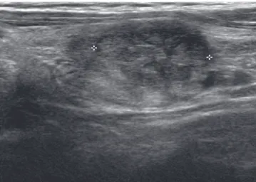
3.2 ng/dL, TSH 0.006 μIU/mL. Then we decided to add methima- zole 10 mg and thyroid ultrasonography was performed. It showed ill defined hypoechoic nodule in the left lobe, 1.7 × 1.8 cm in size, with normal echogenecity in the remaining thyroid gland (Fig. 2).
After that she transferred to local hospital for personal reason, and fine needle aspiration was performed. The result revealed papillary thyroid carcinoma, so she referred to our hospital again, and total thyroidectomy was performed.
Postoperative histological examination revealed papillary carci- noma in the left lobe. Macroscopically, the nodule was 2 × 1.5 sized, and surface was smooth and ill defined, gray-white solid mass. Mi- croscopically, the nodule showed papillary thyroid carcinoma and surrounding thyroid tissue was normal architecture without any his-
tological evidence of Graves’ disease (Fig. 3). Surgical margin was clear and twenty-six resected lymph nodes showed no evidence of metastasis. After the operation the patient showed no complications except mild transient hypocalcemia. The patient took a 150 μg of levothyroxine for 4 months, and the thyroid function changed as follows, T3 1.2 ng/mL, free T4 2.35 ng/dL, TSH 0.034 μIU/mL, anti- thyroglobulin antibody 13.37 IU/mL (0-115), and thyroglobulin was 0.1 ng/mL (5-25).
We did mutation analysis with thyroid tissue sample using real time polymerase chain reaction. We used PNAClamp B-raf Mutation Detection Kit and PNAClamp K-ras Mutation Detection Kit (Pana- gene Inc., Daejeon, Korea) which are using peptide nucleic acid for amplification. And the result showed BRAF mutation, while RAS mu- tation was absent (Fig. 4).
Fig. 1. Thyroid 99mTc scintigraphy shows a hot nodule in the left lobe while re-
maining areas shows minimal activity. Fig. 2. Thyroid ultrasonography reveals a 1.8 × 1.2 × 1.7 cm sized ill-defined hypo echoic nodule in left thyroid lobe.
Fig. 3. Histology of the resected thyroid nodule. A. Histology demonstrates the papillary structure with ill defined margin (H&E stain, × 40, thyroid). B. The large hy- perchromic nuclei with central core and groove are compatible with papillary thyroid carcinoma (H&E stain, × 400, thyroid).
A B
Autonomously Functional Papillary Thyroid Carcinoma
http://dx.doi.org/10.3803/EnM.2012.27.1.59
61
http://www.enm-kes.org
DISCUSSION
Thyroid nodule is common disorder which occurs in about 3-7%
of adults by physical examination, and 6-20% of nodules present autonomously functioning thyroid nodule [1]. When thyroid scinti- graphic imaging reveals hot uptake, it is generally accepted as be- nign feature because malignancy is very rare in hot nodule [2,3].
From the existing guideline, fine needle aspiration biopsy is not recommended for hot nodule, and treatment should be based on thyroid functional status.
However, recent studies have been reporting the several cases of hyperfunctioning nodule which revealed as malignancy [3,4]. The incidence of thyroid carcinoma in a hot nodules is reported to be low in most literatures [2-5]. Mizukami et al. [5] presented that the incidence of hyperfunctioning thyroid cancer was variable from 0.4% to 11.8%, these data were based on ten reports which pub- lished from 1983 to 2003.
The mechanism of producing excessive hormone from thyroid cancer has not been established, but it is assumed that G protein α chain (Gsα) and TSH receptor gene mutation may contribute to the abnormal hormone production [4,6]. The pathogenesis of hyper- functioning thyroid nodule has been revealed that mutated TSH receptor increases the intracellular c-AMP which stimulates the cell growth with excessive hormone production.
BRAF, RAS, and RET mutations are well known oncogenes of papillary thyroid carcinoma, that activate the signaling pathway in thyroid follicular cells and stimulate the tumor progression [6]. Many hormones, cytokines and growth factors control thyroid follicular cell growth through the intracellular signaling system. As RET-RAS- BRAF system is one of main pathway controlling cell cycle, it can stimulate abnormal activation of cell cycle followed by thyroid car- cinoma. And most papillary thyroid cancer has at least one of the RET-RAS-BRAF mutation [7].
Therefore, it can be hypothesized that autonomously hyperfunc- tioning thyroid nodule found to be papillary thyroid carcinoma may contain several combined gene mutations described above. And also several factors including genetic susceptibility, environmental factors, TSH, growth factors, and angiogenic substances either play a distinct and separate role or act synergistically through complex interaction mechanism.
In this case, gene analysis showed BRAF mutation, while RAS mutation was absent (Fig. 4).
The exact mechanisms of hyperfunction of thyroid carcinoma are still unclear. But it is obvious that TSH receptor gene and other oncogenes may be involved. So we need further studies to explore any relationship between oncogenes (RET-RAS-BRAF) leading to thyroid malignancy and other gene mutations (Gsα and TSH recep- tor gene) causing abnormal hormone production.
The limitation of the presenting case was failure to get TSH re- ceptor and Gsα gene analysis. We just assume that BRAF mutation was the oncogene for papillary thyroid carcinoma in this patient.
Granter et al. [8] reported nuclear changes suggestive of papillary thyroid carcinoma in thyroid nodule after 131I treatment. And thyroid carcinomas appeared after radioactive iodine treatment for hyper- thyroidism have been reported [8]. However in other studies no malignant change was observed, either clinically or cytologically after radioactive iodine treatment. The oncogenic effect of radioac- tive iodine treatment has yet to be proven. In our case, since only several months had passed after the 131I treatment, we assume that the autonomous nodule was already malignant at presentation. The latent period of the carcinoma cases after radioactive iodine treat- ment was generally longer than 3 years with mean of 11.4 years [9].
Hyperfunctioning thyroid carcinoma is extremely rare. So it is generally believed that the diagnosis of hot nodule on radionuclide imaging can almost always rule out malignancy in thyroid nodule.
However when nodule shows suspicious malignant features, it is important not to exclude the possibility of malignancy, so we rec- Fig. 4. Gene analysis using real time polymerase chain reaction (PNAClamp B-
raf Mutation Detection Kit) presents amplified B-raf mutation like other sample studies. PNA, peptide nucleic acid.
Fluorescence intensity
Cycle
Kim JH, et al.
http://dx.doi.org/10.3803/EnM.2012.27.1.59 62
http://www.enm-kes.org
ommend to check thyroid ultrasonography and fine needle aspira- tion biopsy when needed [3]. As the number of malignant hot nod- ule is getting increased we should consider setting up the appropri- ate diagnostic tool for it. And, of course, whether the nodule is cold or hot, we need to approach carefully and start the proper manage- ment not to overlook the possibility of the malignancy.
요 약
자율기능성갑상선암은매우드문것으로알려져있다. 갑상선결
절이발견되었을때갑상선스캔상에서기능성결절인열결절의양
상을나타내는경우대개양성종양을시사하며 4% 미만에서악성 종양으로보고된바있다. 이번증례는방사성옥소치료를받은기능
성갑상선결절환자에서유두갑상선암이진단되어이를보고하려
한다. 58세여자환자가 6개월간의 5 kg의체중감량으로인근병원 을찾았다가갑상선결절이촉진되어본원으로전원되었다. 내원시 갑상선호르몬은 T3 1.93 ng/mL, free T4 3.2 ng/dL, TSH 0.006 μIU/
mL로확인되었고갑상선자가항체검사는모두정상범위내로확 인되었다. 99mTc 갑상선스캔을시행한결과 2 × 2 cm 크기의열결절 이왼엽에서발견되었다. 환자는기능성갑상선 결절로진단하에 10 mCi 131I으로방사선옥소치료를시작하였으나 6개월후에도갑
상선기능은호전을보이지않았고추가적으로시행한세침흡인검
사에서유두갑상선암으로진단되어갑상선전절제술을시행받은
사례이다. 이번증례를통해서임상양상과갑상선스캔상에서양성
결절로의심이된다하더라고악성결절의가능성을완전히배재할
수없으며, 갑상선결절의평가에있어기존의진단가이드라인에만
전적으로의존하여평가할시잘못된진단을할수있음을보여준
다. 갑상선암의발병증가에따라갑상선결절에대한관심및검사 가점점증가하는 추세이다. 따라서갑상선결절의정확한평가를
위한진단가이드라인의재정립이필요하다.
REFERENCES
1. American Thyroid Association (ATA) Guidelines Taskforce on Thyroid Nodules and Differentiated Thyroid Cancer, Cooper DS, Doherty GM, Haugen BR, Kloos RT, Lee SL, Mandel SJ, Mazzaferri EL, McIver B, Pacini F, Schlumberger M, Sherman SI, Steward DL, Tuttle RM: Revised Ameri- can Thyroid Association management guidelines for patients with thyroid nodules and differentiated thyroid cancer. Thyroid 19:1167-1214, 2009 2. Ashcraft MW, Van Herle AJ: Management of thyroid nodules. II: scanning
techniques, thyroid suppressive therapy, and fine needle aspiration. Head Neck Surg 3:297-322, 1981
3. Uludag M, Yetkin G, Citgez B, Isgor A, Basak T: Autonomously function- ing thyroid nodule treated with radioactive iodine and later diagnosed as papillary thyroid cancer. Hormones (Athens) 7:175-179, 2008
4. Majima T, Doi K, Komatsu Y, Itoh H, Fukao A, Shigemoto M, Takagi C, Corners J, Mizuta N, Kato R, Nakao K: Papillary thyroid carcinoma with- out metastases manifesting as an autonomously functioning thyroid nod- ule. Endocr J 52:309-316, 2005
5. Mizukami Y, Michigishi T, Nonomura A, Yokoyama K, Noguchi M, Hashi- moto T, Nakamura S, Ishizaki T: Autonomously functioning (hot) nodule of the thyroid gland. A clinical and histopathologic study of 17 cases. Am J Clin Pathol 101:29-35, 1994
6. Paschke R, Tonacchera M, Van Sande J, Parma J, Vassart G: Identification and functional characterization of two new somatic mutations causing con- stitutive activation of the thyrotropin receptor in hyperfunctioning auton- omous adenomas of the thyroid. J Clin Endocrinol Metab 79:1785-1789, 1994
7. Boelaert K: The association between serum TSH concentration and thy- roid cancer. Endocr Relat Cancer 16:1065-1072, 2009
8. Granter SR, Cibas ES: Cytologic findings in thyroid nodules after 131I treatment of hyperthyroidism. Am J Clin Pathol 107:20-25, 1997 9. Tezelman S, Grossman RF, Siperstein AE, Clark OH: Radioiodine-associ-
ated thyroid cancers. World J Surg 18:522-528, 1994