DOI: 10.4046/trd.2011.70.3.191
ISSN: 1738-3536(Print)/2005-6184(Online) Tuberc Respir Dis 2011;70:191-198
CopyrightⒸ2011. The Korean Academy of Tuberculosis and Respiratory Diseases. All rights reserved.
인공호흡기연관 폐렴
성균관대학교 의과대학 내과학교실 삼성서울병원 호흡기내과
전경만Ventilator-Associated Pneumonia
Kyeongman Jeon, M.D.
Division of Pulmonary and Critical Care Medicine, Department of Medicine, Samsung Medical Center, Sungkyunkwan University School of Medicine, Seoul, Korea
Ventilator-associated pneumonia (VAP) is the most frequent nosocomial infection in the intensive care unit (ICU), with an incidence ranging from 8% to 38%. Patients who acquire VAP have higher mortality rates and longer ICU and hospital stays. Because there are other potential causes of fever, leukocytosis, and pulmonary infiltrates, clinical diagnosis of VAP is overly sensitive. The only alternative approach to the clinical diagnosis of VAP is the Clinical Pulmonary Infection Score (CPIS). Employing quantitative cultures of respiratory secretions in the diagnosis of VAP leads to less antibiotic use and probably to lower mortality. With respect to microbiologic diagnosis, however, it is not clear that the use of invasive sampling using bronchoscopy is associated with better outcomes.
Delayed administration of antibiotic therapy is associated with an increased mortality, and inadequate antibiotic therapy is also associated with higher mortality. Therefore, prompt initiation of adequate antibiotic therapy is a cornerstone of the treatment of VAP. The initial antibiotic therapy should be based on the most common organisms in each hospital and the most likely pathogens for that specific patient. When final cultures and susceptibilities are available, de-escalation to less broad spectrum antibiotics should be done. Since clinical improvement usually takes 2 to 3 days, clinical responses to the initial empirical therapy should be evaluated by day 3. A short course of antibiotic therapy appears to be equivalent to a traditional course of more than 14 days, except when treating non-fermenting gram-negative organisms. If patients receive initially adequate antibiotic therapy, efforts should be made to shorten the duration of therapy to as short as 7 days, provided that the etiologic pathogen is not a non-fermenting gram-negative organism.
Key Words: Pneumonia, Ventilator-Associated; Diagnosis; Therapeutics; Review
Address for correspondence: Kyeongman Jeon, M.D.
Division of Pulmonary and Critical Care Medicine, Depart- ment of Medicine, Samsung Medical Center, Sungkyunkwan University School of Medicine, #50, Irwon-dong, Gangnam- gu, Seoul 135-710, Korea
Phone: 82-2-3410-3429, Fax: 82-2-3410-6956 E-mail: [email protected]
Received: Dec. 11, 2010 Accepted: Dec. 11, 2010
서 론
인공호흡기연관 폐렴(ventilator-associated pneumo- nia, VAP)은 기계환기를 시작할 시점에는 폐렴의 발생이 나 잠복기에 있지 않은 환자에서 기관삽관 및 기계환기를
시작한 후 48시간 이후에 발생되는 폐렴으로 정의된다1. 인공호흡기연관 폐렴은 발생 시기에 따라 초기 인공호흡 기연관 폐렴(early-onset VAP)과 후기 인공호흡기연관 폐 렴(late-onset VAP)으로 나눠지며, 이에 따라 다른 예후를 보인다1,2. 즉, 기계환기 적용 후 5일 이전에 발생하는 경 우 비교적 경하고, 항생제에 감수성을 보이는 균주에 의해 발생하여 좋은 예후를 보이며, 이와 반대로 기계환기 적용 후 5일 이후에 발생하는 경우 항생제에 내성을 갖는 균주 에 의한 폐렴으로 높은 사망률을 보인다(Table 1).
1. 인공호흡기연관 폐렴의 발생빈도
인공호흡기연관 폐렴은 중환자실에서 가장 많이 접하 는 병원성 감염으로 기계환기 기간이 증가함에 따라 이환
Review
Table 1. Classification of ventilator-associated pneumonia
Early onset: developed within <5 days Late onset: developed after ≥5 days
Antibiotic sensitive bacteria Multidrug-resistant (MDR) pathogens
- Streptococcus pneumoniae - Pseudomonas aeruginosa
- Haemophilus influenzae - Acinetobacter species
- Staphylococcus aureus - MRSA
Better prognosis Associated with increased mortality and morbidity
MRSA: methicillin-resistant S. aureus .
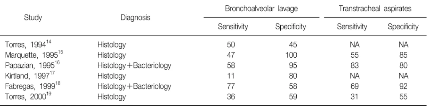
Table 2. Diagnostic utility of BAL in the diagnosis of VAP
Study Diagnosis
Bronchoalveolar lavage Transtracheal aspirates Sensitivity Specificity Sensitivity Specificity
Torres, 1994
14Histology 50 45 NA NA
Marquette, 1995
15Histology 47 100 55 85
Papazian, 1995
16Histology+Bacteriology 58 95 83 80
Kirtland, 1997
17Histology 11 80 NA NA
Fabregas, 1999
18Histology+Bacteriology 77 58 69 92
Torres, 2000
19Histology 36 59 31 55
Values are presented as percents. BAL: bronchoalveolar lavage; VAP: ventilator-associated pneumonia.
율이 증가하게 된다. 현재까지 인공호흡기연관 폐렴의 빈 도에 관한 연구들이 많이 보고되고 있으나 연구들 간에 적용된 인공호흡기연관 폐렴의 정의가 다르고, 중환자실 의 특성 및 병원의 특성이 다르므로 발생빈도는 연구마다 다르게 보고되고 있다1. 미국의 병원감시체계(National Nosocomial Infection Surveillance)에 따르면 전체 중환 자실 환자의 27%에서 발생하는 두 번째로 흔한 병원성 감염으로 보고되고 있고3, 최근 38개의 전향적 연구를 이 용한 계통분석(systematic review)에서는 적게는 8%에서 많게는 38%까지 보고되고 있다4. 특히, 내과계 중환자실 에서는 약 17%의 발생빈도를 보고하고 있다. 이를 기계환 기 기간에 따라 나누면 10일째 6.5%, 20일째 19%, 30일째 28%의 발생빈도를 보여, 기계환기 적용기간이 1일 증가 함에 따라 1%의 발생위험이 증가하게 된다5. 하지만, 캐나 다의 16개의 중환자실의 환자를 대상으로 한 전향적 연구 에서는 이러한 발생위험이 첫 5일까지는 증가하였다가 시 간에 지남에 따라 다시 감소하여, 사용일에 따른 인공호흡 기연관 폐렴의 발생은 초기 5일 동안 3.3%까지 증가한다 고 한다6. 따라서, 기계환기 중인 환자에서 인공호흡기연 관 폐렴에 대한 관심은 초기에 집중하여야 한다.
2. 인공호흡기연관 폐렴의 진단
1) 임상적 소견: 폐렴의 임상적인 진단은 흉부방사선 사 진상 새로운 폐침윤이 있으면서, 체온 38.3oC 이상, 화농 성 기관 및 기관지 분비물, 백혈구 감소 또는 증가(<4,000,
>11,000/mm3)의 3가지 중 2가지 이상을 보일 때로 정의 된다. 하지만, 이런 임상적 증상 및 소견만으로는 인공호 흡기연관 폐렴을 진단하는데 많은 한계가 있다7. 임상적 으로 인공호흡기연관 폐렴을 진단하였을 때 이를 조직학 적으로 확인할 때 적게는 29%, 많게는 71%의 환자에서 잘못된 진단으로 확인되었다8-12. 이러한 임상적 소견의 정 확성은 임상의사의 경험 정도와도 관련이 없었다11. 따라 서, 인공호흡기연관 폐렴의 진단은 임상적 증상 및 소견만 으로는 부족하며 흉부 방사선사진상 새로운 폐침윤 소견 뿐만 아니라 추가적인 진단기준(소견)이 필요하다.
2) 미생물학적 진단: 임상적 진단의 한계를 보완하고자 하부기도 검체를 이용한 미생물학적 진단이 인공호흡기 연관 폐렴의 진단에 이용되고 있다. 하지만, 미생물학적 진단 자체 또한 인공호흡기연관 폐렴을 진단하는데는 제 약이 있다13. Torres 등은 23개의 연구자료를 바탕으로 한 분석에서 기관지세척술(bronchoalveolar lavage, BAL)을 이용한 미생물학적 진단은 평균 민감도가 73±18%이고,
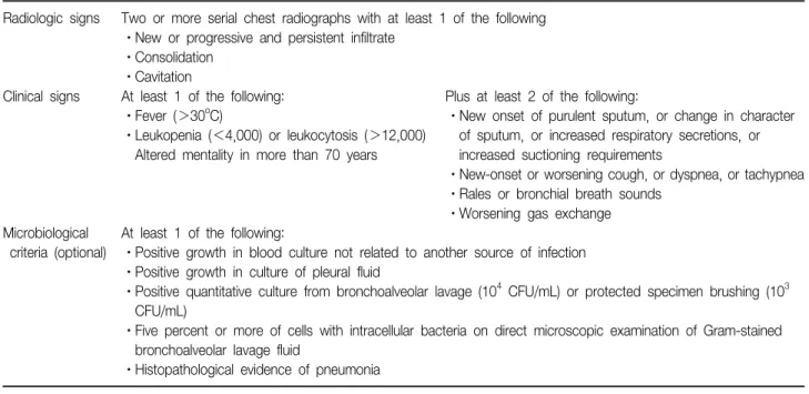
Table 3. Diagnostic flow of pneumonia from CDC, 2002
Radiologic signs Two or more serial chest radiographs with at least 1 of the following ㆍNew or progressive and persistent infiltrate
ㆍConsolidation ㆍCavitation
Clinical signs At least 1 of the following: Plus at least 2 of the following:
ㆍFever (>30
oC) ㆍNew onset of purulent sputum, or change in character ㆍLeukopenia (<4,000) or leukocytosis (>12,000) of sputum, or increased respiratory secretions, or Altered mentality in more than 70 years increased suctioning requirements
ㆍNew-onset or worsening cough, or dyspnea, or tachypnea ㆍRales or bronchial breath sounds
ㆍWorsening gas exchange Microbiological At least 1 of the following:
criteria (optional) ㆍPositive growth in blood culture not related to another source of infection ㆍPositive growth in culture of pleural fluid
ㆍPositive quantitative culture from bronchoalveolar lavage (10
4CFU/mL) or protected specimen brushing (10
3CFU/mL)
ㆍFive percent or more of cells with intracellular bacteria on direct microscopic examination of Gram-stained bronchoalveolar lavage fluid
ㆍHistopathological evidence of pneumonia CDC: Centers for Disease Control and Prevention.
평균 특이도가 82±19%라고 보고하였다. 또한 연구자료 간의 민감도 및 특이도에 대한 변이는 각각 42∼100%, 55∼100%로 연구마다 차이가 크며, 이는 대상 환자의 특 성, 기계환기 적용 기간의 차이, 또는 인공호흡기연관 폐 렴의 확진 방법의 차이에 따른 것으로 해석된다. 그러나 조직학적으로 확진된 연구들만 분석하여도, 민감도와 특 이도는 만족스럽지 않다(Table 2)14-19. 또한 기관흡인 (transtracheal aspirate, TTA)을 이용한 미생물학적 진단 과 비교할 때에도 큰 차이를 보이지 않는다15,16,18. 이후의 후속연구들을 대상으로 한 메타분석에서도 BAL 또는 protected specimen brushing (PSB)를 이용한 침습적 미 생물학적 진단과 TTA를 이용한 비 침습적 미생물학적 진 단을 비교하였으나 차이를 보이지 않았고, 결과적으로 사 망률 감소에도 기여하지 못하고 있다20. Canadian Critical Care Trial Group은 이러한 결과들을 대규모 임상시험을 통해 확인해 보고자 미국 및 캐나다의 28개 중환자실의 740명의 환자들을 대상으로 비교임상시험을 시행하였으 나, BAL을 이용한 진단이나 TTA를 이용한 진단에서 28일 사망률 및 항생제 사용기간의 차이는 없었다21. 따라서, 인공호흡기연관 폐렴의 미생물학적 진단에 기관지내시경 을 통한 BAL 또는 PSB가 반드시 필요하지는 않으며, 비교 적 간단하고 쉽게 시행할 수 있는 TTA를 이용한 미생물 검사로도 충분하다.
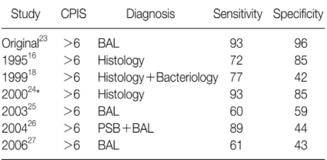
3) 임상적 진단 기준(점수체계): 미국질병관리본부(Cen- ter for Disease Control and Prevention, CDC)에서는 흉 부방사선 소견, 임상적 증상 및 징후, 그리고, 미생물검사 결과를 바탕으로 인공호흡기연관 폐렴의 진단에 적용할 수 있는 임상적 진단기준을 마련하였다(Table 3). 하지만, 조직학적으로 확진된 환자들을 대상으로 한 후향적 분석 에서 CDC 진단기준에 이용되는 임상 소견들만으로는 진 단적 정확성이 떨어짐이 확인되었고, 흉부 방사선 소견만 이 진단에 영향을 주는 소견으로 확인되었다22. 또한, CDC 진단기준에서 미생물학적 검사 결과는 임상적 소견 이 부족한 경우에만 부가적으로 사용된다. Pugin 등23은 인공호흡기연관 폐렴의 진단에서 체온, 백혈구 수, 기관분 비물의 양상 및 정도, 동맥혈 산소화 정도, 흉부방사선 소 견 및 TTA를 이용한 그람염색 및 배양결과를 바탕으로 한 Clinical Pulmonary Infection Score (CPIS)를 적용하고 자 하였다(Table 4). 즉, CPIS 점수가 6점보다 높을 경우 BAL을 이용한 미생물학적 진단과 비교할 때 상관관계가 높았다(AUROC, 0.95). 후속 연구에서도 CPIS를 이용한 인공호흡기연관 폐렴의 진단은 높은 민감도(60∼93%)와 특이도(42∼85%)를 보이며(Table 5)16,18,24-27
, 특히, 인공 호흡기연관 페렴 가능성이 높은 환자에서는 93%의 민감 도와 85%의 특이도를 보인다24. 하지만, CPIS 점수체계도 관찰자간에 차이가 있어 표준화된 진단기준으로 받아들
Table 4. Clinical Pulmonary Infection Score (CPIS)*23
Sign Points
Temperature,
oC
36.5∼38.4 0
38.5∼38.9 1
≤36 or ≥39 2
Blood leukocytes, cells/μL
4,000∼11,000 0
<4,000 or >11,000 1
>500 band forms 2
Oxygenation, PaO
2/FiO
2>240 or ARDS 0
≤240 and no evidence of ARDS 2
Pulmonary radiography
No infiltrate 0
Diffuse (or patchy) infiltrates 1
Localized infiltrate 2
Tracheal secretions score (quantifying amount of tracheal secretions)
†<14 0
≥14 1
Purulent sputum 2
Culture of tracheal aspirate
Pathogenic bacteria cultured minimal or no growth 0
Pathogenic bacteria cultured moderate or more growth 1
Moderate or greater growth of pathogenic bacteria consistent with that seen on Gram stain 2
*Total score of >6 points suggests ventilator-associated pneumonia,
†Score calculated by quantifying amount of tracheal secretions on a subjective 0∼4 scale multiple times per day, then summing all of a patient' score for the day.
ARDS: acute respiratory distress syndrome.
Table 5. Characteristics of CPIS in the diagnosis of VAP
Study CPIS Diagnosis Sensitivity Specificity
Original
23>6 BAL 93 96
1995
16>6 Histology 72 85
1999
18>6 Histology+Bacteriology 77 42
2000
24* >6 Histology 93 85
2003
25>6 BAL 60 59
2004
26>6 PSB+BAL 89 44
2006
27>6 BAL 61 43
Values are presented as percents. *Patients already suspected of having VAP.
CPIS: clinical pulmonary infection score; VAP: ventilator-asso- ciated pneumonia.
이긴 어렵다28,29. 따라서, 미국흉부학회/미국감염학회 (ATS/IDSA)에서는 흉부 방사선 소견 및 임상적 소견을 바 탕으로 인공호흡기연관 폐렴이 의심되는 경우 하기도 검 체를 통한 미생물 검사를 시행하고, 경험적 항생제치료를
시작할 것을 권고하고 있으며, 2∼3일 뒤 인공호흡기연관 폐렴의 진단에 대해 다시 평가할 것을 권고하고 있다2.
3. 인공호흡기연관 폐렴의 치료
1) 신속하고 적절한 초기 치료: 인공호흡기연관 폐렴의 치료에서 초기 항생제 치료의 지연은 높은 사망률과 관련 이 있으므로30, 가능성이 높은 원인 균주에 대한 초기 경험 적 항생제의 조기 투여는 인공호흡기연관 폐렴의 치료에 있어 아주 중요하다1,2. 또한, 부적절한 항생제 역시 높은 사망률과 관련이 있으므로31-33, 이러한 초기 경험적 항생 제는 가능성 있는 인공호흡기연관 폐렴의 원인 균주 모두 를 포함하여야 하며, 이를 위해서는 인공호흡기연관 폐렴 의 원인 균주에 대한 역학적인 정보가 필요하다34. 또 항 생제 내성 패턴 및 기계환기 적용 기간과 이전 항생제 치 료력 등의 임상적 정보도 중요하다1,2.
2) 인공호흡기연관 폐렴의 원인 균주 및 경험적 항생제 치료: 과거부터 인공호흡기연관 폐렴의 원인균에 대한 정
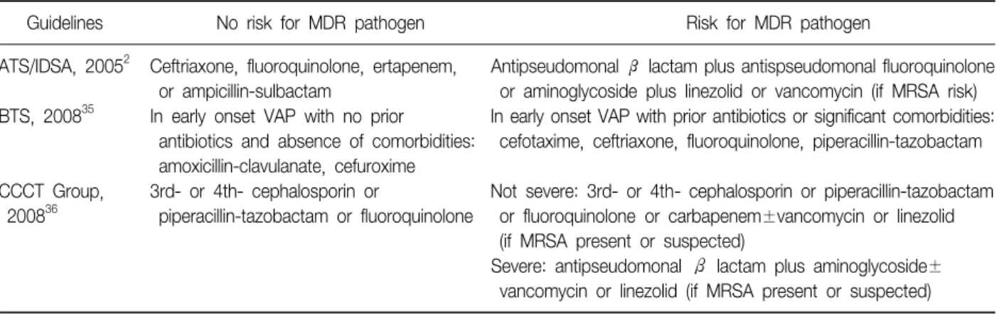
Table 6. Guidelines for empirical antimicrobial therapy for VAP
Guidelines No risk for MDR pathogen Risk for MDR pathogen
ATS/IDSA, 2005
2Ceftriaxone, fluoroquinolone, ertapenem, Antipseudomonal ß lactam plus antispseudomonal fluoroquinolone or ampicillin-sulbactam or aminoglycoside plus linezolid or vancomycin (if MRSA risk) BTS, 2008
35In early onset VAP with no prior In early onset VAP with prior antibiotics or significant comorbidities:
antibiotics and absence of comorbidities: cefotaxime, ceftriaxone, fluoroquinolone, piperacillin-tazobactam amoxicillin-clavulanate, cefuroxime
CCCT Group, 3rd- or 4th- cephalosporin or Not severe: 3rd- or 4th- cephalosporin or piperacillin-tazobactam 2008
36piperacillin-tazobactam or fluoroquinolone or fluoroquinolone or carbapenem±vancomycin or linezolid
(if MRSA present or suspected)
Severe: antipseudomonal β lactam plus aminoglycoside±
vancomycin or linezolid (if MRSA present or suspected) VAP: ventilator-associated pneumonia; MDR: multi-drug resistant; ATS: American Thoracic Society; IDSA: Infectious Disease Society of America; MRSA: methicillin-resistant S. aureus; BTS: British Thoracic Society; CCCT Group: Canadian Critical Care Trial Group.
보는 서구의 자료를 이용할 수밖에 없었다. 대표적으로 미국에서 시행된 24개의 연구에서 보고된 인공호흡기연 관 폐렴의 원인 균주로는
Pseudomonas aeruginosa
가 24%로 가장 많으며, 그 뒤로Staphylococcus aureus
가 20%를 보인다1. 그 외 Enterobacteria 14%,Hemophilus
spp. 10%,Strepcoccus
spp. 8%,Acinetobacter
spp. 8%로 보고되었다1. 하지만, 이런 분포는 인공호흡기연관 폐 렴의 발생시점에 따라 다르며(Table 1), 이전 항생제 치료 력이나 병원입원력 등도 다제내성(multi-drug resistant, MDR)균의 위험인자로 알려져 있다2. 따라서, ATS/IDSA 에서는 이런 다제내성균의 위험에 따라 경험적 항생제를 다르게 권고하고 있다2. 즉, 다제내성균의 위험이 없을 경 우 ceftriaxone, fluorquinolone, ampicillin/sulbactam, er- tapenem 등의 항생제 단독요법을 권고하고 있으나, 다제 내성균의 위험이 있는 경우 cefepime, ceftazidime 등의 antipseudomonal cephalosporin이나 imipenem, mer- openem같은 antipseudomonal carbapenem, 또는 β- lactam/β-lactamase inhibitor를 antipeudomonal fluo- roquinolone이나 aminoglycoside와 함께 쓰는 병합요법 을 권고하고 있으며, MRSA의 위험이 있는 경우 linezolid 나 vancomycin을 추가할 것을 권고하고 있다(Table 6)2. 영국이나 캐나다의 경우에도 각 국가의 원인 균주 분포에 따라 조금씩 다른 권고안을 내놓고 있다(Table 6)35,36. 아 시아국가의 경우 서구자료와 다르게
Acinetobacter
spp.의 빈도가 아주 높으며(16∼38%),
S. aureus
의 경우 대부 분 메티실린 내성균(methicillin-resistantS. aureus
, MRSA) 으로 보고되고 있다37. 그 외 약제내성균의 분포가 서구와 달라 초기 인공호흡기연관 폐렴의 경우에도 antipseudo-monal cephalosporin이나 antipseudomonal carbapenem 또는 β-lactam/β-lactamase inhibitor의 단독, 또는 fluo- roquinolone이나 aminoglycoside의 병합요법을 권고하고 있으며, 높은 MRSA의 빈도를 고려하여 초기 인공호흡기 연관 폐렴에서도 linezolid나 vancomycin을 추가할 것을 권고하고 있다37. 국내의 경우 최근 보고된 전국병원감염 감시체계(Korean Nosocomial Infection Surveillance Sys- tem, KONIS)의 자료에 따르면
S. aureus
가 39%로 가장 빈도가 높으며, 그 뒤로A. baumanii
22%,K. pneumonia
11%,P.aeruginosa
10%라고 보고되고 있으나, 인공호흡 기연관 폐렴의 진단기준이 다르며, 참여병원들의 병상규 모 및 기계환기 적용 정도가 다르므로 본 자료를 모든 병 원에 공통적으로 적용하기는 어렵다38. 하지만 경험적 항 생제 치료에 대한 국내의 권고지침이 따로 없는 상황에서 다른 아시아국가들의 원인 균주 양상과 크게 다르지 않으 므로 아시아국가를 대상으로 한 권고지침을 따라야 할 것 으로 생각된다37.3) 항생제 치료 기간: 과거부터 미국흉부학회에서는 인 공호흡기연관 폐렴의 경우 최소 14∼21일 동안 치료할 것을 권고해 왔다39. 이는 중등도, 항생제 치료에 대한 반 응 및 원인 균주에 따라 다르게 권고되어 다엽성 폐렴, 영양부족, 공동, 그람음성균에 의한 괴사성 폐렴, 또는
P.aeruginosa
나A. baumanii
가 동정된 경우 장기치료를 권고하고, MSSA나H. influenza
의 경우는 7∼10일의 단 기치료를 권고하였다39. 하지만, 최근 수정된 지침에서는 가능한 짧은 치료기간을 권고하고 있다2. 이는 프랑스에 서 시행된 대규모 임상연구 결과를 바탕으로 한 것으로, 402명을 대상으로 한 전향적 무작위 비교연구에서 적절한Figure 1. Algorithm of diag- nosis and treatment of VAP. VAP: ventilator-asso- ciated pneumonia; CPIS:
clinical pulmonary infection score.
초기 경험적 항생제치료를 받은 인공호흡기연관 폐렴의 경우 8일 단기치료와 15일 장기치료 간에 사망률, 기계환 기 기간 및 중환자실 체류기간에서 차이가 없었다40. 하지 만,
P.aeruginosa, A. baumanii, S. maltophilia
같은 non- fermenting 그람음성균의 경우 중복감염이나 재발이 높으 므로 이런 균주에 대해서는 단기치료를 적용할 수 없다40. 또한, 대부분의 인공호흡기연관 폐렴은 임상적 호전이 2∼3일째 나타나, 항생제치료 후 6일 정도 경과되면 모든 임상적 소견들이 안정화되므로41,42, 임상적 호전을 보이는 경우 적극적으로 단기치료를 적용할 때 항생제 치료기간 을 의미 있게 줄일 수 있다고 한다43.
결 론
아직까지 인공호흡기연관 폐렴의 진단 및 치료에 해결 해야 할 부분이 많은 실정이지만, 이상의 결과를 바탕으로 인공호흡기연관 폐렴의 진단 및 치료적 접근을 다음과 같 이 정리해 보고자 한다(Figure 1). 인공호흡기연관 폐렴의 진단은 흉부방사선 소견, 임상적 소견 및 미생물학적 소견 을 바탕으로 한 종합적인 임상상을 바탕으로 하고, 인공호 흡기연관 폐렴이 의심되는 경우 하기도 검체를 이용한 미 생물 검사를 시행하며, 다제내성균의 위험 정도에 따라 적절한 경험적 항생제를 조기에 투여하여야 한다. 경험적 항생제치료를 시작한 후 2∼3일이 경과하면 임상적 호전
정도를 다시 확인하고 임상적 호전을 보이는 경우 원인균 주에 따라 가능한 de-escalation 또는 단기치료를 적용하 여야 한다.
참 고 문 헌
1. Chastre J, Fagon JY. Ventilator-associated pneumonia.
Am J Respir Crit Care Med 2002;165:867-903.
2. American Thoracic Society; Infectious Diseases Society of America. Guidelines for the management of adults with hospital-acquired, ventilator-associated, and heal- thcare-associated pneumonia. Am J Respir Crit Care Med 2005;171:388-416.
3. Richards MJ, Edwards JR, Culver DH, Gaynes RP.
Nosocomial infections in medical intensive care units in the United States. National Nosocomial Infections Surveillance System. Crit Care Med 1999;27:887-92.
4. Safdar N, Dezfulian C, Collard HR, Saint S. Clinical and economic consequences of ventilator-associated pneu- monia: a systematic review. Crit Care Med 2005;33:
2184-93.
5. Fagon JY, Chastre J, Domart Y, Trouillet JL, Pierre J, Darne C, et al. Nosocomial pneumonia in patients re- ceiving continuous mechanical ventilation. Prospective analysis of 52 episodes with use of a protected speci- men brush and quantitative culture techniques. Am Rev Respir Dis 1989;139:877-84.
6. Cook DJ, Walter SD, Cook RJ, Griffith LE, Guyatt GH, Leasa D, et al. Incidence of and risk factors for ven- tilator-associated pneumonia in critically ill patients.
Ann Intern Med 1998;129:433-40.
7. Metlay JP, Fine MJ. Testing strategies in the initial man- agement of patients with community-acquired pneu- monia. Ann Intern Med 2003;138:109-18.
8. Andrews CP, Coalson JJ, Smith JD, Johanson WG Jr.
Diagnosis of nosocomial bacterial pneumonia in acute, diffuse lung injury. Chest 1981;80:254-8.
9. Bell RC, Coalson JJ, Smith JD, Johanson WG Jr. Multi- ple organ system failure and infection in adult respira- tory distress syndrome. Ann Intern Med 1983;99:293-8.
10. Fagon JY, Chastre J, Hance AJ, Guiguet M, Trouillet JL, Domart Y, et al. Detection of nosocomial lung in- fection in ventilated patients. Use of a protected speci- men brush and quantitative culture techniques in 147 patients. Am Rev Respir Dis 1988;138:110-6.
11. Fagon JY, Chastre J, Hance AJ, Domart Y, Trouillet JL, Gibert C. Evaluation of clinical judgment in the identi- fication and treatment of nosocomial pneumonia in ventilated patients. Chest 1993;103:547-53.
12. Petersen IS, Aru A, Skødt V, Behrendt N, Bols B, Kiss K, et al. Evaluation of pneumonia diagnosis in in- tensive care patients. Scand J Infect Dis 1999;31:299- 303.
13. Torres A, El-Ebiary M. Bronchoscopic BAL in the diag- nosis of ventilator-associated pneumonia. Chest 2000;
117(4 Suppl 2):198S-202S.
14. Torres A, el-Ebiary M, Padró L, Gonzalez J, de la Bellacasa JP, Ramirez J, et al. Validation of different techniques for the diagnosis of ventilator-associated pneumonia. Comparison with immediate postmortem pulmonary biopsy. Am J Respir Crit Care Med 1994;
149:324-31.
15. Marquette CH, Copin MC, Wallet F, Neviere R, Saulnier F, Mathieu D, et al. Diagnostic tests for pneumonia in ventilated patients: prospective evaluation of diagnostic accuracy using histology as a diagnostic gold standard.
Am J Respir Crit Care Med 1995;151:1878-88.
16. Papazian L, Thomas P, Garbe L, Guignon I, Thirion X, Charrel J, et al. Bronchoscopic or blind sampling tech- niques for the diagnosis of ventilator-associated pneu- monia. Am J Respir Crit Care Med 1995;152:1982-91.
17. Kirtland SH, Corley DE, Winterbauer RH, Springmeyer SC, Casey KR, Hampson NB, et al. The diagnosis of ventilator-associated pneumonia: a comparison of his- tologic, microbiologic, and clinical criteria. Chest 1997;
112:445-57.
18. Fàbregas N, Ewig S, Torres A, El-Ebiary M, Ramirez J, de La Bellacasa JP, et al. Clinical diagnosis of ventilator associated pneumonia revisited: comparative validation using immediate post-mortem lung biopsies. Thorax 1999;54:867-73.
19. Torres A, Fàbregas N, Ewig S, de la Bellacasa JP, Bauer TT, Ramirez J. Sampling methods for ventilator-associ- ated pneumonia: validation using different histologic and microbiological references. Crit Care Med 2000;28:
2799-804.
20. Shorr AF, Sherner JH, Jackson WL, Kollef MH. Invasive approaches to the diagnosis of ventilator-associated pneumonia: a meta-analysis. Crit Care Med 2005;33:46- 53.
21. Canadian Critical Care Trials Group. A randomized trial of diagnostic techniques for ventilator-associated pneu- monia. N Engl J Med 2006;355:2619-30.
22. Klompas M. Does this patient have ventilator-associated pneumonia? JAMA 2007;297:1583-93.
23. Pugin J, Auckenthaler R, Mili N, Janssens JP, Lew PD, Suter PM. Diagnosis of ventilator-associated pneumo- nia by bacteriologic analysis of bronchoscopic and nonbronchoscopic "blind" bronchoalveolar lavage fluid. Am Rev Respir Dis 1991;143:1121-9.
24. Bregeon F, Papazian L, Thomas P, Carret V, Garbe L, Saux P, et al. Diagnostic accuracy of protected catheter sampling in ventilator-associated bacterial pneumonia.
Eur Respir J 2000;16:969-75.
25. Fartoukh M, Maitre B, Honoré S, Cerf C, Zahar JR, Brun-Buisson C. Diagnosing pneumonia during me- chanical ventilation: the clinical pulmonary infection score revisited. Am J Respir Crit Care Med 2003;168:
173-9.
26. Luyt CE, Chastre J, Fagon JY. Value of the clinical pul- monary infection score for the identification and man- agement of ventilator-associated pneumonia. Intensive Care Med 2004;30:844-52.
27. Croce MA, Swanson JM, Magnotti LJ, Claridge JA, Weinberg JA, Wood GC, et al. The futility of the clin- ical pulmonary infection score in trauma patients. J Trauma 2006;60:523-7.
28. Schurink CA, Van Nieuwenhoven CA, Jacobs JA, Rozenberg-Arska M, Joore HC, Buskens E, et al. Clini- cal pulmonary infection score for ventilator-associated pneumonia: accuracy and inter-observer variability. In- tensive Care Med 2004;30:217-24.
29. Zilberberg MD, Shorr AF. Ventilator-associated pneu- monia: the clinical pulmonary infection score as a sur- rogate for diagnostics and outcome. Clin Infect Dis
2010;51 Suppl 1:S131-5.
30. Iregui M, Ward S, Sherman G, Fraser VJ, Kollef MH.
Clinical importance of delays in the initiation of appro- priate antibiotic treatment for ventilator-associated pneumonia. Chest 2002;122:262-8.
31. Kollef MH, Ward S. The influence of mini-BAL cultures on patient outcomes: implications for the antibiotic management of ventilator-associated pneumonia. Chest 1998;113:412-20.
32. Leroy O, Meybeck A, d'Escrivan T, Devos P, Kipnis E, Georges H. Impact of adequacy of initial antimicrobial therapy on the prognosis of patients with ventilator-asso- ciated pneumonia. Intensive Care Med 2003;29:2170-3.
33. Teixeira PJ, Seligman R, Hertz FT, Cruz DB, Fachel JM.
Inadequate treatment of ventilator-associated pneumo- nia: risk factors and impact on outcomes. J Hosp Infect 2007;65:361-7.
34. Koenig SM, Truwit JD. Ventilator-associated pneumo- nia: diagnosis, treatment, and prevention. Clin Microbi- ol Rev 2006;19:637-57.
35. Masterton RG, Galloway A, French G, Street M, Armstrong J, Brown E, et al. Guidelines for the man- agement of hospital-acquired pneumonia in the UK: re- port of the working party on hospital-acquired pneu- monia of the British Society for Antimicrobial Chem- otherapy. J Antimicrob Chemother 2008;62:5-34.
36. Muscedere J, Dodek P, Keenan S, Fowler R, Cook D, Heyland D, et al. Comprehensive evidence-based clin- ical practice guidelines for ventilator-associated pneu- monia: diagnosis and treatment. J Crit Care 2008;23:
138-47.
37. Song JH; Asian Hospital Acquired Pneumonia Working Group. Treatment recommendations of hospital-acquir-
ed pneumonia in Asian countries: first consensus re- port by the Asian HAP Working Group. Am J Infect Control 2008;36(4 Suppl):S83-92.
38. Kwak YG, Cho YK, Kim JY, Lee SO, Kim HY, Kim YK, et al. Korean nosocomial infections surveillance sys- tem, intensive care unit module report: data summary from July 2008 through June 2009 and analysis of 3-year results. Korean J Nosocomial Infect Control 2010;15:14-25.
39. Hospital-acquired pneumonia in adults: diagnosis, as- sessment of severity, initial antimicrobial therapy, and preventive strategies. A consensus statement, American Thoracic Society, November 1995. Am J Respir Crit Care Med 1996;153:1711-25.
40. Chastre J, Wolff M, Fagon JY, Chevret S, Thomas F, Wermert D, et al. Comparison of 8 vs 15 days of anti- biotic therapy for ventilator-associated pneumonia in adults: a randomized trial. JAMA 2003;290:2588-98.
41. Dennesen PJ, van der Ven AJ, Kessels AG, Ramsay G, Bonten MJ. Resolution of infectious parameters after antimicrobial therapy in patients with ventilator-asso- ciated pneumonia. Am J Respir Crit Care Med 2001;
163:1371-5.
42. Luna CM, Blanzaco D, Niederman MS, Matarucco W, Baredes NC, Desmery P, et al. Resolution of ven- tilator-associated pneumonia: prospective evaluation of the clinical pulmonary infection score as an early clin- ical predictor of outcome. Crit Care Med 2003;31:676- 82.
43. Micek ST, Ward S, Fraser VJ, Kollef MH. A randomized controlled trial of an antibiotic discontinuation policy for clinically suspected ventilator-associated pneumo- nia. Chest 2004;125:1791-9.