Vol.21 No.1 p120-126, June 2004
1)
서 론
책임저자:김용대, 대구시 남구 대명동 317-1, 영남대학교 의과대학 이비인후과학교실 Tel: (053) 620-3784, Fax: (053) 628-7884, E-mail: [email protected]
비강 및 부비동에서 발생하는 부비강미분화 암종은 1986년 Frierson 등
1)이 처음 기술한 후
부비강미분화암종 3예
김용대․곽동석․이형중․신재흔․배창훈․송시연
영남대학교 의과대학 이비인후-두경부외과학교실Sinonasal Undifferentiated Carcinoma
Yong-Dae Kim, Dong Suk Kwak, Hyung Joong Lee, Jae Heun Sin, Chang Hoon Bai, Si Yeon Song
Department of Otorhinolaryngology-Head and Neck Surgery, College of Medicine, Yeungnam University, Daegu, Korea
-Abstract-
Sinonasal Undifferentiated Carcinoma (SNUC) is a very rare, highly aggressive malignant tumor of the nasal cavity and paranasal sinuses. SNUC tends to present with advanced-stage disease, often with intracranial invasion. It requires an aggressive multimodality therapy that includes surgical resection. A cure rate of less than 20% is generally reported in the literature, with most patients dying within 1 year of onset of the disease. Three patients diagnosed as SNUC were treated at the Yeungnam University Medical Center between the years 2000 and 2003 were analyzed retrospectively. All patients presented with the disease very advanced. The three cases were given chemotherapy or chemotherapy with radiotherapy.
Two patients died of the disease, surviving only 6 and 11 months following treatment, respectively. We did a follow-up on just the one remaining case with incomplete controlled disease for 27 months. The overall prognosis of SNUC is very poor. We consider that more intensive multimodality therapies are recommended for all patients with SNUC.
Key Words: Sinonasal undifferentiated carcinoma, Paranasal sinus, Nasal cavity
현재까지 매우 드물게 보고되는 종양이다. 초 기에는 코막힘, 비출혈, 시력저하, 복시, 통증 등의 증상이 거의 나타나지 않으므로 진단이 늦어지는 매우 침습적인 암종이다.
1-5)부비강미분화암종은 진단 당시에 이미 안구 나 뇌를 침범할 정도로 진행된 경우가 많으 며,
1, 4, 5-7)항암 치료나 방사선 치료 혹은 수술 적 치료를 적극적으로 시행함에도 불구하고 불 량한 예후를 가진다.
1-4, 6, 8, 9)조직학적으로 악 성림프종, 악성흑색종, 횡문근육종, 림프상피종, 고등급 샘낭암종, 신경내분비암종, 후각신경모 세포종 등과 유사하므로 정확한 감별이 이루어 져야 한다.
1-3, 8, 9)국내에서는 1987년 Rha
10)가 상악동에 발생 한 2예, 1998년 Cho 등
11)이 비중격에 발생하여 비강저 점막을 침범한 2예를 보고하였으나 예 후 추적관찰에 대한 기록이 미비한 실정이다.
저자들은 2000년부터 2003년까지 영남대학교 의과대학 부속병원에서 경험한 비강 및 부비동 에 발생한 부비강미분화암종 3예를 문헌고찰과 함께 보고하는 바이다.
증례 1
34세 남자가 내원 1개월 전부터 지속되는 좌측 안구돌출과 전두동 부위 부종을 주소로 내원하였다. 과거력상 14년간 하루 2갑의 흡연 력이 있었다. 현병력상 좌측 코막힘, 비루, 안 와 통증을 호소하였으며 후비루, 무후각증, 재 채기, 비소양증, 복시, 안구운동장애 등의 증상 은 없었다.
전비경 및 비내시경 소견상 좌측 비강에 폴 립모양 종괴가 관찰되었고 비중격이 좌측으로 편위되어 있었다. 외견상 좌측 안구 내측면과 전두부 부종, 좌측 안구 돌출이 관찰되었다. 부
비동 전산화단층촬영상 양측 사골동의 종괴가 비강과 전두부 피하조직, 양측 전두개 오목까 지 침범한 소견과 양측 사상판, 좌측 안와벽, 그리고 좌측 상악동벽의 골파괴 소견이 관찰되
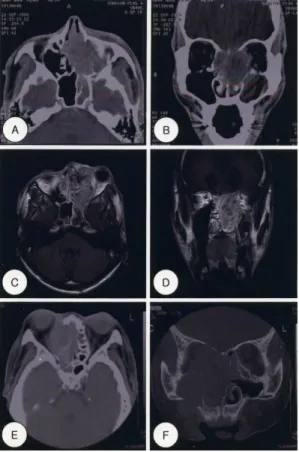
Fig. 1. Axial and coronal views of paranasal sinus (PNS) CT scans and MRI. A and B: Axial and coronal views of PNS CT scans of case 1 show mass lesion originated from left ethmoid sinus.
This mass lesion invades nasal cavity and
maxillary sinus, and destructs medial and superior
wall of left maxillary sinus, medial wall of orbit
and both cribriform plate. C and D: T1-weighted
axial and coronal MRI images of case 1 show
inhomogeneous mass originated from the left nasal
cavity and it invades left maxillary sinus and
orbit. E and F: Axial and coronal views of PNS
CT scans of case 2 show massive destruction of
nasal cavity, nasal septum, maxillary sinus,
ethmoid sinus and orbital structure.
었다(Fig. 1A, 1B). 자기공명영상상 불균일한 종괴가 좌측 비강에서 발생하여 좌측 상악동, 좌측 안와, 전두 부위 피하조직 및 양측 전두 엽 아래 부위까지 침범해 있었다(Fig. 1C, 1D).
원격전이 소견은 없었다.
환자는 국소마취하에 좌측 비내시경적 조직 검사를 받았다. 조직 소견에서 다형성 혹은 역 형성 세포들이 모여 불규칙한 모양의 둥지 혹 은 기둥 구조를 이루며 뚜렷한 핵소체, 핵의 과 다염색성, 괴사 등의 소견이 관찰되었다(Fig. 2A).
면역조직화학검사상 AE1/AE3에는 양성(Fig. 2B), S-100 protein과 leukocyte common antigen, neuronspecific enolase에는 음성반응을 보여 부 비강미분화암종으로 진단되었다. 진단 후 환자 는 항암화학요법(cyclophosphamide/5-fluorouracil)
을 1차례 시행 중 스스로 퇴원하였고 11개월 후 사망하였다.
증례 2
75세 여자가 1년 전부터 지속되는 양측 코 막힘을 주소로 내원하였다. 과거력상 1994년 우측 경부 원발 불명암으로 근치 경부 절제술 후 방사선치료를 수차례 받았으며, 2000년 우 측 비루관 폐색으로 눈물주머니코안연결술을 시행받은 병력이 있었다.
현병력상 코막힘, 비루 및 후비루, 후각저하 를 제외한 두통, 재채기, 비소양감, 안면통, 안 구운동장애 등의 증상은 없었다. 전비경 및 비 내시경 소견상 양측 비강 내에 불규칙한 표면 을 가지면서 쉽게 출혈되는 종괴가 관찰되었
Fig. 2. Histopathologic findings of sinonasal undifferentiated carcinoma. A: The mass shows proliferation of anaplastic cells, arranged in irregular shaped nests or trabeculae. The tumor cells have round hyperchromatic nuclei with prominent nucleoli. Mitotic figures are frequently identified and tumor necrosis is found (H&E, ×200). B: The tumor cells show strong positivity for AE1/AE3 (ABC method,
×200).
고, 양측 중비도에 점액농성비루가 관찰되었다.
수술 전 촬영한 부비동 전산화단층촬영에서 우측 상악동에 연부조직 음영의 종괴가 관찰되 었고, 이 종괴는 우측 상악동의 전벽을 침범하 여 우측 협부까지 커져 있었다. 그리고 우측 상악동의 하벽과 우측 안와 하내측의 광범위한 골파괴 소견과 비중격 및 우측 안와 상벽의 국 소적인 골파괴 소견을 보였으며, 종괴가 우측 안구를 돌출시키고 있는 소견이 보였다(Fig.
1E, 1F). 간초음파, 갑상샘초음파, 골스캔, 흉부 X-ray상 원격전이 소견은 없었다.
처음 시행한 천자조직검사에서 응고괴사의 소견만 보여 수술실에서 광범위한 절제생검을 시행할 예정이었으나, 출혈이 심하고 고령으로 인해 환자 및 보호자가 수술적 치료를 거부하 여 환자는 국소마취하에 우측 내시경적 부분조 직검사를 받았다. 수술 할 당시 양측비강 내에 는 폴립모양 종괴가 관찰되었고 우측 중비도 내에는 불규칙 표면을 가지고 괴사된 조직을 가진 종괴가 관찰되었으나, 환자의 협조가 되 지 않아 조직을 완전히 제거하기 곤란하였다.
조직 검사상 부비강미분화암종으로 진단되었 다. 진단 후 방사선치료를 받았으며(6000 cGY), 3개월 후 시행한 부비동 전산화단층촬영상 우 측 상악동과 비강 내 종괴의 크기는 감소한 상 태에서 현재 27개월간 추적관찰중이다.
증례 3
63세 여자가 내원 1개월 전부터 지속되는 좌측 안면부 통증을 주소로 내원하였다. 과거 력상 특이소견은 없었고, 현병력상 반복되는 비출혈, 비루, 좌측 안면부 부종을 제외한 코막 힘, 후비루, 재채기, 비소양감, 시력저하, 복시, 안구운동장애 등의 증상은 없었다.
비내시경하 좌측 상악동내 조직검사상 부비 강미분화암종으로 진단되었고 진단 후 광범위 한 절제술을 시행하려 했으나 환자의 거부로 시 행하지 못하고 항암요법과 방사선치료, 괴사조직 절제술을 시행하였으나 6개월 후 사망하였다.
고 찰
부비강미분화암종은 1986년 Frierson 등
1)이 비강, 상악동, 사골동에서 발생한 환자 8예를 처음 기술하였는데 그 중 6명은 안와벽을 침범 하였고 평균생존율은 4개월밖에 되지 않을 정 도로 매우 침습적인 암이라 하였다. 그 후 Righi 등
12)이 11예, Gallo 등
3)이 13예를 보고하였고, 2002년 Jeng 등
13)이 부비강미분화암종과 Epstein- Barr virus와의 관계에 대해서 발표하기까지 몇 차례 보고가 있었지만, 국내에서는 1987년 Rha
10)가 상악동의 2예, 1998년 Cho 등
11)이 비 중격과 비강저 점막의 일부를 침범한 2예를 보 고한 후 거의 보고된 예가 없어, 병태생리 및 진단과 치료에 대해 정확히 알려진 바가 없다.
부비강미분화암종은 비출혈, 코막힘, 두통 등의 증상을 거의 나타내지 않으나,
1-5)생기더 라도 양성 비질환의 증상과 유사해서 감기나 부비동염으로 오인되기도 하며,
9)안구돌출, 안 와주위 부종, 복시 등의 증상이 몇 개월 내에 나타날 정도로 급속히 진행하는 경우가 많다.
4)심지어 뇌척수액을 통해 경막내골수외로 원격 전이를 일으켜 신경학적 증상도 일으킨다.
7)본 증례들에서도 환자들은 코막힘이나 비루 등의 증상보다는 안면부 혹은 전두 부위 통증 및 부 종, 안구돌출 등 진행된 경우의 증상으로 내원 한 경우였다.
발생 원인은 아직 정확히 알려져 있지 않으
나, 흡연이 중요한 역할을 하며, 유전적 소인과 지역적 요인뿐만 아니라 환경적인 요인도 관계 가 있다.
1, 14)최근 보고에 의하면 망막모세포종 으로 방사선치료를 시행한 환자에게 발생할 수 있다고 하였고,
5)RB-1 유전자의 자연 변종 혹 은 방사선에 의한 변종이 중요한 역할을 한다 고 하였다.
15)특히 아시아에서는 Epstein-Barr virus가 발생 원인이 될 수 있다는 보고가 있 다.
13, 14, 16)본 증례들 중 1예에서는 과거 방사 선치료 병력, 1예에서는 과도한 흡연의 병력이 있었으나 Epstein-Barr virus와의 관계는 확실 하지 않으며 뚜렷한 발병원인을 알 수 없었다.
부비강미분화암종은 비강, 사골동, 상악동 순으로 발생한다.
13, 17)본 증례들 중 2명은 상 악동, 1명은 비강에서 발생하였다.
조직학적으로는 악성림프종, 악성흑색종, 횡 문근육종, 림프상피종, 고등급 샘낭암종, 신경 내분비암종, 후각신경모세포종 등과 유사하며, 특히 신경내분비암종, 후각신경모세포종과는 감 별하기 힘들다.
1-3, 8, 9)Jeng 등
13)은 비인두형 미분화암종은 소포성 핵, 융합적 양상, 방추형 세포, 괴사 조직이 없다는 점에서 부비강미분 화암종과 구별된다고 하였다. 부비강미분화암 종은 큰 세포들이 둥지, 판구조, 기둥모양을 이 루고, 세포질에 비해 핵의 비율이 높고, 핵은 과다염색성을 띠고 둥글거나 타원형의 다형성 형태를 나타내며 세포질은 호산성의 특징을 가 진다.
1-4, 12, 18)본 증례들의 조직 소견도 위와 유사한 소견을 보이고 있었다. 그리고 이런 세 포들은 광범위하게 괴사되거나 종종 혈관을 침 범하기도 한다.
1-4, 9)그에 반해 신경내분비암종 과 후각신경모세포암종의 세포는 신경내분비성 핵염색질을 가지며, 국소적인 괴사, 적은 혈관 침범의 특징을 가진다.
1, 4)특히 Homer-Wright
로제트, 세포간 원섬유, 은친화성 과립, 편평 분화 혹은 샘분화 등은 후각신경모세포종에는 있지만 부비강미분화암종에는 없는 소견이다.
1, 4)면역조직화학적으로 AE1/AE3 항체에 양성 반응, S-100 protein과 leukocyte common antigen에는 음성반응, epithelial membrane antigen과 neuron-specific enolase에는 다양한 반응을 나타낸다.
1, 4)본 증례들 중 2예에서 AE1/AE3에 양성이었으나, S-100 protein과 leukocyte common antigen, neuron-specific enolase, synaptophysin은 음성이었다. 나머지 한 예에서는 epithelial membrane antigen에는 양성, S-100 protein, leukocyte common antigen, 그리고 Epstein-Barr virus에서는 음성반응을 나타내었다.
부비동 전산화단층촬영 소견으로는 부비강 미분화암종과 다른 암종을 구별하기가 힘들지 만 진단을 위해서는 뇌를 포함하는 두경부 전 산화단층촬영과 자기공명영상이 필요하다.
4, 19)전산화단층촬영상에서는 종괴의 경계가 불분명 하며 석회화 소견이 없고 다양한 조영증강을 보인다.
19)특히 광범위한 골파괴 소견을 보이 는 경우가 많다.
2, 3, 6, 15, 18)전 머리뼈 우묵, 날개입천장 오목, 부인두강 해면정맥동까지 침 습되어 있는 경우도 있다.
19)본 증례들에서도 경계가 불분명한 종괴가 상악동이나 비강에서 발생하여 주위 부비동이나 연부조직을 침범한 양상과 주위의 안와 벽과 상악동 벽의 광범위 한 골파괴 소견이 관찰되었다.
Houston 등
18)은 자기공명영상상 T-1 강조
영상에서는 근육과 같은 신호강도를, T-2 강
조영상에서는 근육과 같거나 높은 신호강도를
나타내며,
4, 18)gadolinium 조영 자기공명영상
상에서는 다양한 신호강도를 나타낸다.
18, 19)부비강미분화암종은 진단 당시에 이미 안구 나 뇌를 침범해 있을 정도로 진행된 경우가 많 으며, 항암화학요법나 방사선 치료 혹은 수술 적 치료를 적극적으로 시행함에도 불구하고 극 히 예후가 불량하여 아직까지 적절한 치료법이 없다.
1-4, 6, 8, 9, 12, 18)1986년 Frierson 등
1)은 모든 환자에게 방사선치료를 시행한 후 수술적 절제 나 항암화학요법을 시행함에도 불구하고 평균 4개월의 생존율을 가진다고 하였다. 그 외 항 암화학요법과 방사선치료를 동반한 복합치료를 시행하더라도 생존율이 12-15개월 정도이다.
5, 12)한편 두개내 침범이나 원격전이가 없이 증상 이 일찍 나타나서 진단되는 경우에는 수술적 절제 전에 항암화학요법 (cyclophocphamide/
vincristine/doxorubicin)을 시행하여 진단 후 36개월 동안 병의 재발증거가 없다는 보고도 있다.
2)부비동에서 발생한 환자보다 비강 내에 서 발생한 경우에 예후가 더 좋으며, 경부전이 나 안와를 침범한 경우에 예후가 더 불량하다 고 하였다.
3)본 증례들에서는 환자들의 거부로 수술적 처치를 시행하지 못하고 방사선 치료 혹은 항암화학요법을 시행하였고 2예의 생존율 은 6개월과 11개월이었다. 따라서 부비강미분 화암종의 치료로는 방사선요법, 항암화학요법, 그리고 수술적 절제술을 적극적으로 함께 사용 하는 것이 생존율을 높일 수 있을 것이며 이에 대해 앞으로 더 많은 연구가 이루어져야 할 것 이다.
참 고 문 헌
1. Frierson HF Jr, Mills SE, Fechner RE, Taxy JB, Levine PA. Sinonasal undifferentiated carcinoma. An aggressive neoplasm derived
from schneiderian epithelium and distinct from olfactory neuroblastoma. Am J Surg Pathol 1986 Nov;10(11):771-9.
2. Deutsch BD, Levine PA, Stewart FM, Frierson HF Jr, Cantrell RW. Sinonasal undifferentiated carcinoma: a ray of hope. Otolaryngol Head Neck Surg 1993 Jun;108(6):697-700.
3. Gallo O, Graziani P, Fini-Storchi O. Undifferen- tiated carcinoma of the nose and paranasal sinuses. An immunohistochemical and clinical study. Ear Nose Throat J 1993 Sep;72(9):588- 90, 593-5.
4. Kerrebijn JD, Tietze L, Mock D, Freeman JL.
Sinonasal undifferentiated carcinoma. J Otolaryngol 1998 Feb;27(1):40-2.
5. Levine PA, Frierson HF Jr, Stewart FM, Mills SE, Fechner RE, Cantrell RW. Sinonasal undifferentiated carcinoma: a distinctive and highly aggressive neoplasm. Laryngoscope 1987 Aug;97(8 Pt 1):905-8.
6. Sharara N, Muller S, Olson J, Grist WJ, Grossniklaus HE. Sinonasal undifferentiated carcinoma with orbital invasion: report of three cases. Ophthal Plast Reconstr Surg 2001 Jul;17(4):288-92.
7. Ghosh S, Weiss M, Streeter O, Sinha U, Commins D, Chen TC. Drop metastasis from sinonasal undifferentiated carcinoma: clinical implications Spine. 2001 Jul 1;26(13):1486-91.
8. Houston GD. Sinonasal undifferentiated carcinoma:
report of two cases and review of the literature. Oral Surg Oral Med Oral Pathol Oral Radiol Endod 1998 Feb;85(2):185-8.
9. Gorelick J, Ross D, Marentette L, Blaivas M.
Sinonasal undifferentiated carcinoma: case series and review of the literature. Neurosurgery 2000 Sep;47(3):750-4; discussion 754-5.
10. Rha KS. A Clinical Study of Cancer of the Maxillary Sinus. Chungnam Med J 1987;14:
460-5.
11. Cho SH, Kim HT, Kim MS, Sun DI, Koo Y.
A Clinical Study of Malignant Neoplasms of the Nasal Septum. Korean J Otolaryngol 1998;
41(1):68-72.
12. Righi PD, Francis F, Aron BS, Weitzner S, Wilson KM, Gluckman J. Sinonasal undifferen- tiated carcinoma: a 10-year experience. Am J Otolaryngol 1996 May-Jun;17(3):167-71.
13. Jeng YM, Sung MT, Fang CL, Huang HY, Mao TL, Cheng W. et al. Sinonasal undifferen- tiated carcinoma and nasopharyngeal-type undifferentiated carcinoma: two clinically, biologically, and histopathologically distinct entities. Am J Surg Pathol 2002 Mar;26(3):
371-6.
14. Lopategui JR, Gaffey MJ, Frierson HF Jr, Chan JK, Mills SE, Chang KL. et al. Detection of Epstein-Barr viral RNA in sinonasal undifferentiated carcinoma from Western and Asian patients. Am J Surg Pathol 1994 Apr;
18(4):391-8.
15. Greger V, Schirmacher P, Bohl J, Bornrmann
A, Hurter T, Passarge E. et al. Possible involvement of the retinoblastoma gene in undifferentiated sinonasal carcinoma. Cancer 1990 Nov 1;66(9):1954-9.
16. Gallo O, Di Lollo S, Graziani P, Gallina E, Baroni G. Detection of Epstein-Barr virus genome in sinonasal undifferentiated carcinoma by use of in situ hybridization. Otolaryngol Head Neck Surg 1995 Jun;112(6):659-64.
17. Cerilli LA, Holst VA, Brandwein MS, Stoler MH, Mills SE. Sinonasal undifferentiated carcinoma: immunohistochemical profile and lack of EBV association. Am J Surg Pathol 2001 Feb;25(2):156-63.
18. Houston GD, Gillies E. Sinonasal undifferen- tiated carcinoma: a distinctive clinicopathologic entity. Adv Anat Pathol 1999 Nov;6(6):317-23.
19. Phillips CD, Futterer SF, Lipper MH, Levine PA. Sinonasal undifferentiated carcinoma: CT and MR imaging of an uncommon neoplasm of the nasal cavity. Radiology 1997 Feb;202 (2):477-80.