© 2011 The Korean Nutrition Society DOI 10.4163/kjn.2011.44.3.196
녹차 카테킨, Epigallocathechin Gallate (EGCG)의 흰쥐췌장종양 선 세포 AR42J의 MAP Kinase 세포 신호전달
기전을 통한 Neurogenin 3 발현에 미치는 영향
김 성 옥
1· 최 원 경2§
텍사스의과대학교 실리 암센터,1 김천대학교 식품영양학과2
Effect of EGCG on Expression of Neurogenin 3 via the MAP Kinase Signaling Pathway in AR42J Cells, a Rat Pancreatic Tumor Cell Line
Kim, Sung Ok1
ㆍ
Choe, Won Kyung2§1Department of Surgery and the Sealy Center for Cancer Cell biology, University of Texas Medical Branch, Galveston, Texas 77555, USA
2Department of Food Nutrition, Gimcheon University, Gimcheon 740-704, Korea
ABSTRACT
Epigallocatechin gallate (EGCG), or epigallocatechin 3-gallate, is the ester of epigallocatechin and gallic acid, and is a type of catechin. EGCG may be therapeutic for many disorders including diabetics and some types of cancer. Howev- er it is unknown whether EGCG can induce transdifferentiation of pancreatic cells in pancreatitis. The aim of this study was to investigate the effects of EGCG on the expression of pancreatic regenerating related markers in pancreatic AR42J cells, a model of pancreatic progenitor cells. AR42J cells, differentiated with betacellulin and activin A, were cultured with/without EGCG in a time-dependent manner. Cell growth rate, levels of mRNA, and protein expression were examined with the MTT assay, quantitative PCR, and Western blots, respectively. The results showed that AR42J cell growth rates were inhibited by EGCG in a dose-dependent manner. mRNA and protein expression of amylase, in- sulin and neurogenin 3 (ngn 3) increased in AR42J cells treated with EGCG. Additionally, we demonstrated that the signal transduction pathway of mitogen-activated protein (MAP) kinase is active in EGCG-treated AR42J cells. ERK and JNK phosphorylation decreased in cells treated with EGCG but not p38 phosphorylation. Activation of the p38 MAP kinase pathway was confirmed by specific MAP kinase pathways inhibitors: U0126 for ERK, SP600126 for JNK, and SB203580 for p38. Activated p38 phosphorylation was inhibited by the specific p38 inhibitor SB203580 but p38 phosphorylation was inhibited with increased EGCG treatment. The ERK and JNK MAP kinase pathways were not affected by EGCG treatment. Although further studies are needed, these results suggest that EGCG affects the in- duction of pancreatic cell regeneration by increasing the ngn 3 protein and mRNA expression and activating the p38 MAP kinase pathway. (Korean J Nutr 2011; 44(3): 196 ~ 202)
KEY WORDS: EGCG, AR42J, neuroginin 3, MAP kinase pathway.
접수일:2011년 3월 16일 / 수정일:2011년 4월 17일 채택일:2011년 5월 29일
§To whom correspondence should be addressed.
E-mail: wkchoe@gmail.com
서 론
EGCG (Epigallro Catechine Gallate)는 녹차의 생리활성 성분으로 알려진 폴리페놀 물질1,2) 성분으로 항암, 항 혈전, 항산화, 항염증 작용을 사람과 동물 그리고 실험관내 실험에
서 연구보고 되고 있다.2-4) 급·만성 췌장염는 알코올, 흡연, 노화 등의 많은 물리적 요인5-11)의 지속적인 노출은 스트레 스, 비만 등과 연관되어 제2형 당뇨병의 중요한 발병원인으로 알려져 있다. 또한 당뇨병 발병에 매우 중요한 요인 중의 하나 로 인슐린을 생성하는 베타 세포수의 감소이다. 그러나 EG- CG의 항 당뇨 생리기능의 촛점이 혈당, 항산화 및 체지방조 절 등에 있었고 보다 근본적인 췌장 세포수의 증가, 즉 췌장 선 세포 (acinar cell)의 분화 및 당뇨병 유발과 관련한 급·만 성췌장염, 췌장암으로 인한 췌장세포의 기능 상실 및 급격히 감소된 세포 수에 미치는 영향의 연구는 여전히 미흡하다.
최근 췌장 acinar 세포는 췌장 islet조직 또는 duct조직으 로 transdifferntiation 할 수 있다는 연구보고가 있었고 이와 밀접한 표식 유전자로 뉴로기닌 3 (Neurogenin 3, ngn 3)의 발현과 매우 밀접한 관련이 있는 것으로 보고되어12-16) 있지 만 그 기전에 대한 연구 또한 부족하다.
Ngn 3는 세포의 운명을 결정하는 전사조절 단백질인 ba- sic Helix-loop-helix (bHLH) superfamily 단백질중 하나로 췌장 랑게르한스의 islet 세포 분화에 관련되어져 있다고 보 고되어 있다.13,15) 다른 연구보고에 따르면 췌장세포에서의 ngn 3 발현의 억제 및 감소는 모든 islet 세포들 (alpha-, beta-, delta- islet 세포, pancreatic polypeptide 세포)의 결여를 초 래한다고 보고한다.17) 췌장 선세포의 분화 전사조절 인자인 ngn 3 발현을 위한 세포내 전사신호전달 기전의 확인을 위 해 세포내 세포신호 전달 기전들 중 특히 유전자 발현 (gene expression), 유사분열 (mitosis), 세포 분화 (differentiation), 성장 (proliferation)과 세포 생존 (survival)/사멸 (apoptosis) 등의 다양한 세포활동 조절과 밀접한 MAP Kinase (mitogen- activated protein kinases)기전인12,15-17) ERK, p38, JNK MAP Kinase 기전에 미치는 EGCG의 영향을 알아보았다. 따라서 본 연구에서는 최근 당뇨병 치료 및 예방에 있어 췌장 베타 세포나 선세포의 분화를 유도하는 치료방법이 대두되고18-22) 있어 췌장에서 모종의 역할을 하는 유전자로 알려져 있는 ngn 3의 발현17)에 EGCG가 미치는 영향과 그 세포내 전사전 달 신호기전을 알아보고자 본 실험을 수행하였다.
연 구 방 법
세포배양 및 기타시약
본 실험에 사용한 흰쥐 췌장종양 세포인 AR42J 세포는 ATCC (American Type Culture Collection, USA)에서 구입 하여 사용하였다. 세포배양은 F-12K 배지 (Gibco BRL, USA) 에 20% fetal bovine serum (FBS, Gibco BRL, Grand Island, NY, USA), 세포의 분화를 위해 betacellulin과 activin A를 첨가하여 37℃, 5% CO2 조건 하에서 배양하였다. MAP Ki- nase 기전 저해제들, U0126, SP600126, SB203586은 Sigma Chemical Company (St. Louis, MO, USA), activin A, beta- cellulin는 R&D System (Minneapolis, MN, USA)에서 각 각 구입하여 본 실험에 사용하였다. 나머지 실험에 사용된 시 약들은 Sigma Chemical Company (St. Louis, MO, USA)에 서 구입하여 사용하였다.
세포증식 실험
본 실험에 사용한 AR42J 세포는 F12-K 배지에 betacel-
lulin과 activin A를 첨가하여 37℃, 5% CO2 조건 하에서 세 포 배양하여 다양한 농도의 녹차 카테킨을 처리하여 48시간 배양 후 배지를 제거하고 tetrazolium bromide salt (MTT, Amresco, Solon, OH, USA)를 0.5 mg/mL 농도가 되게 배 지로 희석하여 2 mL씩 분주하고 3시간 동안 CO2 incubator 에서 배양시킨 다음 MTT 시약을 깨끗하게 제거하고 DMSO 를 1 mL씩 분주하여 well에 생성된 formazin을 모두 녹인 후 96 well plate에 200 μL씩 옮겨서 ELISA reader (Mo- lecular Devices, Sunnyvale, CA, USA)로 540 nm에서 흡 광도를 측정하였다. 측정은 모두 세 번의 독립적인 실험으로 하였으며, 그에 대한 평균값과 표준 오차를 Statistical anal- ysis system (SAS) 프로그램 (v 9.0)으로 구하였다.
RT-PCR
세포증식 실험결과를 바탕으로 EGCG가 AR42J 세포사 멸 (nesrosis, apoptosis) 유도로 인한 세포수의 감소가 없는 1uM EGCG 농도를 시간 의존적으로 처리한 후 PBS로 세척 하고 TRIzol reagent (Invitrogen)를 4℃에서 1시간 동안 처 리하여 total RNA를 분리하였다. 분리된 RNA를 정량한 후, oligo dT primer와 AMV reverse transcriptase (RT)를 이 용하여 2 μg의 RNA에서 cDNA를 합성하였다. 만들어진 RT product (template cDNA)에 2.5 mm dNTP, 10X buffer, DEPC water, premixed primer (GenoTech, Korea) 및 Taq DNA polymerase를 넣고 Mastercycler gradient (Eppen- dorf, Hamburg, Germany)를 이용하여 polymerase chain reaction (PCR) 방법으로 증폭하였다. RT-PCR을 이용하여 분석을 시도한 유전자의 종류는 Table 1에 나타난 바와 같으 며, 이때 housekeeping 유전자인 glycealdehyde-3-phos- phate dehydrogenase (GAPDH) 유전자를 포함하여 inter- nal control로 사용하였다. 각 PCR 산물들의 양적 차이를 확인하기 위하여 1X TAE buffer로 1% agarose gel을 만들 고 well 당 각각의 primer에 해당하는 PCR 산물에 DNA gel loading solution (5X, Quality Biological Inc.)을 섞어서 loading 한 후 100 V 하에서 확인하여 Kodak Picture works’
photo enhancer를 이용하여 사진 촬영 하여 그 차이를 확
Table 1. Primers sequences used for the PCR study with AR42J cells
Primer Sequence (5’-3’)
Neurogenin 3 Forward-CAGTCACCCACTTCTGCTTC Reverse-GAGTCGGGAGAACTAGGATG Amylase Forward-TTCTCCCAAGGAAGCAGACCT
Reverse-GCCATTCCACTTGCGGATA Insulin Forward-CGAGGCTTCTTCTACACACC
Reverse-GAGGGAGCAGATGCTGGT GAPDH Forward-CGGAGTCAACGGATTTGGTCGTAT
Reverse-AGCCTTCTCCATGGTGGTGAAGAC
인 하였다. 모든 실험은 3회 반복 실험하였다.
단백질발현
mRNA 발현량 조사를 위한 실험과 동일한 조건인 세포사 멸 유도가 없는 1 uM EGCG 농도로 처리된 AR42J 세포들 을 0, 24, 48시간에 각각 PBS로 씻어내고 0.05% trypsin- EDTA로 부유시킨 다음 원심 분리하여 세포를 모았다. 이렇 게 모아진 세포에 단백질 lysis buffer를 첨가하여 4℃에서 1 시간 반응시킨 후 원심 분리하여 그 상층액의 단백질 농도를 Bio-Rad 단백질 정량 시약 (Bio-Rad, Hercules, CA, USA) 으로 정량하여 동량의 Laemmli sample buffer (Bio-Rad)를 섞어서 단백질 시료를 만들었다. 이렇게 만든 단백질을 12%
SDS-폴리아크릴아마이드 겔을 이용하여 전기영동으로 분리 하였다. 분리된 단백질을 함유한 겔을 나일론 막 (Schleicher and Schuell, Keene, NH, USA)에 전이시킨 후, 10% skim milk를 함유한 PBS-T (0.1% Tween 20 in PBS)에 담구어 상 온에서 2시간 정도 반응시켜 비 특이적인 단백질들에 대한 blocking을 실시하고 PBS-T로 15분 세척하였다. 준비된 막 에 1차 antibody [neurogenin 3, insulin (Santa Cruz, CA, USA), pERK, pJNK, pP38 MAP Kinase (Cell signaling, Danvers, MA, USA), α-amlyase, β-actin (Sigma, St. Lou- is, MO, USA)]를 각각 처리하여 상온에서 1시간 또는 4℃에 서 overnight 반응 시킨 다음 PBS-T로 세척하고 처리된 1차 antibody에 맞는 2차 antibody를 사용하여 상온에서 1시간 반응시켰다. 다시 PBS-T로 세척하고 enhanced chemilu- minoesence (ECL) 용액(Amersham Life ScienceCorp.
Arlington Heights, IL, USA)을 적용시킨 다음 암실에서 X- ray film에 감광시켜 특정 단백질의 양을 분석하였다. 단백 질 발현양은 L Process (Version 2.01, Fujifilm, Stanford) 와 Multi Gauge software (Version 2.01, Fujifilm)를 이용하 였고 internal control과 background로 normalize했다. 모 든 실험은 각각 3회 독립적인 실험을 하였다.
통계처리
본 연구의 모든 실험은 독립적으로 3번 이상 실시하였다. 실 험결과는 SAS 프로그램 (v. 9.1)으로 각 실험군의 평균과 편차 를 계산하여 표시하였고 유의성을 p < 0.05 수준에서 Stu- dent’s t-test를 실시하여 대조군에 대한 유의성을 검정하였다.
결 과
녹차 카테킨 (EGCG)의 췌장종양 선세포 AR42J 증식에 미치는 영향
Rat 췌장종양 선세포 AR42J의 성장에 미치는 EGCG의
영향을 조사하기 위해 MTT assay를 실시한 결과 (Fig. 1) EGCG 처리 농도 의존적으로 증식이 억제 되었으며, LC50 의 농도는 10 μM 전후였다. 본 실험에서 EGCG가 췌장세포 수 감소, 즉 세포사멸 (necrosis, apoptosis)유도를 유의적으 로 일으키지 않는 농도인 1 μM EGCG를 0, 24, 48시간 의존 적으로 실험하여 췌장 progenitor 세포 모델인 AR42J 세포 에서 EGCG의 영향을 알아본다.
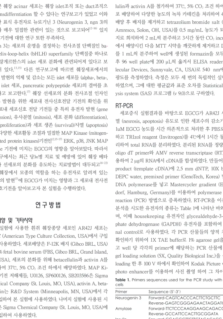
녹차 카테킨 (EGCG)의 ngn 3, α-amylase, insulin의 mRNA 수준과 단백질 발현에 미치는 영향
Ngn 3, α-amylase, insulin mRNA 수준은 RT-PCR을 이 용하였고 단백질 발현은 단백질간 특이적 항원항체 반응을 이용하여 각각 조사 하였다 (Fig. 2. A-F). 췌장조직 재생에 영향을 미치는 것으로 알려진 ngn 3의 mRNA와 단백질 발 현는 EGCG처리 세포에서 시간 의존적으로 유의적 증가를 보였다 (Fig. 2. A, B). 췌장 선 세포의 췌장 islet 세포와 duct 세포 분화 정도를 알아 볼 수 있는 지표인 α-amylase, insu- lin의 mRNA량과 단백질 발현을 조사하였다 (Fig. 2. C-F).
EGCG 처리세포에서 α-amylase, insulin의 mRNA량이 시 간 의존적으로 유의적으로 증가 되었으며 단백질 발현도 동 일한 경향으로 그 발현량이 증가하였다. 따라서 이러한 결과 는 녹차의 주요 생리활성 성분인 EGCG가 췌장 선 세포의 췌장 islet 세포분화와 내분비 세포로의 기능 회복에 영향을 미치는 것으로 사료된다.
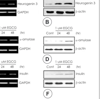
녹차 카테킨 (EGCG)의 MAP Kinase 세포신호 전달기전의 인산화에 미치는 영향
1 μM EGCG 농도를 AR42J 세포에 시간 의존적으로 처 리한 후 췌장 선세 분화의 전사조절인자인 ngn 3의 발현에 영향을 미치는 세포내 전사신호 전달기전들을 조사 하였다.
세포의 다양한 활동 중 세포 분화와 성장에 밀접한 세포 신 호전달기전인 MAP Kinase 관련 기전들인 ERK, JNK 그리
Fig. 1. The inhibition of rat pancreatic tumor cell line, AR42J cells after treatment with EGCG. Cells were plated at 1 × 10⁵ cells per 60 mm culture plate, and incubated for 24 hr. The cells were treated with various concentrations of EGCG for 48 hr. The growth inhibition was measured by the metabolic-dye-based MTT assay. Results are expressed as the means ± S.E. of three in- dependent experiments.
Cont 0.001 0.01 0.1 1 10 100 120
100 80 60 40 20 0
540 nM
EGCG (uM)
고 p38 MAP Kinase 기전 인산화에 미치는 EGCG 영향을 실험하였다. 그 결과 EGCG 처리한 세포에서 비처리 세포에 비해 ERK (Fig. 3A, p < 0.05)와 JNK (Fig. 3B)의 인산화 가 처리시간 의존적으로 억제되었다. 그러나 1 μM EGCG 농 도에서 p38 (Fig. 3C, p < 0.05)의 인산화가 활성 하였다. 이 러한 결과는 3가지 MAP Kinase 세포신호전달 기전 중 p38 기전의 활성화로 ngn 3 발현을 위한 전사신호 전달이 일어 나는 것으로 사료되어진다. 본 실험 결과를 재확인 하기위해 p38 MAP Kinase 기전 저해제인 SB203580과 다른 기전 저 해제인 U0126 (ERK MAP Kinase 기전 저해제), SP600126 (JNK MAP Kinase 기전 저해제)을 각각 세포에 처리하여 세포신호전달 기전을 다시 확인하였다 (Fig. 3D). 그 결과 SB203580 처리세포에서 p38의 인산화가 유의적으로 억제되 었고 다른 기전 저해제 처리 세포에서도 역시 그 활성의 억 제를 보였다. 한편 각각의 저해제와 EGCG를 함께 처리한 결 과 p38 MAP Kinase 기전 인산화가 회복되어 활성의 증가 를 보인 반면, EGCG와 함께 처리한 ERK, JNK 기전 저해 제 처리 세포에서는 그 활성의 억제가 지속되었다. 따라서 AR42J 세포에서 EGCG 처리 시 췌장 선세포의 분화 전사조 절 인자인 ngn 3의 발현을 위한 전사신호전달 기전은 세포
내 p38 MAP Kinase 기전의 활성화를 통하여 이루어지는 것을 재확인하였다.
고 찰
다양한 생리활성을 가진 천연 물질중 하나인 녹차 폴리페 놀성분인 카테킨 (-)-Epigqllocatechin-3-gallate (EGCG)는 암, 심·혈관계질환, 자가 면역반응 질환, 퇴행성질환 그리고 간질환과 관련한 지속적인 연구자들의 관심을 모으고 있다.23) 녹차는 전 세계적으로 가장 많이 음용하는 음료중 하나일 뿐 만 아니라 몇몇 연구에서는 EGCG가 토코페롤, 아스코르
Cont 24h 48h 5
4 3 2 1 0
Denaltometry
*
Cont 24h 48h 7.5
6 4.5 3 1.5 0
Denaltometry
* pERK
1 uM EGCG Cont 24h 48h
β-actin
A
pp38 β-actin 1 uM EGCG Cont 24h 48h
B
Cont 24h 48h 2.5
2 1.5 1 0.5 0
Denaltometry
pJNK 1 uM EGCG Cont 24h 48h
β-actin
pERK pp38 pJNK β-actin DMSO
U0126 SB203580 SP600126 EGCG
C D
Fig. 3. Expression levels of MAP Kinase signaling pathway, pERK, pp38 and pJNK in AR42J cells treated with EGCG for 24, 48 hrs.
A, B, C: after 24, 48 hr incubation with/without EGCG, total pro- teins were lysed and then cellular proteins were separated by SDS-ployacrylamide gels and transferred onto nitrocellulose membranes. The membranes were probed with proper anti- bodies. Proteins were visualized using an ECL detection system.
β-actin was used as internal control. Band intensities in the im- munoblots were quantified by densitometry using L Process (Version 2.01, Fujifilm, Stanford) and Multi Gauge software (Ver- sion 2.02, Fujifilm). Band intensities were normalized relative to the internal control and background. Multiple experiments were combined and data was expressed as % of the control (set as 100%). Statistical Analysis was performed using Student’s T-test;
significance was set at p < 0.05. D: after cells were incubated with/without EGCG for 48 hrs, U0126, SB203580, SP600126 inhibi- ters were treated for 15 mins. Effects of MAP Kinase pathway were monitored by use of proper antibodies. Proteins were visu- alized using an ECL detection system. β-actin was used as inter- nal control.
1 uM EGCG
Heurogenin 3 Cont 24 48 (hr)
GAPDH
1 uM EGCG Cont 24 48 (hr)
Heurogenin 3 β-actin
1 uM EGCG Cont 24 48 (hr)
α-amylase GAPDH
A
Fig. 2. Effects of EGCG treatment on the levels on neurogenin 3, α-amylase and insulin mRNA and protein in AR42J cells. A, C, E:
after 24, 48 hr incubation with EGCG total RNAs were isolated and revers-transcribed. The resulting cDNAs were subjected to PCR with the incubated primers and reaction products were subjected to electophoresis in a 1% agarose gel and visualized by EtBr staining. GAPDH was used as internal control. B, D, F: The cells were lysed and then cellular proteins were separated by SDS-ployacrylamide gels and transferred onto nitrocellulose membranes. The membranes were probed with proper anti- bodies. Proteins were visualized using an ECL detection system.
β-actin was used as internal control.
B
1 uM EGCG Cont 24 48 (hr)
α-amylase β-actin
C 1 uM EGCG
Cont 24 48 (hr) Insulin GAPDH
D
E
1 uM EGCG Cont 24 48 (hr)
Insulin β-actin
F
빅산 보다 더 강력한 항 산화력이 있다고 보고 되어지고 있
다.24-26) 뿐 만 아니라 항 당뇨기전에 대한 EGCG의 영향에
대한 연구도 많은 연구자들에 의해 지속적으로 수행되어 보 고되고 있다.27-30) 그러나 본 연구에서는 흰쥐 췌장 선세포에 인위적 발암을 유도한 췌장종양 세포인 AR42J 세포에 EGCG 처리가 췌장 선세포의 분화를 유도하는 전사조절인자 ngn 3 의 발현에 영향을 미치는지를 알아보고 이와 관련한 세포내 전사신호 전달기전들 중 특히, 세포의 성장, 분화와 밀접한 관련이 있는 MAP Kinase 기전인 ERK, JNK와 p38 MAP Kinase 기전들과의 관련성을 알아보았다. 실험에 사용한 흰쥐 췌장종양 세포인 AR42J 세포는 췌장 progenitor 세포모델로 이미 다수의 연구자들이 췌장분화 관여 물질 연구의 모델로 사용되어 본 실험에서 항 당뇨 생리활성이 보고된 EGCG의 췌장 세포 분화에 미치는 영향을 확인하기의해 세포모델로 적합하다고 사료되어 본 연구에 사용하였다.32-34) 췌장 선 세 포에서 췌장 재생지표로 고려되어지고 있는 ngn 315,35-37)의 mRNA, 단백질 발현 증가에 EGCG의 유의적인 영향을 나 타내었다. 췌장 종양세포의 기능회복 및 분화 간접지표인 α- amylase insulin의 mRNA와 단백질 발현이 EGCG 처리세 포에서 유의적으로 증가하였다. 특히 α-amylase 증가는 급·
만성 췌장염 또는 알코올성 췌장염으로 인한 세포 섬유화38-41) 로 감소되어진 α-amylase분비가 EGCG 처리로 회복 되어질 수 있음을 의미한다고 사료된다. 이러한 결과는 EGCG를 포 함하는 녹차의 섭취가 산화적 스트레스를 억제하여 cerulein 으로 유도된 심각한 급성 췌장염을 줄일 수 있다는 연구결과42) 와 흰쥐 췌장 stellate 세포에서 EGCG는 알코올로 유도된 산화적 스트레스를 억제하여 만성 췌장염으로 인한 췌장의 섬유화를 예방 할 수 있다는 결과26)들이 뒷받침 하듯이 본 실험의 결과처럼 췌장세포의 재생에도 EGCG의 생리활성 효과의 영향이 미치는 것으로 사료된다. 반면에 EGCG의 항 당뇨 작용기전 연구보고는 EGCG가 소장 α-amylase 분비 감소로 혈당을 저하시킨다고 보고도 있다.43)본 실험에서 EGCG는 췌장발달에서 췌장 선세포에서 내분비 세포인 is- let 세포분화 발달을 유도하는 것으로 알려져 있는 전사조절 인자 ngn 3 발현 증가를 유도하며 세포내 전사신호전달 과 정은 명확하게 EGCG의 p38 MAP Kinase 기전 활성으로 유 도되어짐을 p38 MAP Kinase 저해제 (SB203580) 처리실험 으로 재확인 하였다. 이와 같은 결과는 Ogihara 등16)이 보고 한 p38 MAP Kinase의 ngn 3 발현을 위한 세포 신호전달에 매우 중요한 기전이라는 보고와 일치한다. 또한 Umezawa 등40)은 AR42J 세포에 생약성분인 Conophylline을 처리하여 췌도 기능 향상과 베타 세포 증가에 대한 연구에서 AR42J 세포의 인슐린 생성 세포로의 분화에 p38 MAP Kinase 활
성 유도를 보고 하므로써 이 결과들은 본 연구 결과를 뒤받 침 해준다고 사료된다. 결론적으로, 녹차 카테킨인 EGCG를 AR42J 세포에 처리하여 췌장세포 분화에 영향을 미치는 ngn 3 발현 및 세포분비 기능개선에 대한 실험결과 EGCG 의 기존에 알려진 항 당뇨를 비롯한 다양한 생리활성 작용 외에도 췌장조직의 선세포 분화 및 췌장 내분비 세포기능 개 선에 영향을 미치며 그 세포신호전달은 p38 MAP Kinase 기전을 통해 이루어진다고 확인하였다. 그러나 EGCG의 췌 장세포 분화 및 재생에 미치는 영향에 대한 보다 면밀한 연 구가 앞으로 더 진행되어야 할 것으로 사료된다.
요 약
본 연구는 EGCG의 항 당뇨 활성기전으로 췌장종양 선세 포 AR42J의 분화 및 내분비기능 개선에 미치는 영향과 그 세포 신호전달 기전을 확인하였다. 그 결과 첫째, AR42J 세포 에 EGCG 처리 시 췌장종양 선세포의 세포증식이 농도 의존 적으로 감소되었다. 둘째, 세포사멸 유도가 유의적으로 일어 나지 않는 농도인 1uM EGCG를 AR42J 세포에 처리한 결과 ngn 3, α-amylase, insulin은 EGCG처리 24시간에 mRNA, 단백질 발현증가를 나타내었고 48시간에 유의적 증가를 나 타내었다. 셋째, EGCG 처리 시 ERK, JNK MAP Kinase 기 전은 인산화 억제를 나타내었고 반면에 p38 기전의 인산화 는 48시간에 유의적 증가를 하였다. 넷째, p38기전 저해제인 SB203580을 처리하여 EGCG가 MAP Kinase 기전중의 하 나인 p38 기전 인산화 활성의 회복을 나타내어 ngn 3 발현 을 위한 전사 신호전달 기전임을 다시 확인하였다. 따라서 녹 차 생리활성 성분인 EGCG의 췌장종양 선 세포 AR42J 처리 결과 EGCG는 p38 MAP Kinase 기전 활성을 통해 췌장 선 세포의 분화지표인 ngn 3 발현을 증가시키며 췌장내분비 기 능 지표인 α-amylase, insulin 발현증가를 나타내어 세포의 내분비기능 개선에도 영향을 미치는 것으로 사료된다.
Literature cited
1) Suzuki K, Kori S, Morikawa M, Takagi A, Namiki H. Oxida- tive stress-mediated bimodal regulation of polymorphonuclear leukocyte spreading by polyphenolic compounds. Int Immuno- pharmacol 2010; 10(11): 1448-1455
2) Kürbitz C, Heise D, Redmer T, Goumas F, Arlt A, Lemke J, Rimbach G, Kalthoff H, Trauzold A. Epicatechin gallate and catechin gallate are superior to epigallocatechin gallate in growth suppression and anti-inflammatory activities in pancre- atic tumor cells. Cancer Sci 2011; 102(4): 728-734
3) Larsen CA, Dashwood RH, Bisson WH. Tea catechins as in- hibitors of receptor tyrosine kinases: mechanistic insights and
human relevance. Pharmacol Res 2010; 62(6): 457-464 4) Singh R, Akhtar N, Haqqi TM. Green tea polyphenol epigallo-
catechin-3-gallate: inflammation and arthritis. Life Sci 2010;
86(25-26): 907-918
5) Yadav D, Whitcomb DC. The role of alcohol and smoking in pancreatitis. Nat Rev Gastroenterol Hepatol 2010; 7(3): 131-145 6) Yadav D. Recent advances in the epidemiology of alcoholic
pancreatitis. Curr Gastroenterol Rep 2011; 13(2): 157-165 7) Etemad B, Whitcomb DC. Chronic pancreatitis: diagnosis,
classification, and new genetic developments. Gastroenterolo- gy 2001; 120(3): 682-707
8) Lankisch PG, Assmus C, Maisonneuve P, Lowenfels AB. Epi- demiology of pancreatic diseases in Lüneburg County. A study in a defined German population. Pancreatology 2002; 2(5): 469- 9) Lankisch PG, Lowenfels AB, Maisonneuve P. What is the risk 477
of alcoholic pancreatitis in heavy drinkers? Pancreas 2002; 25 (4): 411-412
10) Yadav D, Eigenbrodt ML, Briggs MJ, Williams DK, Wiseman EJ. Pancreatitis: prevalence and risk factors among male veter- ans in a detoxification program. Pancreas 2007; 34(4): 390-398 11) Choudhuri G, Lakshmi CP, Goel A. Pancreatic diabetes. Trop
Gastroenterol 2009; 30(2): 71-75
12) Joglekar MV, Parekh VS, Hardikar AA. New pancreas from old: microregulators of pancreas regeneration. Trends Endocri- nol Metab 2007; 18(10): 393-400
13) Docherty K. Growth and development of the islets of Langer- hans: implications for the treatment of diabetes mellitus. Curr Opin Pharmacol 2001; 1(6): 641-650
14) Schwitzgebel VM. Programming of the pancreas. Mol Cell En- docrinol 2001; 185(1-2): 99-108
15) Zhang YQ, Mashima H, Kojima I. Changes in the expression of transcription factors in pancreatic AR42J cells during differen- tiation into insulin-producing cells. Diabetes 2001; 50(Suppl 1):
S10-S14
16) Ogihara T, Watada H, Kanno R, Ikeda F, Nomiyama T, Tanaka Y, Nakao A, German MS, Kojima I, Kawamori R. p38 MAPK is involved in activin A- and hepatocyte growth factor-mediat- ed expression of pro-endocrine gene neurogenin 3 in AR42J- B13 cells. J Biol Chem 2003; 278(24): 21693-21700
17) Gradwohl G, Dierich A, LeMeur M, Guillemot F. Neurogenin3 is required for the development of the four endocrine cell lin- eages of the pancreas. Proc Natl Acad Sci USA 2000; 97(4): 1607- 18) Edlund H. Transcribing pancreas. Diabetes 1998; 47(12): 1817-1611 19) Wilson ME, Scheel D, German MS. Gene expression cascades 1823
in pancreatic development. Mech Dev 2003; 120(1): 65-80 20) Schwitzgebel VM, Scheel DW, Conners JR, Kalamaras J, Lee
JE, Anderson DJ, Sussel L, Johnson JD, German MS. Expres- sion of neurogenin3 reveals an islet cell precursor population in the pancreas. Development 2000; 127(16): 3533-3542
21) Apelqvist A, Li H, Sommer L, Beatus P, Anderson DJ, Honjo T, Hrabe de Angelis M, Lendahl U, Edlund H. Notch signalling controls pancreatic cell differentiation. Nature 1999; 400(6747):
877-881
22) Jensen J, Heller RS, Funder-Nielsen T, Pedersen EE, Lindsell C, Weinmaster G, Madsen OD, Serup P. Independent development of pancreatic alpha- and beta-cells from neurogenin3-express- ing precursors: a role for the notch pathway in repression of premature differentiation. Diabetes 2000; 49(2): 163-176 23) Trevisanato SI, Kim YI. Tea and health. Nutr Rev 2000; 58(1):
24) Rice-Evans C. Implications of the mechanisms of action of tea 1-10
polyphenols as antioxidants in vitro for chemoprevention in hu- mans. Proc Soc Exp Biol Med 1999; 220(4): 262-266
25) Kao YH, Hiipakka RA, Liao S. Modulation of endocrine sys- tems and food intake by green tea epigallocatechin gallate. En- docrinology 2000; 141(3): 980-987
26) Asaumi H, Watanabe S, Taguchi M, Tashiro M, Nagashio Y, Nomiyama Y, Nakamura H, Otsuki M. Green tea polyphenol (-)-epigallocatechin-3-gallate inhibits ethanol-induced activa- tion of pancreatic stellate cells. Eur J Clin Invest 2006; 36(2):
113-122
27) Sakurai N, Mochizuki K, Kameji H, Shimada M, Goda T.
(-)-Epigallocatechin gallate enhances the expression of genes related to insulin sensitivity and adipocyte differentiation in 3T3-L1 adipocytes at an early stage of differentiation. Nutrition 2009; 25(10): 1047-1056
28) Bursill CA, Abbey M, Roach PD. A green tea extract lowers plasma cholesterol by inhibiting cholesterol synthesis and up- regulating the LDL receptor in the cholesterol-fed rabbit. Ath- erosclerosis 2007; 193(1): 86-93
29) Ryu OH, Lee J, Lee KW, Kim HY, Seo JA, Kim SG, Kim NH, Baik SH, Choi DS, Choi KM. Effects of green tea consumption on inflammation, insulin resistance and pulse wave velocity in type 2 diabetes patients. Diabetes Res Clin Pract 2006; 71(3):
356-358
30) Nagao T, Komine Y, Soga S, Meguro S, Hase T, Tanaka Y, To- kimitsu I. Ingestion of a tea rich in catechins leads to a reduc- tion in body fat and malondialdehyde-modified LDL in men.
Am J Clin Nutr 2005; 81(1): 122-129
31) Imai K, Nakachi K. Cross sectional study of effects of drinking green tea on cardiovascular and liver diseases. BMJ 1995;
310(6981): 693-696
32) Hamamoto K, Yamada S, Hara A, Kodera T, Seno M, Kojima I.
Extracellular matrix modulates insulin production during dif- ferentiation of AR42J cells: functional role of Pax6 transcrip- tion factor. J Cell Biochem 2011; 112(1): 318-329
33) Fukazawa T, Matsuoka J, Naomoto Y, Nakai T, Durbin ML, Kojima I, Lakey JR, Tanaka N. Development of a novel beta- cell specific promoter system for the identification of insulin- producing cells in in vitro cell cultures. Exp Cell Res 2006;
312(17): 3404-3412
34) Kojima I, Umezawa K. Conophylline: a novel differentiation inducer for pancreatic beta cells. Int J Biochem Cell Biol 2006;
38(5-6): 923-930
35) Joglekar MV, Parekh VS, Hardikar AA. New pancreas from old: microregulators of pancreas regeneration. Trends Endocri- nol Metab 2007; 18(10): 393-400
36) Docherty K. Growth and development of the islets of Langer- hans: implications for the treatment of diabetes mellitus. Curr Opin Pharmacol 2001; 1(6): 641-650
37) Schwitzgebel VM. Programming of the pancreas. Mol Cell En- docrinol 2001; 185(1-2): 99-108
38) Carvalho KM, Morais TC, de Melo TS, de Castro Brito GA, de Andrade GM, Rao VS, Santos FA. The natural flavonoid quer- cetin ameliorates cerulein-induced acute pancreatitis in mice.
Biol Pharm Bull 2010; 33(9): 1534-1539
39) Thrower EC, Yuan J, Usmani A, Liu Y, Jones C, Minervini SN, Alexandre M, Pandol SJ, Guha S. A novel protein kinase D in- hibitor attenuates early events of experimental pancreatitis in isolated rat acini. Am J Physiol Gastrointest Liver Physiol 2011;
300(1): G120-G129
40) Umezawa K, Hiroki A, Kawakami M, Naka H, Takei I, Ogata T, Kojima I, Koyano T, Kowithayakorn T, Pang HS, Kam TS.
Induction of insulin production in rat pancreatic acinar carci- noma cells by conophylline. Biomed Pharmacother 2003; 57(8):
341-350
41) Chen LA, Li J, Silva SR, Jackson LN, Zhou Y, Watanabe H, Ives KL, Hellmich MR, Evers BM. PKD3 is the predominant protein kinase D isoform in mouse exocrine pancreas and pro- motes hormone-induced amylase secretion. J Biol Chem 2009;
284(4): 2459-2471
42) Takabayashi F, Harada N. Effects of green tea catechins (Poly- phenon 100) on cerulein-induced acute pancreatitis in rats.
Pancreas 1997; 14(3): 276-279
43) Kao YH, Chang HH, Lee MJ, Chen CL. Tea, obesity, and dia- betes. Mol Nutr Food Res 2006; 50(2): 188-210