I. 서론
골 결손부의 회복을 위해 자가골이나 동종골을 이 식하는 방법은 오래 전부터 이용되어 왔고 현재에도 가장 일반적인 골이식술의 방법이 되고 있다. 하지 만, 자가골의 경우는 공여부위의 이차적 수술이 필요 하고 충분한 양의 골을 얻기가 쉽지 않다는 단점이 있고, 동종골의 경우에도 가능성은 적은 것으로 알려 지고 있지만 일부 전염성질환의 전이가능성이 여전 히 상존해 있다. 이러한 문제들 때문에 충분한 양을 쉽게 얻을 수 있고 질병의 전염가능성이 없는 인공 합성골이 오래전부터 개발되어 사용되고 있다. 대표 적인 인공함성골로서 hydroxyapatite(HA)1-3), trical- cium phosphate(TCP)4, 5)등의 인산칼슘계 이식재와 polymer6-8), bioglass9), calcium carbonate10, 11)등의 이식재들이 사용되고 있고, 이들을 이용한 일부 성공 적인 임상결과들이 보고되고 있다. 하지만 이들에 대한 최근의 평가들을 보면, 조직학적으로 이들은 진 정한 의미의 골유도성은 없는 것으로 확인되고 있으 며 대부분은 결합조직 개재에 의해 골조직과는 분리 되는 단순히‘생적합성이 있는 충전재’(biocompati- ble filler)에 불과한 것으로 인정되고 있다12-14). 따라 서 골친화성이 보다 우수하고 골재생에 맞추어 적절
히 흡수되어 재생골으로 치환될 수 있는 흡수성 골 이식재가 필요한 상황이다.
최근에 들어서 새로운 개념의 조직재생시술의 방 법으로 조직공학의 개념과 기술이 제안되고 있는데, 이것은 재생을 원하고자 하는 조직으로 부터 세포를 분리·배양하고 적절한 생체재료에 접종하여, 증 폭·배양함으로써 인공적으로 조직을 형성하는 기
술이다15-17). 조직공학의 기법을 위해서는 해당조직
세포를 접종·배양할 세포지지체가 필요하며 이는 조직적합성이 뛰어나고 세포접착성이 우수한 생체 재료이어야 한다. 현재 골형성을 위한 세포지지체로 서 연구되고 있는 예로서, 교원질 matrix18, 19), poly(glycolic acid) mesh(PGA)20, 21), poly(lactic-co- glycolic acid) foam(PLGA)22), calcium phosphate ceramics23-25), poly(lactide/glycolide)/hydroxy apatite(PLGA/HA)26)및 polyphosphazenes27)등이 제 시된 바 있지만, 이들 역시 아직은 해결되지 않은 문 제점들을 안고 있어서 부가적인 연구가 필요한 상황 이다.
Calcium metaphosphate(CMP; [Ca(PO3)2]n)는 monocalcium phosphate [Ca(H2PO4)2]를 열분해시켜 얻을 수 있는데 이것은 무기질 고분자로서 4개이상 의 동질이상( α, β, γ, δ)이 있고 모두[-O-P-O-]를 골격 대한치주과학회지 : Vol. 28, No. 4, 1998
생분해성 다공질 Calcium Metaphosphate 블록의 조직적합성에 관한 연구
이용무1·김석영2·신승윤1·구 영1·류인철1·정종평1
1서울대학교 치과대학 치주과학교실
2영남대학교 공과대학 재료금속공학부
이 연구는 서울대학교병원 지정연구비(02-1996-361-0)의 지원에 의한 결과임
으로 하는 쇄상구조를 갖는다. 이 중에서 쇄상의 길 이가 가장 길고 생화학적으로 가장 안정한 β형의 CMP는 열처리 방법에 따라 비정질 혹은 결정질을 얻을 수 있고 제조방법에 따라 혹은 CMP상에 따라 생분해속도를 조절할 수 있다28, 29).
본 연구에서는 CMP의 골이식재 및 조직공학적 골 형성을 위한 세포지지체로서의 응용가능성을 평가 하기 위하여, 다공질의 CMP 블록을 제작하고 이를 가토의 피하, 근육 및 골 내에 이식하고 생체반응을 조직학적으로 관찰하였다.
II. 재료 및 방법
1. 다공질 CMP 블록의 제조무수 Ca(H2PO4)2를 condensation 하여 무결정의 Ca(PO3)2를 얻고 이를 용융하고 냉각시킨 후 분쇄하 여 CMP powder를 얻었다. 다공성의 CMP블록은 이 와 김30)이 기술한 바의 polyurethane(PU) 스펀지를 이용하여 PU를 소환하는 방법으로 sponge 형태의 0.3-1mm의 소공의 크기를 가지는 다공질 CMP블록 (Figure 1)을 제작하였다.
2. 다공질 CMP블록의 가토 조직내 이식
6마리의 웅성 가토(Newzealand White rabbit)에 30mg/㎏의 용량으로 염산 자일리진(Rompun , 한 국바이엘, 한국)과 염산 케타민(Ketalar , 유한양행, 한국)을 혼합하여 복강주사로 마취하고, 두부의 정 수리부분, 등 및 다리의 털을 깎고 potadine과 hibi- tane으로 소독한 후 2% 리도카인으로 국소 침윤마취 를 부가적으로 실시하였다.
(1) 골 내 이식
소독된 두피에 전후방향으로 직선의 절개선을 가 하여 두피와 골막을 절개하였다. 골막을 박리하고 두피와 골막을 젖혀 두개골을 노출시킨 후, 8-mm 직 경의 trephine bur(3i implant innovation, USA)를 이 용하여 뇌막에 손상을 주지 않도록 주의하면서 좌우
의 측두골에 2개의 원형 골 결손부를 형성하였다. 형 성된 골 결손부 중 한 측은 CMP블록을 이식하고 다 른 측은 이식하지 않은 채 골막과 두피를 흡수성 봉 합사와 수술용 실크로 봉합하였다.
(2) 피하 이식
소독된 등의 피부에 2cm 정도의 절개를 가하고 수 술용 가위를 이용하여 피하결합조직을 충분히 dis- section하여 공간을 얻은 후 CMP블록을 이식하고 입 구를 수술용 실크로 봉합하였다. 6마리 가토의 등에 각 2개씩의 CMP블록을 피하 이식하였다.
(3) 근육 내 이식
소독된 다리의 대퇴부에 4 cm 정도의 절개를 가하 고 수술용 가위를 이용하여 피하결합조직을 박리하 고 근육을 노출시킨 후, 다시 근육을 hemostat와 수 술용 가위를 이용하여 근섬유결을 따라 dissection 하여 공간을 얻은 후 CMP블록 근육 내에 이식하였 다. 매식후 입구의 근섬유들을 흡수성 봉합사로 봉 합하고 상부의 피부를 다시 수술용 실크로 봉합하였 다. 6마리의 가토에 2개의 CMP블록을 좌우의 대퇴 부에 각각 이식하였다.
3. 조직학적 관찰
이식 후 4주 및 6주에 각각 3마리씩을 희생하여 두 개골, 피하 및 근육에 이식된 CMP블록을 인접한 조 직과 함께 절제해 낸 후 중성포르말린에 고정하였 다. 통법에 따라 수세 및 탈수하고 supurred low vis- cosity media(Polyscience Inc, USA)로 포매한 후 Exakt cutting and grinding system(Exakt-Apprateb, Germany)으로 절단·연마하여 비탈회연마표본을 제작였다. 제작된 표본은 multiple 염색 및 HE 염색 을 시행한 후 광학현미경하에서 조직학적 관찰을 실 시하였다.
III. 결과
1. 두개골 결손부에서 CMP의 조직반응
수술 4주 및 6주 후의 소견에서 이식하지 않은 대 조군의 결손부는 대부분 섬유성 결합조직으로 치유 되고 있었고 대조군 6주의 소견에서는 결손부 말단 으로부터 일부의 신생골이 자라나고 있었다(Figure 2). CMP 블록이식군에서는 이식 4주 경과후에 결손 부는 대부분 결합조직으로 채워져 있었으나 이식한 CMP주위로 활발한 세포증식과 더불어 골양조직이
침착되는 소견이 관찰되었다(Figure 3, 4). CMP이식 6주후의 결과에서는 결손부 내부에 이식한 CMP와 융합하여 신생골 형성이 활발하게 일어남이 관찰되 었는데, 고배율 관찰결과 이식한 CMP와 직접 접촉 하여 신생골 침착이 일어나고 있었고, 이식한 CMP 는 분해되면서 신생골 침투와 함께 융합되는 소견이 확인되었다(Figure 5, 6).
Figure 1. Photograph of porous CMP block Figure 2. Control group after 6 weeks of healing in rabbit calvarial defect. Figure illustrates the whole defect. Between the wound edges(arrows), the defect is filled with thin loosely organized connec- tive tissues. New bone is localized to the defect border. (multiple stain, original magnification x2.5)
Figure 3. CMP group after 4 weeks of healing rabbit cal- varial defect. Figure illustrates the whole defect.
Between the wound edges(arrows), grafted CMP matrices(open arrows) are seen. Bone regenera- tion is limited to the defect border(multiple stain, original magnification x2.5)
Figure 4. CMP group after 4 weeks of healing in rabbit cal- varial defect. Figure illustrates CMP matrix(C) in the defect. Young osseous tissues(arrows) are deposited on the CMP matrix. m indicates mar- row. (multiple stain, original magnification x25)
2. 피하 결합조직 내에서의 조직반응
CMP 이식 4주후의 소견에서 일부 소수의 염증세 포 침윤이 관찰되기도 하였으나 별다른 이물반응 없 이 조직과 잘 융화하고 있었다(Figure 7). 이식 6주후
의 소견에서는 염증세포는 거의 사라졌고 피하결합 조직과 완전히 융화되어 섬유아세포들이 CMP표면 을 따라 잘 배열하고 있고 부분적으로 구조가 무너 지고 있는 공간으로도 결합조직이 침입하고 있었다 (Figure 8).
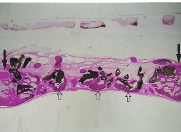
Figure 6. CMP group after 6 weeks of healing in rabbit cal- varial defect. Figure illustrates CMP(C) matrix in the defect. Regenerated bone(*) is apposed directly to CMP matrix. Some regenerated bone(open arrows) infiltrates into and is mingled with CMP matrix being degraded. (multiple stain, original magnification x25)
Figure 7. Tissue response of CMP in rabbit subcutaneous tissue, 4 weeks after implantation. Fibrous con- nective tissue lined along the CMP matrix(C) sur- face. A few inflammatory cells are seen but no significant adverse reaction is observed.
Hematoxylin and eosin stain, original magnifica- tion x50.
Figure 8. Tissue response of CMP in rabbit subcutaneous tissue, 6 weeks after implantation. Any inflamma- tory infiltration or adverse reaction is not seen.
CMP matrix(C) is well adapted in fibrous connec- tive tissue. Hematoxylin and eosin stain, original magnification x50.
Figure 5. CMP group after 6 weeks of healing in rabbit cal- varial defect. Figure illustrates the whole defect.
Between the wound edges(arrows), significant new bone(open arrows) is regenerated and min- gled with grafted CMP matrices.(multiple stain, original magnification x2.5)
3. 근조직 내에서의 조직반응
CMP 이식 4주 후의 소견에서 소수의 염증세포침 윤이 보이기도 하였으나 별다른 이물반응은 나타나 지 않았으며 섬유성 결합조직으로 싸여 있었다 (Figure 9). 이식 6주후의 소견에서는 CMP를 둘러싼 섬유성 결합조직층이 점차 근조직으로 대체되고 있 었고 CMP의 경계가 불명확해지면서 주위조직에 의 해 융화되는 소견을 보이고 있었다(Figure 10).
IV. 고찰
본 연구에서 시험된 CMP는 [-O-P-O-]를 골격으로 하는 쇄상구조를 갖는 무기질 고분자이다. 이는 열 처리방법과 제조공정에 따라 여러 가지 형태로 가공 이 가능하고 흡수속도도 조절이 가능하다28, 29). 이 연 구에서 제작한 다공질의 CMP 블록은 골이식재 뿐만 아니라 조직공학을 위한 세포지지체로서 응용할 목 적으로 개발되었다.
골이식재료는 기본적으로 면역반응이나 염증반응 을 야기하지 않는 생적합성, 재생골로 적절히 치환될 수 있는 흡수성, 골전도성이나 골유도성, 소독가능 성, 방사선 불투과성, 골과 비슷한 강도, 경제성 등이
요구된다31-33). 이러한 이유에서 골과 화학적 성분이
유사한 재료를 찾게 되었고 HA 나 TCP 등의 칼슘인 산계 세라믹재료가 합성골이식재로서 가장 많이 개 발, 사용되고 있다.
이 연구의 결과 CMP 블록은 골조직, 피하결합조 직 및 근조직 내에서 별다른 이물반응이나 염증반응 없이 서서히 흡수되면서 조직과 잘 융화되는 소견을 보이고 있는데, 특히 골 결손부에서 신생골과 직접적 인 접촉에 의해 골조직과 융합되는 소견은 상당히 인상적이다. 이전에 제안되어 사용되고 있는 기존의 골이식재인 HA나 TCP 등이 골 결손부 내에 이식시 결합조직개재에 의한 섬유낭에 싸이는 바의 소견을 비교하면 이 CMP의 골 내 반응은 상당히 다른 양태 를 보여주고 있는데 이것은 이들 재료 보다 CMP가 골친화성 및 골전도성이 월등히 우수하다는 것을 시 사하는 것이라 할 수 있다. 이러한 결과들로 볼 때 CMP가 흡수성 골이식재로서의 여러 가지 요건을 갖 춘 이상적인 재료로 기대된다.
조직공학적 골형성을 위한 세포지지체로서의 요 구조건으로 Ishaug 등은 몇가지 요건을 제시한 바 있 는데, 즉 골아세포에 대한 부착성, 숙주의 혈관조직 이 침투할 수 있고 골아세포의 공간적 증식이 가능 한 다공성의 3차원적 구조, 생분해성이 있어야하고 Figure 9. Tissue response of CMP in rabbit thigh muscle, 4
weeks after implantation. Fibrous connective tis- sue surrounds the CMP matrix(C). A few inflam- matory cells infiltrate but no significant adverse reaction is seen. Hematoxylin and eosin stain, original magnification x50.
Figure 10. Tissue response of CMP in rabbit thigh muscle, 6 weeks after implantation. Thin fibrous connec- tive tissue(*) encapsulates the CMP matrix(C). M marks muscle fiber. Hematoxylin and eosin stain, original magnification x50.
그 분해산물은 부작용 없이 쉽게 대사, 배설되어야 한다22). 골아세포의 3차원적 배양을 위한 세포지지 체로서 교원질18, 19), PGA20, 21), PLGA copolymer22), calcium phosphate ceramic23-25) 및 polyphosp- hazenes26) 등의 여러 가지 재료들이 이용되어 왔지 만, 이들 재료 역시 몇 가지 문제점을 안고 있다. 교 원질의 경우에는 강도가 약하고 흡수속도를 조절하 기 어려운 점이 있고, 대개 이종이나 동종개체로부터 추출하여 사용하기 때문에 면역반응을 야기할 수 있 는 문제점이 있다. PGA mesh의 경우도 강도가 약하 고 100㎛ 정도의 아주 얇은 판상이기 때문에 큰 골 결손부에는 사용이 어려운 단점이 있다. PLGA copolymer의 경우는 소수성이 커서 세포지지체 심 부까지 배지의 확산 및 침투가 어려운 단점이 있다.
기존의 ceramic matrix의 경우는 자연골과 유사한 화 학조성 때문에 생체친화성이 좋은 장점은 있으나, 분 해속도가 너무 느리기 때문에 이식부위에서 형성될 골조직에 물리적 장애요인이 될 수 있다. polyphos- phazene은 현재 새로운 물질로서 시험중인 단계여 서 아직은 더 연구가 필요한 상황이다.
이 연구에서 시험된 CMP블록은 조직내 생체적합 성이 우수하고 골친화성이 뛰어남이 확인되고 있다.
특히 CMP는 제조공정의 변화를 통해 흡수속도를 충 분히 조절할 수 있는 장점이 있어서 기존의 세라믹 재료가 안고 있는 흡수속도가 지나치게 느린 단점을 보완할 수 있다. 이 연구에서는 PU를 소환하는 방법 으로 제작하였는데 PU의 형태 즉 fiber의 굵기나 소 공의 크기를 달리하면 원하는 바의 다공성 구조를 자유롭게 얻을 수 있어서 필요에 따라 세포지지체의 용도로 다양한 형태로의 제조가 가능하다. 또한 미 세 소공을 갖는 과립형의 입자로도 제조가 가능하여 치주병소와 같은 작은 골 결손부를 위한 이식재로도 응용이 가능하다.
이 연구에서는 우선 CMP의 조직반응을 확인하려 는 의도에서 이루어졌기에 장기적인 관찰에 의한 CMP의 흡수과정과 임계크기의 골 결손부34)에서의 재생된 신생골의 정량적인 측정이 이루어지지는 못 하였다. 따라서 골이식재로서의 가능성을 평가하기 위해서는 조절된 동물 실험모델하의 임계결손부에
서 기존의 골이식재와의 비교연구와 장기적인 관찰 에 의한 흡수과정등에 관한 관찰이 계속되어야 할 것으로 보며, 이러한 연구에 기초하여 여러 형태로 가공하여 골이식재로서의 개발가능성이 타진되어야 할 것이다. 또한 조직공학을 위한 세포지지체로서의 가능성을 평가하기 위하여는 효과적인 세포부착, 증 식 및 골조직형성을 위한 소공의 크기조절 및 전체 적 구조의 설정에 관한 계속적인 연구가 필요할 것 으로 생각된다.
V. 결론
이 연구에서는 무수 Ca(H2PO4)2를 condensation 하여 얻은 무결정의 Ca(PO3)2를 이용하여 다공질 CMP블록을 제작하고 골이식재 및 조직공학적 골형 성을 위한 세포지지체로서의 응용가능성을 평가하 기 위하여 이를 가토의 피하, 근육 및 골 내에 매식하 여 생체반응을 조직학적으로 관찰하였다. 관찰결과, CMP는 피하 및 근조직 내에서 별다른 이물반응 없 이 서서히 분해되면서 주위조직과 잘 융화되고 있었 다. 골 결손부에 이식된 CMP는 이식 4주후의 결과에 서 그 주위로 활발한 세포증식과 더불어 골양조직이 침착되는 소견이 관찰되었으며, 6주후에는 결합조직 의 개재 없이 CMP기질에 직접 접촉된 신생골 형성 이 확인되었다. 이러한 결과는 CMP의 골친화성 및 골전도성이 아주 우수함을 나타내는 것으로, 이 재료 가 골이식재로서 뿐만 아니라 조직공학을 위한 세포 지지체로서 적합한 재료로 생각된다.
VI. 참고문헌
1. Froum SJ, Kushner L, Scopp W, Stahl SS:
Human clinical and histologic response to durapatite implants in intraosseous lesions. J Periodontol 1982;53:719-725.
2. Sapokos SW: The use of Periograf in periodon- tal defects-Histologic findings. J Periodontol 1986;57:7-13.
3. Carranza FA Jr, Kenny EB, Lekovic V,
Talamante E, Valencia J and Dimitrijevic B:
Histologic study of the healing of human peri- odontal defect after placement of porous hydroxyapatite implants J Periodontol 1987
;58:682-688.
4. Baldock WT, Hutchens LH, McFall WT, Simpson DM: An evaluation of tricalcium phosphate implants in human periodontal osseous defects of two patients. J Periodontol 1985;56:1-7.
5. Stahl SS, Froum SJ: Histologic evaluation of human intraosseous healing response to the placement of tricalcium phosphate ceramic implants. J Periodontol 1986;57:211-217.
6. Stahl SS, Froum SJ, Tarnow DP: Human clinical and histologic responses to the placement of HTR polymerparticles in 11 intrabony lesion. J Periodontol 1990;61:269-274.
7. Yukna RA: Clinical evaluation of HTR polymer bone replacement grafts in human mandibular Class II molar furcations. J Periodontol 1994;65:342-349.
8. Froum SJ: Human histologic evaluation of HTR polymer and freeze-dried bone allograft. A case report. J Clin Periodontol 1996;23:615- 620.
9. Zamet JS, Dabar UR, Griffiths GS, Bulman JS, Bragger U, Burgin W, Newman HN: Particulate bioglass as a grafting material in treatment of periodontal intrabony defects. J Clin Periodontol 1997;24:410-418.
10. Louise F, Borghetti A: Clinical evaluation of Natural coral implantation in osseous peri- odontal defects. Results after one year. J Parodontol 1991;10:69-76.
11. Yukna RA: Clinical evaluation of coralline car- bonate as a bone replacement graft material in human periodontal osseous defects. J Periodontol 1994;65:177-185.
12. Yukna RA: Synthetic bone grafts in periodon- tics. Periodontol 2000 1993;1:92-99.
13. Yukna RA: Synthetic grafts and regeneration.
In Polson AM. eds. Periodontal regeneration.
Current status and directions. pp103-112, Quentessence Publishing Co., Inc, Chicago, 1994.
14. Garret S. Periodontal regeneration around nat- ural teeth. Ann Periodontol 1996;1:621-666.
15. Cooper ML, Hansbrough JF, Spielvogel RL, Cohen R, Bartel RL, Naughton G: In vivo opti- mization of a living dermal substitute employ- ing employing cultured human fibroblasts on a biodegradable polyglycolic acid or polyglactin mesh. Biomaterials 1991;12:243- 248.
16. Atala A, Vacanti JP, Peter CA, Mandel JP, Retik AB, Freeman MR: Formation of urothelial structures in vivo from dissociated cells attached to biodegradable polymer scaffolds in vitro. J Urol 1992;148:658-662.
17. Vacanti CA, Vancanti JP: Bone and cartilage reconstruction with tissue engineering approaches. Otolaringol Clin North Am 1994;27:263-276.
18. Sudo H, Kodama HA, Amagai Y, itaku Y, Yamamoto S: Mineralized tissue formation by MC3IC3-E1 osteogenic cells embeded in three dimensional gel matrix, In Cell Mediated Calcification and Matrix Vesicles. pp291-296, SY Ali ed. Elsevier Science, Oxford, UK, 1986.
19. Casser-Bette M, Murray AB, Closs EI, Erfle V, Schmidt J: Bone formation by osteoblast-like cells in a three-dimensional cell culture. Calcif Tissue Int 1990;46:46-56.
20. Vacanti CA, Kim W, Upton J, Vancanti MP, Mooney D, Schloo B, Vancanti JP: Tissue engi- neered growth of bone and cartilage.
Transplant Proc 1993;25:1019-1021.
21. Puelacher WC, Vacanti JP, Ferraro NF, Schloo B, Vacanti CA: Femoral shaft reconstruction using tissue-engineered growth of bone. Int J Maxillofac Surg 1996;25:223-228.
22. Ishaug SL, Crane GM, Miller MJ, Yasko AW, Yaszemski MJ, Mikos AG: Bone formation by three-dimensional stromal osteoblast culture in biodegradable polymer scaffolds. J Biomed Mater Res 1997;36:17-28.
23. Goshima J, Goldberg VM, Caplan AI: The ori- gin of bone in composite grafts of porous cal- cium phosphate ceramic loaded with marrow cells. Clin Orthop Rel Res 1991;274-283.
24. Goshima J, Victor MG, Caplan AI: The osteogenic potential of culture-expanded rat marrow mesenchymal cells assayed in vivo in calcium phosphate ceramic blocks. Clin Orthop Rel Res 1991;298-311.
25. Nakahara H, Goldberg VM, Caplan AI:
Cultured-expanded human periosteal-derivd cells exhibited osteochondral potential in vivo. J Orthop Res 1991;9:465-476.
26. Laurencin CT, Attawia MA, Elgendy HE, Herbert KM: Tissue engineered bone-regener- ation using degraddable polymers: The forma- tion of mineralized matrices. Bone 1996:93S- 99S.
27. Laurencin CT, El-Amin SF, Ibim SE, Willough DA, Attawia M, Allock HR, Ambrocio AA: A
highly porous 3-dimensional polyphosp- hazene polymer matrix for skeletal tissue regenration. J Biomed Mater Res 1996;30:133- 188.
28. Kim S: Bioresorbable calcium mataphosphate ceramics: I. Preparation and preliminary in vitro study. Biomaterials Research 1998;2:48- 52.
29. Mclntosh AO, Jablonski WL: X-ray powder patterns of the calcium phosphate, Analytical Chemistry 1956;28:1424-1427.
30. Lee J, Kim S: In Transaction of 5th World Biomaterials Congress, Toronto, May 1996, University of Toronto Press, Tronto, 1966, p53.
31. Bissada NF, Hangorsky U: Alveolar bone induction: Alloplast. Dent Clin N Am 1980;23:739-749.
32. Frame JW, Browne RM, Brady CL:
Hydroxyapatite as a bone substitute in jaws.
Biomaterials 1981;2:19-22.
33. Levine MP, Getter A, Cutright DE, Bhaskar SN:
Biodegradable ceramics in periodontal defects. Oral Surg Oral Med Oral Pathol 1974;38:344-351.
34. Schmitz JOP, Hollinger JO: The critical sized defect as an experimental model for cran- iomandibular nonunions. Clin Orthop 1986;205:299-308.
-Abstract-
Biocompatibility and Bone Conductivity of Porous Calcium Metaphosphate Blocks
Yong-Moo Lee1, Seok-Young Kim2, Seung-Yun Shin1, Young Ku1 In-Chul Rhyu1and Chong-Pyoung Chung1
1Department of Periodontology, College of Dentistry, Seoul National University
2Department of Material Technology, College of Engineering, Yeungnam University
While calcium phosphate ceramics meet some of the needs for bone replacement, they have some limitation of unresorbability and fibrous encapsulation without direct bone apposition during bone remodelling. To address these problem, we developed a new ceramic, calcium metaphosphate(CMP), and report herein the biologic response to CMP in subcutaneous tissue, muscle and bone. Porous CMP blocks were prepared by condensation of anhydrous Ca(H2PO4)2to form non-crystalline Ca(PO3)2. Macroporous scaffolds were made using a polyurethane sponge method. CMP block possesses a macroporous structure with approximate pore size range of 0.3-1mm. CMP blocks were implanted in 8 mm sized calvarial defect, subcutaneous tissue and muscle of 6 Newzealand White rabbits and histologic observation were performed at 4 and 6 weeks later. CMP blocks in subcutaneous tissue and muscle were well adapted without any adverse tissue reaction and resorbed slowly and spontaneously. Histologic observation of calvarial defect at 4 and 6 weeks revealed that CMP matrix were mingled with and directly apposed to new bone without any intervention of fibrous connective tissue.
CMP blocks didn't show any adverse tissue reaction and resorbed spontaneously also in calvarial defect. This result revealed that CMP had a high affinity for bone and was very biocompatible. From this preliminary result, it was suggested that CMP was a promising ceramic as a bone substitute and tissue engineering scaffold for bone formation.
Key Words: biocompatibility, bone conductivity, calcium metaphosphate, bone substitute, tissue engineering scaffold