I. 서 론
Brånemark 임플란트가 완전무치악 환자에 처음 시 술된후 30여년간 임상에서 널리 이용되고 있다. 초기 에는 골유착 개념에 관한 가설과 이를 입증하는 많은 연구가 진행되었으며(6, 12, 13) 이후 구강내 식립된 임플 란트의 성공여부를 판단하기 위한 객관적인 평가기준 을 세우기 위하여 임상연구가 계속 이루어졌다. 1978 년 NIH-Hazard Consensus(60)에서는 임플란트의
“Benefit and risk”를 주제로 골유착성 임플란트를 제 외한 여러 종류의 임플란트에 관하여 논의되었는데 임 플란트 성공의 기준에 대한 언급과 임플란트는 5년간 75%가 지속되어야 한다고 언급되었으며 Schnitman 과 Schlman(6)은 그들의 연구에서 최초로 5개 항목으 로 이루어진 임플란트 성공기준을 언급하였으나 이러 한 기준들은 과학적인 연구자료가 부추난 상태였다.
1986년 Albrektsson, Zarb, Wington과Erin(7)이 임플란트의 체계적인 성공기준을 발표하였다. 첫째, 임상적으로 개개의 임플란트는 동요도가 없어야 하며 둘째, 방사선 사진상 임플란트 주위에 어떤 방사선 투 과상도 없어야 하며 세째 임플란트 시술 1년후부터는 연간 수직골 소실량이 0.2 mm이하가 되어야하고 네 째, 동통, 감염, 신경학적 장애, 하치조신 경관의 침범 등 지속적이고 비가역적인 증상들이 없어야 한다고 하
였다.
1977년부터 1989년까지 발표된 논문들을 분석한 결 과(5)임플란트의 성공에 대한 광범위한 기준으로 동통, 강한 고정, 탐침깊이, 골소실, 출혈지수, 임플란트 주 위의 염증반응, 타진, 방사선학적 평가를 제시하고 있 다. 이들 논문에서는 Silness와 Loe의 치은지수, 치태 지수를 사용하였고 일부 논문에서는‘Discomfort’라 는 주관적 항목을 조사하였으며‘환자의 만족도’를 언 급하기도 하였고 대부분 동요도와 방사선 사진 분석에 의한 생존률을 발표하였다. James(30)는 부착력이 약한 접합상피가 임플란트와 골상반 연조직 사이에 존재하 므로 치아 주위보다 임플란트에서 변연골에 더 쉽게 탐침될 수 있다고 하였고 Schnittman(56) 등은 치주낭 탐침기 사용을 권장하였으며 Bergman등(9)은 골유착 성 임플란트의 상태를 측정함에 있어서 치은지수, 치 태지수, 탐침깊이를 이용하고 보철물의 교합, 심미성 도 평가하였으며, 방사선 검사로는 임플란트와의 계면 에 간극의 존재여부를 조사하였다. Lekholm(4) 등은 골유착된 임플란트에서는 2-6 mm의 치주낭 깊이를 가지며 부분 무치악에서는 주변 치아보다 임플란트 주 위에 큰 치주낭 깊이를 나타낸다고 하였다. Misch(15)는 풍융한 임플란트 상부구조로 인해 탐침이 곤란하며 출 혈지수가 임플란트 성공여부에 관해 자연치 만큼 신뢰 도가 없다고 하고, Cox(38) 등은 치은지수, 출혈지수와
- 대한 치과 보철학회지 Vol. 34 No. 1, 1996 -
부분무치악 환자에서 기능하중노출이 골유착성 임플란트 주위의 치조골 소실에
미치는 영향에 관한 방사선학적 연구
연세대학교 치과대학 보철학교실
양순봉ㆍ한동후ㆍ양자호
같은 치주조직을 평가하는 지수는 불표요하다고까지 하였다. 또한 탐침깊이는 임플란트 식립전에 존재하던 국소적인 질환의 유무와 기존의 조직두께와 연관을 갖 으며 그러나 아직 합리적이고 과학적인 기준이 없는 상태에서 쉽게 관찰되며 임플란트 주위 치은의 염증상 태를 말해주고 조직성상, 출혈, 삼출물, 변연골의 변화 여부를 감지하는데 이용되고 있으나(69) 이러한 검사가 약한 부착구조에 손상을 줄수 있고 술자에 따라, 숙련 도에 따라 상이한 결과를 가져올 수 있다.
방사선학적 검사도 임플란트의 건강도 측정에 이용 되는 중요한 진단 척도 중 하나다. Adell등(3), Quirynen등(53), Jemt등(37), Fugazzotto등(21), Gotfredson(23)의 Smith등(68)의 연구에서 Brånemark 임플란트를 대상으로 하여 수술후의 골소실량을 방사 선 사진을 이용하여 조사한 연구들을 발표하였다. 이 들은 초기 1년간 적게는 0.6 mm부터 많게는 1.9 mm 의 골소실을 보이고 1 후부터는 매년 0.05 mm에서 0.2 mm의 골소실을 보인다고 하였으나 방사선 사진 의 한계성과 함께 임플란트는 실제 치아의 위치보다 하방에 심겨진 경우가 많아 근육의 위치관계로 인해 평행한 치근단 방사선 사진을 얻기가 어렵고 이렇게 왜곡된 방사선 사진에서는 치조골소실 여부에 대한 해 석이 어렵게 된다. 치조골의 변연부는 치밀골이므로 해면골이 40% 이상 감소되어야 방사선 사진으로 변화 를 감지할수 있기 때문에 임플란트 주위에 방사선 투 과상이 관찰되지 않는다고 하여 임플란트와 골사이의 계면에 반드시 골조직이 있다고 할 수는 없다(22). 따라 서 변연골의 조기상실은 임플란트 치경부의 응력에 대 한 증거로써 임플란트의 협측에 발생하나 방사선 사진 상에는 근심과 원심부위만 관찰되고, 설측과 구개측은 두껍고 높은 치조골능이 위치하므로 임상적인 문제에 대한 초기 표시로써 임플란트 건강도를 판정하는 것은 매우 어려운 방법이다. 또한 골변화가 미세하게 진행 되기 때문에 노출조건, 현상과정에서 발생하는 명도 대비의 차이, 촬영의 재현성을 얻기 힘들다는 이유로 그 신뢰도가 낮다. 그러나 Ziedses des plantes(77)가 개발한 subtraction radiograph 방법을 Jeffcoat(31,
32, 33), Wenzel(72) 등이 치과치료에 처음 도입하여 치주
조직의 변화를 평가하는데 널리 쓰이고 있으며,
Brägger(10, 11)는 컴퓨터를 이용한 digital subtraction 방법으로 임상과 연구에서 임플란트에 이용하고 있다.
기존의 임플란트가 완전무치악에 주로 시술된 반면 최근에는 부분무치악의 치료에 더 많은 임플란트가 이 용되고 있고(43, 73),이에 대한 성공률도 무치악 경우와 유사하게 보고되고 있으나 비교적 단기간 동안의 연구 결과이고 아직도 위험성이 지적되고 있으며(49) 골조직 의 형태와 질이 상이하고 섭취하는 음식물의 종류와 저작습관이 서양인과 다른 국내에서의 연구자료가 미 비한 상태이다.
본 연구의 목적은 서양인과 조건이 상이한 우리나라 환자, 특히 부분무치악 환자에서 저작기능노출이 골유 착성 임플란트 주위 치조골에 미치는 영향을 알아보기 위하여 연세대학교 치과대학 치과병원에 내원하여 임 플란트를 시술받은은 부분무치악 환자에서 보철물 연 결시부터 정기적으로 촬영된 구내 방사선 사진으로 디 지탈 방법(26)을 이용하여 보철물 장착 후 2년간의 치조 골 변화를 관찰하여 다음과 같은 결과를 얻었기에 이 를 보고하는 바이다.
II. 재료 및 방법
가. 재료
1991년 6월부터 1994년 12월까지 연세대학교 치과 대학 치과병원에 내원하여 임플란트 보철 치료를 시행 한 부분무치악 환자를 대상으로 하였다(Table 1). 임플 란트란 Brånemark system�(Nobelpharma, Gotenberg, Sweden)과 IMZ system�(Friatec, Mannheim, Germany)을 이용하였다. 특이한 전신질 환이 없는 18세부터 66세까지의 환자로 평균 연령은 48.3세이었다. 보철술식 및 정기적 검진은 숙련된 술 자 1인에 의해 일관적으로 시행되어 술자간의 유의차 를 최소화하려고 노력했으며 검진시 전문가에 의한 치 태조절 및 구강위생교육을 시행하여 양호한 구강내 상 태를 유지하도록 도왔다. 또한 출자의 판단에 의해 환 자의 식이종류, 저작방식, 근육양상, 습관 등을 고려하 여 교합면 재료를 선택하고 정기적인 내원시마다 비정 상적인 기능 여부 및 교합에 대한 평가를 시행하였고
이같이 환자의 경우는 취침시 교합안정장치를 제작하 여 장착하도록 하였다. 1 차수술시 자가골 이식및 상악 동 거상술을 이용하여 임플란트를 식립한 경우는 연구 대상에서 제외하였다. 본 연구에는 보철물 장착시, 12 개월후 및 24개월후에 정기적인 검진시 내원하여 구내 방사선 사진을 촬영한 환자 중 누락된 측정 값 (missing value)이 없는 37개 임플란트에 한정하였 다.
나. 연구 방법
정기적 검진을 위해 내원시, 보철물을 장착한 시점 을 기준으로 하여 12개월후, 24개월후에 촬영한 구내 방사선 사진을 이용하였다.
1) 방사선 사진의 디지탈 이미지화(Digitalization) 방 사 선 사 진 을 slide 입 력 가 능 한 slide scanner(Polaroid Co., Massachusetts, U.S.A.)를 이용하여 해상도 1018 dpi, 256 gray scale로 입력한 후,개인용 컴퓨터(486 DX2 IBM호환)에서 computer graphic design분 야 에 서 이 용 되 는 Adobe photoshop(Adobe Co., version 2.51)을 이용하여 각 각의 방사선 사진을 디지탈 이미지화 하였다.
2) 임플란트-치조골 접합부에서 치조골 가저부 판 독및 기준점의 설정
디지탈 이미지에서, 임플란트-치조골 경계부의 흡 수된 치조골 양상 중 최하방기저부의 이미지를 해당
임플란트의 치조골 높이로 기준을 설정하였다.(Fig. 1, 2) Brånemark 임플란트에서는 연마된 치경부의 상부 를 기준점으로 하고 IMZ 임플란트에서는 방사선 사진 에서 방사선 투과성으로 판독이 용이한 임플란트와 내 부가동성가구(Polyoxymothylene Intra Mobile Element)간의 경계부를 골계측의 기준점으로 설정하 고 각 방사선 사진에서 이렇게 설정된 두 점을 프로그 램 상의 도구를 이용하여 디지탈 이미지 상에 표시하 였다.
3) 치조골 소실의 계측
방사선 사진의 특성상 확대, 축소를 감안하여 이미 알려진 기준점을 각각 설정하여 비례식으로 계산하였 다. Brånemark system에서는 임플란트의 연마된 치 경부(1.00 mm)를 기준으로 하였고 IMZ system에서 는 각 임플란트의 직경(3.30또는 4.00 mm)을 기준으 로 하였다(그림 1, 2). 치조골 소실 부위로 설정된 두 점간의 거리 측정은 Borland사의 Turbo-C 프로그램 을 이용하여 임플란트 근, 원심면의 치조골 소실을 계 측하였다.
4) 통계 처리
본 연구에 앞서 남녀간, 근원심 골흡수의 차이, 길이 측정에 이용되는 프로그램내의 오차에 따른 유의차가
Fig. 1. Schematic drawing illustrating measurement of margianl bone change in Brånemark implant.
Table 1. Number of implants in each jaws according to implant system
이 없음을 확인하고 이들 변수의 영향을 배제하였다.
따라서 임플란트 종류, 상하악을 구분지어 통계처리하 여 이들의 영향을 평가하고자 하였다. 모든 측정값은 각각 6회씩 측정하고 각 임플란트 근, 원심면의 평균값 을 구하고, 후 최대값과 최소값을 버린후 통계처리하 여 관찰자내 유의차가 없도록 노력 하였다. Wilcoxon Signed Rank test를 이용하여 각 시기별 측정값의 유 의차를 검사하였고, 임플란트 종류간의 유의성 검증에 는 Mann-Whitney test를 이용하였다.
III. 연구 성적
가. 보철물 장착후 골소실량
1. Brånemark 임플란트의 보철물 장착후 골소실량 보철물 장착시를 기준으로 하여 12 개월후에는 1.831 mm, 24개월 후에는 1.833 mm의 골소실을 보였으며 보철물 장착 12개월 이후 1년간 진행된 골소실량은 0.002mm이었다.(p<0.05)
2. IMZ 임플란트의 보철물 장착후 골소실량 보철물 장착시를 기준으로 하여 12개월후에는 1.578 mm, 24 개월후에는 2.097 mm의 골소실은 보였으며, 보철물 장착 12개월 이후 1년간 진행된 골소실량은 0.519 mm 이었다.(p<0.05) 보철물 장착후 전체 임프란트의 골소 실량은 12 개월후 1.755 mm이고 24개월후에는 1.921 mm였으며, 보철물 장착 12개월이후 1년간 진행된 골 소실량은 0.166 mm이었다(p<0.05)(Table 2, Fig. 3).
나. 임플란트 두 종류간의 골소실량 비교 12개월후 골 소실량 비교시 IMZ 임플란트가 Brånemark 임플란트에 비해 유의성 있게 적었으며 보철물 장착 12개월 이후 1년간 진행된 골소실량은 Brånemark 임플란트가 적었다.(p <0.05)
다. 상하악간의 골소실량 비교
상악에 시술한 임플란트 수가 적어 통계학적으로 비 교할 수가 없었다.
Table 2. Marginal bone level change around loaded fixtures.
Fig. 2. Schematic drawing illustrating measurement of margianl bone change in IMZ implant.
라. 임플란트 주위의 치조골 흡수 양상
임플란트 치경부 주위의 골변화는 전형적인 쐐기 모 양을 나타내며 초기 1년간 급격한 골흡수를 나타내나 1 년 이후에는 Brånemark 임플란트인 경우 임플란트 첫번째 나사부위에서 안정됨을 볼 수 있었고 IMZ 임플 란트에서는 연마된 치경부 하방 TPS(Titanium Plasma Spray)된 부위에서 안정됨을 알 수 있었다(사 진 1, 2, 3, 4, 5, 6). 그러나 보철물의 기계적 결함(나 사의 풀림, 휘어짐, 파절, resin 내부 가동성기구의 파 열 등)이 발생한 임플란트의 경우에는 수평골 흡수가 동반된 수직골 흡수 양상을 볼 수 있었다.(Fig. 6, 7) 몇몇 경우에는 치조골 소실이 안정된 후 임플란트 주 변골의 방사선 불투과성이 증가된 양상도 관찰 할 수 있었다.
IV. 고 찰
우리나라 부분무치악 환자에서 보철물 장착후 골유 착성 임프란트 주위의 치조골에 미치는 영향을 평가하 기 위하여 Brånemark 임프란트와 IMZ임프란트에 보 철물 장착시를 기준으로 하여 12개월, 24개월후 정기 적으로 촬영된 구내방사선 사진을 컴퓨터를 이용한 방 법으로 골변화량을 측정하였다. Hollender등(27)은 방
사선 검사시 실제 변연골 높이와 방사선사진상에서 얻 은 값의 차이가 0.3 mm를 넘지않게 하여야 하며 이를 위해 방새선 수평조사각도를 9�이내에서 유지하여 동 일한 상을 얻어야 한다고 하였다. 그러나 동일한 상을 얻지 못한 경우에도 제시하기도 하였다.
본 연구에서는 변연골 측정시 digital image 자체를 프로그램 상에서 측정하고자하는 부위의 pixel(이미지 의 최소단위)을 측정함으로서 다른 연구에서 발생하는 측정자 내 오차를 제거하여 이를 보관하였다. 방사선 사진을 표준화하기 위하여 panoramic radiography 를 이용하기도 하는데 Molander등46)의 연구에서와 같 이 ±1 mm 이상의 차이를 보이며 변연골 소실등의 미 세한 변화인 경우 측정자의 판독능력이 저하된다는 통 계학적 연구에 따라 본 연구에서는 구내 방사선 사진 만을 이용하였다.
Sewerin(64, 65)은 Brånemark 임플란트과 IMZ 임플 란트의 구조가 방사선 사진 촬영시에 밀도와 모양에 미치는 영향을 분석하고 그 장단점을 논의하면서 나선 구조(implant thread)를 이용한 판독의 유용성에 대 하여 논하였다. 그래서 기존의 연구에서는 주로 임플 란트의 전체길이를 비례기준으로 삼았으나 하부의 수 직적으로 형성된 절단부와 하단부 구멍에 의한 이미지 의 변형을 제외시키기 위하여 Brånemark 임플란트는 임프란트 shoulder를 기준으로 하였고, IMZ 임플란트 에서는 연마된 치경부의 임플란트 직경을 기준으로 하 여 비례계산하는 program을 이용하였다.
Brånemark 임플란트를 중심으로하여 어러가지 임 플란트에 대한 비교연구방법(15)으로 치조골 상실에 대 한 많은 연구가 진행되었다. Adell(1, 2, 3) 등, Henry등
(25), Jemt등(34), Lekhlolm 등(30), Quirynen등(54), Steenberghe등(60) Brånemark system의 연구에서는 초기 1년간 0.6-1.9 mm의 급격한 골소실을 보인 후 안정되어 향후 1년간 골흡수는 0.05-0.2 mm의 범위 에서 15 년간 성공적인 보철물의 안정성(7)을 보인다고 보고하였다. Adel(3), Quirynen등(54, 55)의 보고에서 기 술통계값에서 하악보다 상악의 골소실량이 많지만 통 계학적 유의차는 없다고 하였다. 이에 비하여 IMZ system은 상대적으로 보고된 바는 적으나 3년간의 연 Fig. 3. Marginal bone changes of loaded implants
throughout the 2-year observation period for the two types of implants and total implants.
구에서 높은 성공률과 안정된 골흡수 양상을 보고하고 Brånemark 임플란트와 성공률을 비교하면서 부분무 치악, 완전무치악, 단독 치아의 보철로 나누어 만족할 만한 결과를 보고한 연구(40)도 있다. 이러한 치조골 소 실은 여자에서보다 남자에서, 하악에서보다 상악에서 기술통계학상 많은 골소실량이 보고 되었지만(34) 대부 분 통계학적 유의차가 없다고 하였고(3, 48, 53) 치태의 골 흡수에 미치는 영향에 대하여 의견이 다양하다.(62, 70) 치태세균의 분포를 비교할때, 임플란트를 식립한 환자 의 부분 무치악에서 완전무치악에 비해 유의차 있는 결과(52)를 나타낸 반면 부분 무치악 환자에서 치아와 임플란트 사이의 세균분포 차이는 유의차가 없어 골소 실에 영향을 미치지 않는다고 보고하였다(39). 그러나 치아가 같은 구강내의 티타늄 임플란트의 세균군집을 위한 공급자로서의 역할을 한다는 데에는 의견의 일치 를 나타내어 이는 자연치아에서 기인한 치태세균이 임 플란트 주위염을 유발할 가능성이 있음을 시사한다(20). 임플란트 형태의 설계에 따른 골소실에 대하여도 self-tapping fixture, standard fixture, comical fixture 순으로 골흡수가 증가되지만(53, 66)이러한 설계 와 무관하게 임플란트 주위의 과다한 골소실은 비정상 적인 기능에 의한 과부하로 보는 견해도 있다(37). 또한 교합면 재료의 영향47)에 대하여 레진이 충격흡수 역할 을 한다고 알려져 있지만(29, 67) 생체외, 생체내 실험에 서 통계학적 유의차는 없다고 하였다. 그러나 순간적 으로 강한 힘이 작용될 경우에는 resin이 이러한 충돌 력(impact force) 완화에 효과적이라고 하였다(18).
본 연구에서는 Brånemark 임플란트의 경우 보철욜 장착후 초기 12개월에는 1.831mm, 24 개월후 관찰한 결과 1.833 mm의 골소실을 보이며 쐐기모양의 전형 적인 골소실양상을 나타내었다. IMZ 임플란트에서는 초기 보철물 장착시를 기준으로 하여 12개월후에는 1.578 mm, 24개월후에는 2.097 mm의 골소실은 보 였다. 보철물 장착후 초기 12개월동안 IMZ 임플란트 보다 Brånemark임플란트에서 골소실이 많은 이유는 임플란트의 구조적 특성상 수술시 Brånemark 임플란 트 식립시 countersinking시에 가해지는 외상과 이후 임플란트 식립시 조이는 과정에서 발생되는 압력으로
인한 골소실로 추정된다. 또한 보철물 장착을 기준으 로 12개월이 지난 이후에 진행되는 골소실에서 Brånemark 임플란트가 IMZ 임플란트에 비해 적은 이유는 나선형 임플란트의 나선구조가골소실의 진행 을 막는 역할을 할 것으로 추측된다. 각각의 임플란트 측정값을 살펴보면 골흡수량이 반대로 나타나 골의 증 가를 나타내는 경우를 볼 수 있는데 이는 Adell(3), Quirynen등(53)의 연구에서처럼 주로 상악의 최후방지 대치에서 나타나 Adell등은 이를“corticalization”에 의한 방사선 불투과성의 증가에 기인한 것(50)으로, 그 원인이 실제 골체적의 증가인지 무기질 성분의 증가 인지는 알려져 있지 않다(24).
IMZ 임플란트 생산자는 내부 ∙가동성기구가 교합 압을 흡수, 분산시키는 장점(29, 44, 58)이 있고 Setz등(63) 은 감지되지 않는 미세한 기계적 결함을 보상한다고 하였다. 그러나 사용한 대부분의 경우에 사전부하 (preloading)를 부여해 줄 수 없었기 때문에 반복적인 나사의 풀림, 휘어짐, 파절, 내부가동성기구의 파열 등 지속적인 보철물의 기계적결함이 나타나고 이에 따라 급격한 변연골 소실이 발생됨을 관찰하였다. 그러나 초기부터 내부가동성기구를 대신하여 티타늄 IMC(Titanium IntraMobile Connenctor)를 장 착한 경우는 Brånemark 임플란트와 유사한 골흡수 양상을 보여 안정된 보철물 상태를 보여주었다. 이러한 보철 물의 기계적결함에 의한 골흡수는 Quirynen(54)등이 보고한대로 상대적으로 IMZ가 높은 임플란트 표면거 칠기를 가져 티타늄의 높은 표면 에너지와 함께 치태 침착에 많은 영향을 미치는 까닭도 포함시켜 고려해야 한다. 이렇게 방사선 사진상 초기 골흡수가 과다하게 진행될 경우 이는 보철물의 기계적 결함의 전주곡이 된다는 이전의 연구(59)와 동일한 결과를 얻었다.
두가지 종류의 임플란트에서 초기 골소실은 연마된 치경부 하방까지 진행되는데(59, 75, 76)이는 Wolff(74)의 stress concentration 개념으로 설명되고 이후 여러 의견이 있어왔지만(19)골의 재형성에 응력이 일정부분 영 향 을 미 친 다 는 데 공 통 점 을 가 지 고 있 다 . Haraldson(24)에 따르면 치조골 상실은 수술시 발생하 는 외상으로 설명된다. 골막절개로 인해 혈류가 차단
되고 치조골 변연부가 절단되고, 임플란트 식립시 조 이는 과정에서 발생된 변연부 응력이 치밀골에 집중되 어 연한 해면골에 영향을 미치지 못하여 변연골 부위 에‘Zone of Anemia’가 형성되어 초기변연골 소실이 일어나게 된다. 본 연구에서는 보철물을 연결하여 저 작기능이 발휘된 시점을 기준으로 변연골 소실량을 측 정하였으므로 주로 저작압에 의한 골소실 결과라고 생 각되며 Albreksson과 Zarb의 연구에서 임플란트 식 립후 1년간 2mm의 골소실과 1년후 연간 0.2mm 이내 로 안정되어야 한다는 것과 비교하여 유사한 결과를 나타내었다. 다소 골 소실량이 많은 경우도 있어 평균 골소실량에 대한 표준 편차가 큰 이유는 우리나라 환 자의 상악골과 하악골 자체의 질과 양의 차이 및 섭취 음식의 차이, 저작 습관의 차이에 의한 것이라 사료된 다. 골조직은 적절한 시기가 되면 임플란트를 통해 전 달된 힘에 대한 반응으로 골구조의 변화 및 석회화작 용(mineralization)을 진행하게 된다. 이는 수술 후 진 행되는 부하와 관련된 골개조과정으로 느린 속도(5)로 진행되며 평형상태에 도달할때 까지 최소 1년에서 수 년까지 지속된다고 한다.(36) 이렇게 골흡수의 증거를 응력의 측면에서 볼때 임플란트의 설계를 고려하지 않 을 수 없다. Brunsky(14)는 IMZ 임프란트와 같은 원주 형 에 서 는 전 단 응 력 이 작 용 하 고 , 작 용 하 고 , Brånemark등의 나사형에서는 압축응력, 인장응력이 주로 작용한다고 하였고 Clelland(16)등은 형태에 관계 없이 치경부에 응력집중이 나타난다고 하였다. 그러나 나사형 임플란트에서 응력에 의한 골소실이 첫번째 나 사에서 안정되는 것과 같이 원주형 임플란트에서 연마 된 치경부 하방에서 골소실이 안정되는 것을 설명해 주지는 못하였다. 섬유성 식이섭취가 상대적으로 많은 동양인에게서 IMZ임플란트에서 보철물의 기계적결함 의 원인을 이러한 다양한 응력 및 강도에 관한 측면에 서 조사해 보는 것도 의의가 있으리라 사료된다. 본 연 구에 있어서 앞선 골소실량은 조사한 연구(1-3, 25, 34, 38,
54, 69)와 차이를 보이는 이유를 다음과 같이 생각해 볼
수 있다. 첫째, 2차원적인 골소실 양상에서 협설측의 충첩된 이미지에서 다른 연구에서와는 달리 최하방에 위치한 점을 기준으로 삼아 측정한 것이 특히 많은 초
기 골소실 양상을 보였다고 여겨진다. 둘째, 디지탈이 미지화시켜 크고 확대된 화면상에서 기준점을 설정함 으로서 방사선 판독에서 간과되는 미세한 국소적인 골 흡수 양상을 설정함으로써 위와 같은 결과를 가져올 수 있다고 생각된다. 그러나 이와 같은 방법을 이용함 으로써 이후 연구에서의 재현성과 지속성, 객관성을 확보하는데 의의를 찾을 수 있다고 할 수 있다. 셋째, 본 연구에서 변수로 고려하지 않은 항목중 각 환자가 섭취하는 음식물의 종류, 저작습관, 저작시 양상 등 서 양인과 다른 점도 연구성적에 영향을 줄 수 있는 요인 으로 생각된다.
본 연구에서 임플란트는 보철물 장착후 초기 12개월 동안 치조골 소실을 보인후 안정되는 양상을 보여 우 리나라 환자에게서도 중장기적인 안정된 보철술식으 로 이용될 수 있다고 사료되며 또한 치조골 소실에 영 향을 줄 수 있는 보철물의 기계적 결함에 대한 광범위 한 임상 연구가 필요하리라 사료된다.
V. 결 론
연세대학교 치과대학 치과병원에 내원한 부분무치 악 환자에서 Brånemark 임플란트와 IMZ 임플란트에 보철물을 장착한 시점을 기준으로 하여 12개월, 24개 월 후의 치조골 변화를 방사선 사진을 이용하여 비교 한 결과 다음과 같았다.
1. 전체 임플란트에서 골소실량은 보철물 장착시점 를 기준으로 하여 12개월 후에는 1.755 mm, 24 개월 후에는 1.921 mm이었다.(p<0.05) 2. Brånemark 임플란트는 보철물 장착시를 기준
으로 하여 12개월 후에는 1.831 mm, 24개월 후 에는 1.833mm이었다.(p<0.05)
3. IMZ 임플란트는 보철물 장착시를 기준으로 하 여 12개월 후에는 1.578 mm, 24개월에는 2.097 mm이었다.(p<0.05)
4. 임플란트 두 종류간의 골소실량을 보철물 장착 시를 기준으로 하여 비교시, 12개월 후의 골소실
량은 IMZ 임플란트가 Brånemark 임플란트에 비해 적었으며, 보철물 장착 12개월 후 1년간 진 행된 골소실량은 Brånemark 임플란트가 적었 다.(p>0.05)
5. IMZ 임플란트에서 사용중 반복적으로 보철물의 기계적 결함이 발생한 경우 수평골 흡수를 동반 한 수직골 흡수가 일어났다.
이상의 결과로 임플란트 주위의 치조골 소실은 보철 물 장착후 1년간 진행되고 그 이후 안정됨을 보이므로, 보철물 장착후 1년간은 사용시 주의가 필요하다고 사 료된다.
참고문헌
1. Adell, R. : Clinical result of osseointegrated implants supporting fixed prosthesis in edentulous jaw, J, Prosthet. Dent.,50 : 251, 1983.
2. Adell, R., et al. : A 15-year study of osseointegral.ed implants in the treatment of the edentulous jaw, Int J Oral Surg., 6 : 387, 1981.
3. Adell, R., et al. : Marginal tissue reactions at the osseointegrated titanium fixtures. I. A three- year longitudinal prospective study, Int. J, Oral Surg.,15 : 39-52,1986.
4. Albrektsson, T. : Frakturl kningsteorier, Astra- Syntex, 1982 : 325-341.
5. Albrektsson, T., Branewark, P.-I., Mansion, H- A. & Lindstr m J. : Osseointe-grated titinium implants. Requirements for ensuring a long- lasting direct bone-to-implant anchorage in man. Acta Orthop. Scand., 1981 : 52 : 155-170.
6. Albrektsson T., Sennerby L. : State of the art in oral implants, J. Clin. Periodontology, 18(6) : 474-81, 1991 Jul.
7. Albrektsson,T., Zarb,G., Worthington, P., Eriksson, A. : The long-term efficacy of currently used dental implants. A review and
proposed criteria for success. Int J Oral Maxillofac Imp, 1 : 11-25, 1986.
8. Al-Sayyed A., Deporter DA., Pilliar RM., et al. : Predictable crestal bone remodelling around two porous-coated titanium alloy dental implant design ; a radiographic study in dogs, Clin.Oral Implants Research, 5(3) : 131-41, 1994 Sep.
9. Bergman, B. : Evaluation of the result of treatment with osseointegrated implants by the Swedish National Board and Health Welfare, J, Prosthet. Dent. 50 : 114-115, 1983.
10. Brägger U., Pasquali L. : Color conversion of alveolar bone density changes in digital subtraction images, J. Clin. Periodontol, 16 : 209-214, 1989.
11. Brägger U. et al. : Digital subtraction radiography for the assessment of change in peri-implant bone density, Int. J. Oral Maxillofac. Implants, 6 : 160-166, 1991.
12. Brånemark, P-I., et al. : Ossointegrated implants in the treatment of the edentulous jaw.
Experience from a 10-year period, Scand. J.
Plast. Reconstruc. Surg. 16(Suppl.), 1977.
13. Brånemark, P.-I., Breine, U., Adell, R., Hassen B.-O., Lindstr m, J., Ohlsson, A. : Intraosseous anchorage of dental prostheses I. Experimental studies, Scand. J. Plast Reconstr Surg, 3 : 81-93, 1969.
14. John B. Brunsky : Biomechanics of oral implants : future research directions, J. Dent. Edu. 52 : 775-787, 1988.
15. Carl E. Misch : Contemporary implant dentistry, St Louis, C.V.Mosby, Co. 1982. pp 29-42.
16. Clelland N.L. et al. : Use of axiosymmetric finite element method to compare maxillary bone variables for loaded implant, J. Prosthodontics, 2 : 183-189, 1993.
17. Davis, D., Rimrott, R. & Zarb,G. : Studies on frameworks for osseointegral.ed prostheses :
Part 2. The effect of adding acrjlic resin or porcelain to form the occlusal superstructure. Int J Oral Maxillofac Implants, 3, 275-280, 1988.
18. De Bruyn H., Collaert B., Linden U. : A comparative study of the clinical efficacy of Screw-vent implants versus Brånemark fixtures installed in periodontal clinic, Clin.Oral Implants Research, 3(1) : 31-41, 1992 Mar.
19. Deines DN., Eick JD., Cobb CM., Bowles CQ..
Photoelastic stress analysis of natural teeth and three osseointegrated implant designs, Int J Periodontics and Restorative Dentistry, 13(6) : 540-9, 1993 Dec.
20. Esposito M., Ekestubbe A., Grondahl K. : radiologic evaluation of mar-ginal bone loss at tooth surface facing single Brånemark implants, Clin.Oral Implants Research, 4(3) : 151-7, 1993 Sep.
21. Fugazgotto PA., Gulbransen HJ., Wheerler SL., Lindsay JA. : The use of IMZ osseointegrated implants in partially and completely edentulous patients ; Success and failure rates of, 2, 023 implant cylinders up to 60% months in function, Int J Oral & Maxillofacial Implants, 8(6. : 617-21, 1993.
22. Goaz P., White SC : Oral Radiology : Principles and interpretation. St. Louis,1982, CV Mosby.
23. Gotfredson K., et al. : Marginal tissue response adjacent to Astra Dental implants supporting overdentures in the mandible, Clin.Oral Implants Research, 4(2) : 83-9, 1993 Jun.
24. Gurgan C., Grondahl K., Wennstrom JL. : Radiographic detectability of bone loss in the bifurcation of mandibular molars ; an experimental study, Dento-Maxillo-Facial Radiology, 23(3) : 143-8, 1994 Aug.
25. Haraldson, T. : A Photoelastic study of some biomechanical factors affecting the anchorage of osseointegrated implants in the jaw, Scand. J,
Plast. Reconstr. Surg., 14 : 209, 1980.
26. Hildeholt CF., BArtlett TQ., Brunsden BS., et al. : Bitewing-based alveolar bone densinometry : digital imaging resolution requirement, Dento- Maxillo-Facial Radiology, 23(3. : 19-34, 1994 Aug.
27. Henry P.J., Bower R.C., Woolridge J.A. : Radiographic evaluation of marginal bone height around titanium implants, J. Dent. res., 67 : 629, 1989.
28. Hollender L., Rockier B. : Radiographic evaluation of ossointegration of jaws.
Dentomaxillofac. Radiol., 9 : 91-95, 1980.
29. Holmes DC., Hagaman CR., Aquiline SA. : Deflection of superstucture and stress concentrations in the IMZ implant system, Int J Prosthodontics,7(3) : 239-46,1994 May-Jun, 30. Ismail, Y., Kukunas,S., Pipho, D. & Ibiary, W. :
Comparative study of various occlusal materials for implant prosthodontics. J Den Research 68, 962, 1989.
31. James R.A., Schultz R.L. : Hemidesmosomes and the adhesion of junctional epithelial cells to metal implants-Preliminary report, J. Oral.
implant., 4 : 294, 1974.
32. Jan Ahlquist et al. : Osseointegrated implants in edentulous jaws : A 2-year longitudinal study, Int J Oral Maxillofac. Implants, 5 : 155-163, 1990.
33. Jeffcoat M.K.et al. : Quantitative digital subtraction radiography for the assessment of peri-implant bone change, Clin Oral Implant Research, 3 : 22-27, 1992.
34. Jeffcoat M.K. : Application of Digital Radiography to Implantology., J Dent Symposia, 30-33, 1993.
35. Jeffcoat M.K. : Radiographic methods for the detection of progressive alveolar bone loss, J Periodontology,63 : 367-372, 1992.
36. Jemt T. : Fixed implant-supported prostheses in the edentulous maxilla ; A 5-year follow-up report, Clin Oral Implants Research, 5(3) : 142-7, 1994 Sep.
37. Jemt T., Lekholm U. : Oral implant treatment in posterior patially edentulous jaw ; a 5-year follow up report, Int J Oral Maxillofacial Implants, 8(6) : 635-40, 1993.26.
38. J.F.Cox et al. : The longitudinal clinical efficacy of osseointegrated dental implants, Int J Oral Maxillofac. Implants, 2 : 91-100, 1987.
39. Kempfle, B. : Kontrolle der Heilungsdauer Von Frakturen mil radioaktiven Isotopen, Fortschr Kiefer rind gesichtschir, 1975 : 19 : 27-29.
40. Lars W. Linquist et al. : Bone around fixtures in edentulous patients treated with mandibular fixed tissue integrated prosthesis, J Prosthet Dent.,59 : 59-63,1988.
41. Leklolm, U., et al. : Marginal tissue reactions at osseointegrated titanium fixtures. II. A cross- sectional retrospective study, Int J Oral Surg. 15 : 53-61, 1986.
42. Leonhardt A., Adolfsson B,, Lekolm U., Wikstrom M. : A longitudinal microbiological study on osseointegrated titanium implants in partially edentulous patients, Clin Oral Implants Research,4(3) : 113-20, 1993 Sep.
43. Lill W., Thornton B., Reichsthaler J. : Statistical Analyses on the success potential of osseointegrated implants ; a retrospective single dimension statistical analysis, J Prosthetic Dent., 69(2) : 176-85, 1993 Feb.
44. McGlumphy EA., Campagni WV., Peterson LJ. : A comparison of the stress transfer characteristics of a dental implant with a rigid or a resilient internal element, J Prosthet Dent., 62(5) : 586-93, 1989 Nov.
45. Meskin L., Brown L. : Prevalence and Patterns of tooth loss in U.S. employed adults and senior
populations, J Dent Educa., 52 : 686-691, 1988.
46. Molander B., Ahlqwist M., Grondhal HG. : Agreement between panoramic and intra-oral radiography in the assessment of marginal bone height, Dento-Maxillo-Fatrial Radiology,20(3) : 155-60,1991 Aug.
47. Morgan MJ., James DF., Pilliar RM. : Fractures of the fixture component of an osseintegrated implant, Int J Oral Maxillofacial Implants, 8(4) : 409-414, 1993.
48. Naert I. et al. : A study of 589 consecutive implants supporting complete fixed prosthesis.
Part II. Prosthetic aspect, J Prosthet Dent. 68 : 949-956, 1992.
49. Naert I. et al. : A six-year prosthodontic study of 509 consecutively insearted implants for the treatment of partial edentulism, J Prosthet Dent., 67 : 236-245, 1992.
50. Newman M., Leckholm V., Brånemark P-I : A clinical evaluation of fixed bridge restoration supported by the combination of teeth and osseointegrated titanium implants, J Clin Perodontol., 13 : 307-312, 1986.
51. Quirynen M. et al. : A study of 589 consecutive implants supporting complete fixed prosthesis Part I. Periodontal aspect, 68 : 655-663, 1992.
52. Quirynen M. et al. : Fixture design and overload influence of marginal bone loss and fixture sucess in the Brånemark system, Clin Oral Implants research, 3 : 104-111, 1992. 0
53. Quirynen M.et al. : Periodontal aspects of osseointegrated fixtures supporting a partial bridge. An up to 6-year retrospective study, J Clin Periodontology, 19 : 118-126, 1992.
54. Quirynen M., Bollen CM., Willium G., Van Steenberghe D. : Comparison of surface characteristics of six commercially pure titanium abutments, Int J Oral Maxillofacial Implants, 9(1) : 71-6, 1994.
55. Quirynen M., Listgarten MA. : Distribution of bacterial morphotypes around natural teeth and titanium implants ad modum Branewark, Clin Oral Implants Research, 1(1) : 8-12, 1990 Dec.
56. Recommendations, in Schnitmann P.A., Schulman L.B.(eds) : Dental implants. Benefit and risk : An NIH-Harvard Consensus Development Conference. Bethesda, Md, US department of Health and Human Services Publication No.(NIH)81-1531, 1980 pp326-339.
57. Reddy MS., Mayfield-Donahoo TL., Jeffcoat Mk.
: A semi-automated computerassisted method for measuring bone loss adjacent to dental implants, Clin Oral Implants Research, 3(1) : 28- 31, 1992 Mar.
58. Rieder CE., Parel SM. : A survey of natural tooth abutment intrusion with Implant-connected fixed partial dentures, Int J Periodontics and Restorative Dentistry, 13(4) : 334-47, 1993 Aug.
59. Roberts W.E., GarettoL.P., Dacastro LA. : Remodelling of devitalized bone threatens periosteal margin integrity of endowseous titanium implants with threader or smooth surfaces : Indications for provisionbal loading and axially directed occlusion, J Indiana Dental Associal.ion,68 : 19-24, 1989.
60. Schnitmann PA., Schulman L.B. : Dental implants : Benifit and risk : An NIH-Harvard Consensus Development Conference. Bethesda, Md, US department of Health and Human Services Publication No.(NIH)81-1531, 1980 pp326-339.
61. Schnitman, PA., Shulman, L.B. : Recommendation of the consensus development conference on dental implants. J Am Dent Asso, 98 : 373-377, 1979.
62. Schou S., Holmstrup P., Hjorting-Hansen E., Lang NP. : Plaque-induced marginal tissue reactions of osseointegrated oral implants ; a
review of the literature, Clin Oral Implants Research, 3(4) : 149-61, 1992 Dec.
63. Setz J., Kramer A., Benzine U., Weber H. : Complete dentures fixed on dental implants ; chewing patterns and implant stress, Int J Oral Maxillofacial Implants, 4(2) : 107-11, 1989 summer.
64. Sewerin IP. : Estimation of angulation of Brånemark titanium fixtures from radiographic thread images, Clin Oral Implants Research, 2(1) : 20-3, 1991 Jan-Mar.
65. Sewerin IP. : Comparison of radiographic image charateristics of Brånemark and IMZ implants, Clin Oral Implants Research, 2(3) : 151-6, 1991 Jul-Sep.
66. Siegele D. et al. : Numerical investigations of the influence of implant shape on stress distribution in the jaw bone, Int J Oral Maxillofacial implants,4 : 333-340, 1989.
67. Skalak R. : Aspects of biomechanical considerations in tissue integrated prosthesis.
Osseointegration in clinical dentistry, eds.
Brånemark, P.-I., Zarb, & Albrektsson T., pp.117-128.
68. Smith D., Zarb G. : Criteria for success of osseointegrated endosseous implants, J Prosthet Dent., 62 : 567-575, 1989.
69. Steflik D.E., Mckinney R.V., Koth D.1. : Ultrastructural(TEM) observations of the gingival response to the single crystal ondosteal implant, J Dent Res., 61 : 231, 1982.
70. Tammisalo T., Vahatalo K. : Comparison of periapical and Detailed Narrow-Beam radiography for diagnosis of periodontal pathology, Dento-Maxillo-Facial Radiology, 23(2) : 97-101, 1994 May.
71. Trey Pylant et al. : A retrospective evaluation of endosseous titanium implants in the partially edentulous patient, Int J Oral Maxillofacial
Implants, 7 : 195-202, 1992.
72. Van Steenberghe D. et al. : Periodontal indices around natural and titanium abutments : a longitudinal multicenter study, J Periodontology, 64 : 538-541, 1993.
73. Van Steenberghe D. et al. : The rehabilitation of oral defects by osseointegrated implants, J Clin Periodontol., 18 : 488-493, 1991.
74. Wenzel A., warrer K., Karling T. : Digital subtraction radiography in assessing bone changes in periodontal defects following guided tissue regeneration, J Clin Periodontol., 19 : 208- 213, 1992.
75. Wientraub J., Burt B. : Oral health status in the United States : Tooth loss and edentulism, J.
Dent. Educ., 49 : 368-376, 1993.
76. Wolfe LA. : Stress analysis of endosseous implants using the Boundary Integral Equation(81E) method, J. Biomed. Eng., 15 : 319- 323, 1993.
77. William R. Laney, Dan E. Tolman(eds) : Tissue integration in Oral, Orthopedic and Maxillofacial reconstruction, Quintessence books, 1990, p 65.
78. Woo S.L.-Y et al. : A comparison of cortical bone atropy secondary to fixation with large differences in bonding stiffness, J, Bone Jt.Surg.
58 : -A : 190-195, 1976.
79. Ziedses des plantes, B.G. : Subtraction, stuttgart, 1961, Georg Thieme verlag.
논문사진부도 설명
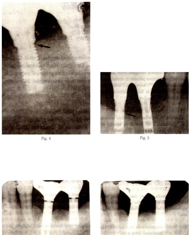
Fig. 4. Radiograph of the Brånemark implants supporting at 30-month examination after delivery of prosthesis in mandiblar left molar. The marginal bone surrounding the fixture showed wedge shape but, marginal bone loss stabilized first thread(arrow).
Fig. 5. Radiograph of the IMZ implants supporting at 30-month examination after delivery of prosthesis in mandiblar right molar. The marginal bone surrounding the fixtyre showed horizontal bone loss pattern but, marginal bone stabilized below polished neck of the implants(arrow).
Fig. 6. Radiograph of the IMZ implants supporting at 12-month examination after delivery of prosthesis in left mandiblar molar. This happened mechanical failure such as rupture of the IMC intramobile element, screw loosening, screw bending repeatedly. Radiograph showed vertical bone loss pattern with moderate horizontal bone loss.
Fig. 7. Radiograph of the same patient in fig 6. At 24-month examination. IMC intramobile element and IMC titanium insert were changed with Titanium IMC due to mechanical failure. No other prosthetic problem occurred thereafter. Radiograph showed stabilized marginal bone level without advanced bone compared with 2-month examination.
Fig. 4
Fig. 6 Fig. 7
Fig. 5
사진부도
=Abstract =
RADIOGRAPHIC EVALUATION OF BONE CHANGE AROUND OSSEOINTEGRATED IMPLANTS IN PARTIALLY EDENTULOUS JAWS WITH FIXED
TISSUE-INTEGRATED PROSTH ESES
Yang Soon-Bong, Han Dong-Hoo
Department of prosthodontics, College of Dentistry, Yonsei University