대퇴골 원위부에 발생한 내연골종에 대한 내시경적 소파술과 calcium sulfate를
이용한 치료 - 1예 보고 -
성애병원 정형외과, 광명 성애병원 정형외과*
송경섭 ・전승주・전호승・박병문 ・조인기
양성 골종양의 치료에 있어서 고식적인 관혈적 소파술은 종양조직의 철저한 제거를 위해서 피질골에 결손을 남기 게 되고, 자가골 이식술은 공여부의 합병증 가능성과 특히 여성에 있어서 미용상의 문제점이 있다. 이에 저자들은 미혼의 젊은 여자의 대퇴골 원위부에 발생한 내연골종에 대해 내시경적 소파술과 골이식 대체물로서 c a l c i u m s u l f a t e를 이용함으로써, 고식적인 관혈적 소파술에 따른 피질골의 결손을 최소화할 수 있었고, 종양조직을 철저 히 제거할 수 있었으며, 자가골 이식술의 단점을 피할 수 있었기에 그 결과를 보고하고자 한다.
색인 단어: 대퇴골 원위부, 내연골종, 내시경적 소파술, Calcium sulfate
Endoscopic Curettage and Defect Filling with Calcium sulfate in Enchondroma of the Distal Femur
- A Case Report -
Kyeong Seop Song, M.D.
1, Seung Ju Jeon, M.D., Ho Seung Jeon, M.D., Byeong Mun Park, M.D.
1, In Kee Cho, M.D.
Department of Orthopaedic Surgery, Sung Ae General Hospital, Seoul, Korea Department of Orthopedic Surgery, Kwang Myung Sung-ae General Hospital, Seoul, Korea
1For the treatment of benign bone tumor, classical open curettage and autogenous bone graft has been generally accepted. However, significant cortical defect is resulted from classical open curet- tage for complete removal of tumor tissue and many complications are possible in autogenous bone graft. Especially in young female patients, cosmetic problem is significant as a result of auto- genous bone graft. Therefore we treated the enchondroma of distal femur in unmarried young female patient with endoscopic curettage and calcium sulfate as bone graft substitute for the pur- pose of avoiding the significant cortical defect and donor site problems of autogenous bone graft.
* Address for Correspondence : Seung Ju Jeon, M.D.
Department of Orthopaedic Surgery, Sung Ae General Hospital.
451-5 Shingil-Dong, Youngdeungpo-Gu, Seoul, Korea
Tel : 82-2-840-7232, Fax : 82-2-840-7755, E-mail : [email protected]
자가골 이식술이다2 ). 고식적인 관헐적 소파술은 종 양조직의 철저한 제거를 위해서 피질골에 큰 결손을 남기게 되고, 자가골 이식술은 공여부의 합병증 및 미용상으로 환자의 불만족을 초래할 수 있다. 따라 서 저자들은 미혼의 젊은 여자의 대퇴골 원위부에 발생한 내연골종에 대해 내시경을 이용한 소파술과 calcium sulfate를 이용한 치료를 시행하고 그 결 과를 보고하고자 한다.
증례 보고
2 4세 미혼의 여자 환자로 약 2년 전부터 특별한
상 T1 weighted image 에서는 저신호강도, T2 weighted image 에서는 고신호강도를 보이는 약 2 . 5×2 . 8×3.4 cm 크기의 종물이 보였으며, 이 종물은 조영제에 의해 조영증강되는 소견을 보였 고, 주위에 연부조직 종물은 관찰되지 않았다( F i g . 2-A, B, C, D). 골동위원소 검사 상 좌측 대퇴골 원위부 내측에 국한된 단발성의 병변을 보였다 (Fig. 3).
수술은 척수마취 하에 대퇴 원위부 내측에 약 2 . 5 c m의 종절개를 시행하고, 1×1 cm 의 피질골창을 만들어 조직을 얻은 후, frozen biopsy를 통해 악 성종양을 배제하였다. 기존의 피질골 창을 통하여
Fig. 1. (A, B) Preoperative plain radiographs of the left knee show the osteolytic lesion of distal femur. This lesion is medially eccentric and the cortex is thin but cortical continuity is preserved.
A B
Fig. 2. (A, B, C, D) Magnetic resonance images of the left knee. T1-weighted image shows low signal intensity (A), and T2-weighted image shows high signal intensity (B). The mass is measured by 2.5×2 . 8×3.4 cm. Heterogenous enhancement is shown in enhanced images (C, D) and soft tissue mass is not seen on any MR images.
Fig. 3. The single hot uptake lesion is seen in whole body bone scan image at the medial aspect of the left distal femur.
A B
C D
보행을 시행하고, 이후 점진적인 체중부하를 허용하 (Fig. 12).
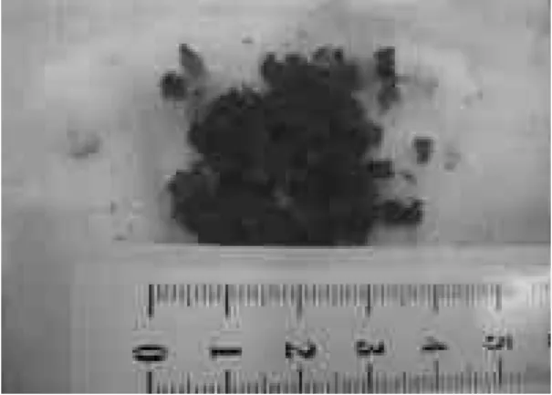
Fig. 4. The curetted fragments of tumor are whitish and glistening. The nature of tumor material is mix- ture of chondroid and fibrous tissue.
Fig. 5. Calcium sulfate (OsteosetⓇ: Wright Medical Co.
USA) was used as bone graft substitute in the form of tablet.
Fig. 6. Endoscopic images in surgical field. (A) The whitish soft tissue is seen on endoscopic image before complete curettage. (B) After through curettage, normal bone is seen without any tumor remnant.
A B
Fig. 7. Pathologic photograph shows benign cartilage tis- sue with lobulated appearance and single nucleus is seen in each chondrocyte (H&E stain, X 100).
Fig. 8. Immediate postoperative radiographs show the calcium phosphate granules in the bone defect which is resulted from curettage of tumor mass.
Fig. 9. Radiographs at 3 weeks after surgery show the beginning of calcium sulfate resorption.
Fig. 10. Complete resorption is seen on the radiographs at 6 weeks after surgery.
Fig. 11. The last follow up radiographs in one year after surgery show the trabeculation at the bone defect site on the AP image and previous operative cortical win- dow is seen as a osteolytic lesion on the lateral image.
Fig. 12. At the last follow up, the knee joint function is normal and the linear scar is measured by 2.5 cm.
및 주위 연부조직의 손상을 최소화 하는 장점이 있 다. 이 내시경적 소파술은 연부조직까지 종양조직이 확장되지 않은 경우, 내시경 삽입과 시야 확보가 가 능할 정도의 병변 크기를 갖는 경우, 관절 주위 종 양에 의해 붕괴된 관절의 재건을 필요로 하지 않는 경우에 적용이 되는 것으로 알려져 있다6 ).
저자들의 경우, 최소한의 절개와 작은 피질골 창 을 통한 내시경적 소파술을 시행함으로써 피질골의 결손을 최소화화면서, 병변조직을 효과적으로 제거 하였고, 병변조직의 잔존여부를 명확하게 확인할 수 있었다(Fig. 6-A, B).
한편, 종양 조직의 제거 후 발생한 골 결손 부위 에 대한 치료로써는 자가골 이식술, 동종골 이식술, 골 이식 대체물 등이 사용되고 있는데, 자가골 및 동종골 이식술의 단점을 보완하기 위하여 h y d r o x- yapatite, tricalcium phosphate, calcium sulfate 등의 골이식 대체물들이 임상적으로 사용 되고 있다1 ). 이중 calcium sulfate는 결손 부위의 모양에 맞게 채워 넣을 수 있고, 쉽게 흡수되며 골 형성을 방해하는 대사산물을 만들지 않으며, 생체적 합성이 뛰어나서 이물 반응을 거의 일으키지 않는 장점이 있다. 또한 방사선 비투과성이고 생체내로 거의 흡수되므로 단순 방사선 검사로 흡수 및 골형 성 정도를 확인할 수 있는 장점이 있다4 , 7 , 8 )
. Calcium sulfate는 대개 5 ~ 7주내에 흡수되는 것 으로 보고되고 있는데8 ), 저자들의 경우 수술 후 3주 째 흡수가 시작되어 6주째 완전히 흡수됨을 확인할 수 있었다(Fig. 9, 10). Calcium sulfate는 역 학적 특성상 압박력에는 잘 견디나 신장력에는 약하 며, 쉽게 깨지기 때문에 골 생성이 이루어지기까지
형성을 확인할 수 있었다(Fig. 11).
최근에는 환자들의 요구가 높아져서 미용상의 만 족도도 중요한데, 특히 여성의 경우에는 미용상의 요구가 증가되고 있다. 저자들의 경우 종양부위에는 내시경을 이용함으로써 최소 절개술로 종양을 효과 적으로 제거하였고, calcium sulfate를 사용함으 로써 공여부의 문제를 피할 수 이 방법이 추천할 만 한 치료방법으로 생각되었다.
참고문헌
01) Bucholz RW, Carlton A and Holmes RE : Hydroxyapatite and tricalcium phosphate bone graft substitute. Orthop Clin N Am, 18(2): 323- 334, 1987.
02) Damien CJ: Bone graft and bone graft substitute.
A review of current technology and application. J Appl Biomat, 2: 187-208, 1991.
03) Hadijivavlou AG, Simmons JW, Youg J, Nicodemus CL, Esch O and Simmons DJ : Plaster of Paris as an osteochondroductive mater - ial for interbody vertebral fusion in mature sheep.
Spine, 25: 10-16, 2000.
04) Han JS, Yoon KH and Ha JH : The Use of Calcium Sulfate as a Treatment of Benign Bone Tumor, J Korean Bone & Joint Tumor Soc, 9: 31- 37, 2003.
05) Joseph ML and Mathias PGB : Bone grafting and new composite biosynthetic graft materials.
Instructional Course Lecture, AAOS, 47: 525-534, 1988.
06) Kim DC and Kim JW: Endoscopic Curettage
Without Bone Grafting for Enchondromas in the Hand. J Korean Soc Surg Hand, 9: 278-282, 2004.
07) Oh CW, Ihn JC, Kim PT, Park IH and Kim D H: The use of Calcium Sulfate as a Bone Substitute, J Korean Orthop Assoc, 33: 1859- 1866, 1998.
08) Peltier LF : The use of plaster of paris to fill defects in bone. Clin Orthop, 21: 1-31, 1961.
09) Wulle C: On the treatment of enchondroma. J Hand Surg Br, 15: 320-330, 1990.
10) Yamazaki Y, Oida S, Akinoto Y and Shoida S:
Response of the mouse femoral muscle to an implant of a composite of bone morphogenic pro - tein and plaster of paris. Clin Orthop, 234: 240- 249, 1988.