• 검색 결과가 없습니다.

Acute Epididymitis in Children: 10-Year Retrospective Study of Single Center

N/A
N/A
Protected

Academic year: 2021

Share "Acute Epididymitis in Children: 10-Year Retrospective Study of Single Center"

Copied!
6
0
0

로드 중.... (전체 텍스트 보기)

전체 글

(1)

173

•교신저자: 이상돈, 부산대학교 의과대학 비뇨기과학교실 부산시 서구 아미동 1-10번지

Tel: 051-240-7348, Fax: 051-255-7133 E-mail: [email protected]

Acute Epididymitis in Children: 10-Year Retrospective Study of Single Center

Jung Man Kim

1

, Chang Yell Lee

1

, Sang Don Lee

1,2

From the

1

Department of Urology, College of Medicine, Pusan National University, and

2

Medical Reserch Institude, Pusan National University Hospital, Busan, Korea

[Abstract]

Purpose: We studied to describe clinical aspects, to assess the value of diagnostic tests and to determine conspicuous characteristics of acute epididymitis between children and adult.

Materials and Methods: A total of 205 patients with acute epididymitis over a 10-year period were divided into 3 groups: group A (prepuberty; 49 patients), group B (puberty; 42 patients) and group C (adult; 114 patients). We analysed retrospectively the medical records: clinical features, laboratory tests, radiological examinations, accompanying diseases and treatment.

Results: The mean ages were 6.5±2.7, 14.1±2.8, and 43.7±13.4 years in the group A, B, and C (p<0.001), respectively. Prevalence of symptoms including swelling, pain, tenderness and fever were not different among these 3 groups. Serum ESR and CRP levels were significantly higher in group C compare to group A and B (p=0.008 and p<0.001). On urinalysis, pyuria was presented 4 (8.2%), 5 (11.9%), and 19 patients (16.7%) in each group (p=0.341). However, there were only 4 positive urine cultures (1.8%):

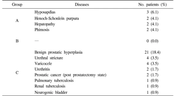
group A; 2 E. coli, group C; 2 Pseudomonas. All of patients performed by Doppler ultrasonography (CDUS) and testicular scan demonstrated the increased vascularity and isotope uptake, respectively. The concomitant diseases causing acquired epididymitis were predominent in the group C. The epididymectomy and scrotal exploration were performed in 0 (0.0%), 1 (2.4%), 1 (0.9%) patients, and in 4 (8.2%), 0 (0.0%), 2 (1.8%) patients in each group, respectively.

Conclusions: Compare to the adults, for the diagnosis of acute epididymitis, CDUS and/or testicular scan were very useful, however serum ESR and CRP level, urinalysis and urine culture seem to be not useful in children. This study suggests that if there is no evidence of infections, empirical antibiotics treatment should not be recommended. (Korean J UTII 2007;2:173-178)

Key Words: Acute epididymitis, Children, Adult

INTRODUCTION

While testicular torsion and appendix testicular

torsion are considered the most common causes of

acute scrotum, acute epididymitis has been recongnized

to have an important role and distressing problem in

acute scrtoum in boys; it accounts for up to 35% of

pediatric patients of acute scrotum.

1

If the adequate

treatment is not provided in the early phase, severe

(2)

complications such as infertility, testicular abscess, chronic pain, infarction and necrosis can occur.

1-4

A lot of studies on acute epididymitis have been performed.

However, the etiology and management of this condition in boys have not been clearly defined. There are a few studies on conspicuous characteristics of acute epididymitis in children, whereas many adult studies are available.

Herein, we studied to describe clinical aspects, to assess the value of diagnostic tests and to determine conspicuous characteristics of acute epididymitis between children and adult.

MATERIALS AND METHODS

From march 1996 to May 2005, a total of 205 patients with acute epididymitis were divided into 3 groups: group A (prepuberty; before the age of 10 in 49 patients), group B (puberty; 11~20 years old in 42 patients) and group C (adult; older than 21 years old in 114 patients). Diagnosis was based on symptoms, physical findings, color Doppler ultrasonography (CDUS) and/or testicular scan; patients with chronic epididymitis, tubercular epididymitis, idiopathic acute scrotal swelling, appendix testis torsion, and testis torsion were excluded from this study. The patients were analysed retrospectively through the medical records following as: age, clinical symptoms, serum ESR and CRP level, urinalysis, urine culture, radiologic tests, accompanying diseases and treatment.

This study was approved by the ethics committee of Pusan National University Hospital and we received informed consent from all patients or patient's parents prior to their inclusion in this study.

Statistical analyses of age, blood examination, urinalysis, and urine culture were carried out with Kruskal-Wallis test and chi-square test. Fisher's exact test was used for analysis of clinical symptoms. For all of tests, p<0.05 was considered to indicate statistical significance.

RESULTS

The mean ages in group A, B, C and for all patients were 6.5±2.7 (4 months~10 years), 14.1±2.8 (11~19 years), 43.7±13.4 (21~75 years) and 28.8±19.8 years (p<0.001).

The total incidence of bilaterality (right/left/both) for acute epididymitis in each group was 25/24/0 (51.0/49.0/0%), 14/23/5 (33.3/54.8/11.9%) and 40/62/12 (35.1/54.4/10.5%) in group A, B, and C, respectively; therefore, there was a significantly higher incidence of acute epididymitis on the left or both sides in older ages (p=0.046).

In consideration of clinical symptoms, each group had 47 (95.9%), 39 (92.9%), 100 patients (87.6%) with swelling (p=0.261); pain was reported in 49 (100%), 42 (100%), 107 patients (93.9%) (p=0.075), tenderness in 46 (93.9%), 42 (100%), 108 patients (95.2%) (p=0.335), and fever (temperature ≥38℃) in 7 (14.3%), 10 (23.8%), 17 patients (14.9%) (p=0.367), respectively.

For the laboratory assessment, leukocytosis (WBC>10

4

/mm

3

) was observed in 18 (36.7%), 20 (47.6%), 21 patients (18.4%) in each group, respectively; there was a significantly high prevalence of leukocytosis in children (p<0.001). In each group, the average serum ESR and CRP levels were 19.1±26.0, 22.4±10.8, 45.6±33.1mm/hr. (total average of 29.2±28.8mm/hr.) and 0.4±0.6, 2.6±3.0, 3.4±4.1 mg/dl (total average of 2.0±3.2mg/dl), respectively.

Both ESR and CRP levels were significantly low in

group A (p=0.008 and p<0.001, respectively). On

urinalysis, pyuria (WBC>5/HPF) was present in 4

(8.2%), 5 (11.9%), 19 patients (16.7%) in each group,

respectively; there was higher prevalence of pyuria in

older ages, but this was not statistically significant

(p=0.341). For urine culture, there were only 4

patients reported in total and 2 were in group A

(4.1%, E. coli) and 2 in group C (1.8%,

(3)

Table 2. Concomitant diseases

Group Diseases No. patients (%)

A

Hyposapdias 3 (6.1)

Henoch-Schonlein purpura 2 (4.1)

Hepatopathy 2 (4.1)

Phimosis 2 (4.1)

B ― 0 (0.0)

C

Benign prostatic hyperplasia 21 (18.4)

Urethral stricture 4 (3.5)

Varicocele 4 (3.5)

Urethritis 2 (1.7)

Prostatic cancer (post prostatectomy state) 2 (1.7)

Pulmonary tuberculosis 1 (0.9)

Renal tuberculosis 1 (0.9)

Neurogenic bladder 1 (0.9)

Table 1. Summary of laboratory findings

Laboratory findings Group (%)

p-value*

A B C

WBC >5/HPF on urinalysis 4 (8.2%) 5 (11.9%) 19 (16.7%) 0.341

Positive result on urine culture 2 (4.1%) 0 (0.0%) 2 (1.8%)

WBC count (mm

3

) 9515±2956 10174±3011 8243±2725 <0.001

ESR (mm/hr) 19.1±26.0 22.4±10.8 45.6±33.1 0.008

CRP (mg/dl) 0.4±0.6 2.6±3.0 3.4±4.1 <0.001

* Kruskal-Wallis test,

Chi-square test Pseudomonas) (Table 1).

CDUS and testicular scan were performed in 48 (97.9%), 39 (92.9%), 103 patients (90.4%) and 25 (51.0%), 26 (61.9%), 25 patients (21.9%) in each group, respectively; all patients showed the increased vascularity and isotope uptake. CDUS demonstrated reactive hydrocele in 38 (79.2%), 31 (79.5%), 73 (70.9%) in each group, respectively.

The concomitant diseases were hypospadias (3 patients, 6.1%), Henoch-Schonlein Purpura, hepatopathy and phimosis (2 patients, 4.1%, respectively) in group A, and benign prostate hypertrophy (21 patients,

18.4%), urethral stricture and varicocele (4 patients, 3.5%, respectively), urethritis and prostatic cancer (2 patients, 1.7%, respectively) in group C (Table 2).

The treatment approach included conservative mana- gement and epididymectomy which were performed in 45 (91.8%), 41 (97.6%), 111 patients (97.4%) and in 0 (0.0%), 1 (2.4%), 1 (0.9%) patients due to severe inflammations with abscess formation, respectively.

Scrotal exploration was carried out in 4 (8.2%), 0

(0.0%), 2 (1.8%) patients in each group, respectively.

(4)

DISCUSSION

Acute epididymitis is a rare but important diagnostic entity in the child who presents with acute scrotal inflammation. Acute epididymitis should be diagnosed promptly and must be differentiated from hernia, testicular torsion, hydrocele, and testicular tumors. Distinguishing acute epididymitis from testicular torsion is often difficult bases solely on signs and symptoms; however, with the use of CDUS and/or testicular scan, the accuracy of diagnosis has increased. Because it is difficult to perform testicular scans at night, CDUS, which is non-invasive and simple to use, has proved to be the diagnostic tool of choice. In this study, patient's symptoms and physical findings were included as well as the diagnostic values of laboratory and radiologic tests; the sensitivities of CDUS and testicular scan for diagnosis were 100% for diagnosis in all groups. Therefore, they were thought to be highly valuable tools for the diagnosis of acute epididymitis regardless of age.

Measuring the serum CRP level was useful in differentiating acute epididymitis from testicular torsion and testicular tumors in the acute scrotum and therapeutic outcome could be estimated by decrease in the CRP level.

5

However, in this study, because the level of serum CRP and ESR decreased proportionate to the age of the patients, it was less useful in diagnosing acute epididymitis in children.

The etiology and management of acute epididymitis in children are still subject to controversy.

6

While several etiologies have been considered, such as ascending infection, urethrovasal reflux, and bacterial and nonbacterial infection, they are actually documented only in a minority of patients.

6

The cause of acute epididymitis in children has been suspected to be an ascending urinary tract infection caused by organisms similar to those found in adults; thus antibiotics based on common causative organisms, in

adults, have been used for treatment. Ascending urinary tract infections are explained by the theory that bacterial infection starts in the lower urinary tract and ascends into epididymis causing inflammation;

evidences of inflammation is noted in positive urine eventhough it is acutally documented only in a minority of pediatric patients. The most common causative organism found in acute epididymitis is E. coli; in addition, Staphylococci, Proteus and Haemophilus influenza are commonly identified.

7,8

Melekos and Asbach

9

stated, in adult patients, that typical urinary tract infection are frequent in patients over 40 years old, whereas infection with Chlamydia was common before the age of 40. These reports were mainly focalized on adults; the sources of infection for pediatric patients remain to be identified. In this study, the main causative organism was E. coli; in children and Pseudomonas in adults.

According to Oh et al,

10

the incidence of urinary tract infections in acute epididymitis was found to be 42.5% in adults and only 3.6% in childhood. Lau et al

11

and Merlini et al

12

also reported that the incidence of an urinary tract infection was 10% and 12% in children, respectively. In this study, pyuria was observed in 8.2% of prepuberty, 11.9% of adolescents, and 16.7% of adults. This suggests that the younger the patients, the lower the incidence of pyuria.

Other than ascending infection, the phenomenon of

urethral-ejaculatory duct reflux has been implicated in

the etiology of acute epididymitis in children.

13

This

condition has been described mainly in children who

have undergone instrumentation, chronic infection or

bladder outlet obstruction such as posterior urethral

valves, urethral strictures, posterior urethral masses,

duplicated urethra with obstruction, bladder neck

obstruction, neurogenic bladder and impeforated

anus.

13,14

According to Lewis et al

1

and Siegel et al,

13

38% and 47% of acute epididymitis patients presented

with an underlying urogenital anomaly. On the other

hand, Luzzi and O'brien

15

presented that urological

(5)

factors predisposing to acute epididymitis are more likely to be found in older patietns and those with bacteriuria. Contrast to adults, underlying anatomical abnormalities in children are identified rarely.

16

However, anatomical abnormalities as predisposing factors are much commoner in infants whereas, in older children, most acute epididymitis is idiopathic.

15

In this study, urinary difficulties or obstructive urinary diseases like BPH, urethral stricture, neurogenic bladder were mainly related to acute epididymitis in adults (24.6%), whereas they were not seen in children.

For the primary therapeutic approach, as in other reports,

7-10

the treatment of acute epididymitis is generally a conservative approach including bed rest for limited activity and cold packs; providing management of scrotal edema along with pain relief, which may require admission to the hospital. Conservative treatments for pediatric acute epididymitis are considered to reduce clinical symptoms caused by the inflammatory reaction; use of antibiotics should be considered for routine urinary tract infections as well as complicated infections. However, there are reports that the satisfactory outcome has been observed without antibiotic treatments or with a little role for antibiotics in pediatric patients.

11,17,18

Recently it was suggested that antibiotics are indicated only in a minority of children with epididymitis, especially in light of the fact that urine culture as well as other fluid cultures are generally sterile in epididymis.

6,20

In this study, only 1.8% of the patients had a positive urine culture.

These findings lead to questions about the importance of antibiotic use in all of patients regardless of evidence of urinary tract infection. Serum ESR and CRP levels were low in pediatric acute epididymitis compared to levels evaluated in adolescents and adults.

This further supports the fact that the use of antibiotics does not appear to be essential for management;

inappropriate use of antibiotics may unfortunely cause problems such as the emergence of drug resistant

organisms.

19

Thus, in the absence of definite inflammatory reactions or bacterial infection in acute epididymitis, reconsideration of the empirical use of antibiotics is recommended.

CONCLUSIONS

CDUS and/or testicular scans had high diagnostic values in all of groups, whereas serum ESR and CRP levels, urinalysis, and urine cultures demonstrated insignificant values especially in children. Furthermore, the empirical of antibiotics should be reconsidered for children with acute epididymitis that has no evidences of inflammatory reactions or bacterial infections.

REFERENCES

1. Lewis AG, Bukowski TP, Javis PD, Wacksman J, Sheldon CA. Evaluation of acute scrotum in the emergency department. J Pediatr Surg 1995;30:277-81 2. Mevorach RA, Lerner RM, Dvorotsky PM, Rabin-

owitz R. Testicular abscess: diagnosis by ultra- sonography. J Urol 1986:136;1213-6

3. Hoppner W, Strohmeyer T, Hartmann M, Lopez- Gamarra D, Dreikorn K. Surgical treatment of acute epididymitis and its underlying disease. Eur Urol 1992;22:218-21

4. Berger RE. Sexually transmitted disease: the classic disease. In: Walsh PC, Retik AB, Vaughan ED Jr., Wein AJ, editors. Campbell's Urology, 7th ed.

Philadelphia: WB saunders; 1998;663-83

5. Doehn C, Fornara P, Kausch I, Buttner H, Friedrich HJ, Jocham D. Value of acute-phase proteins in the differential diagnosis of acute scrotum. Eur Urol 2001;

39:215-21

6. Somekh E, Gorenstein A, Serour F. Acute epi- didymitis in boys: evidence of a post-infectious etiology. J Urol 2004;171:391-4

7. Harnisch JP, Berger RE, Alexander ER, Monda G, Holmes KK. Aetiology of acute epididymitis. Lancet 1977;16:819-21

8. Lin YC, King DR, Birken GA, Barson WJ. Acute

(6)

scrotum due to Haemophilus influenza type b. J Pediatr Surg 1988;23:183-4

9. Melekos MD, Asbach HW. Epididymitis: aspects con- cerning etiology and treatment. J Urol 1987;138:83-6 10. Oh DK, Kim SJ, Ahn HS. Experiences of 313 pa-

tients of acute scrotum: properties of acute epi- didymitis and differential diagnosis of testicular torsion. Korean J Urol 2002;43:624-30

11. Lau P, Anderson PA, Giacomantonio JM, Schwarz RD. Acute epididymitis in boys: are antibiotics in- dicated? Br J Urol 1997;79:797-800

12. Merlini E, Rotundi F, Seymandi PL, Canning DA.

Acute epididymitis and urinary tract anomalies in children. Scand J Urol Nephrol 1998;32:273-5

13. Siegel A, Snyder H, Duckett JW. Epididymitis in in- fants and boys: underlying urogenital anomalies and ef- ficacy of imaging modalities. J Urol 1987;138:1100-3

14. Weber TR. Hemophilus influenzae epididymo-orchitis.

J Urol 1985;133:487

15. Luzzi GA, O'brien TS. Acute epididymitis. BJU Int 2001;87:747-55

16. Gislason T, Noronha RF, Gregory JG. Acute epi- didymitis in boys: a 5-year retrospective study. J Urol 1980;124:533-4

17. Hutson JM, Dewan PA. Acute epididymitis in boys:

are antibiotics indicated? Br J Urol 1997;80:970-1 18. Foley SJ, Kashif KM, Holmes SA. Acute epididymitis

in boys. Br J Urol 1998;81:179-80

19. Culp LA, Carson CC. Antibiotics resistance in the genitourinary system. Contemp Urol 1998;10:62-75 20. Cappele O, Liard A, Barret E, Bachy B, Mitrofanoff

P. Epididymitis in children: is further investigatin nec- essary after the first episode? Eur Urol 2000;38:

627-30

수치

Table  1.  Summary  of  laboratory  findings

참조

관련 문서

Background: The aim of this study was to examine the influence of body mass index (BMI) on the development of acute kidney injury (AKI) in critically ill patients in

Since elderly and high-risk patients comprise a significant proportion of the patient group undergoing CEAs, differences in co- morbidity and mortality were evaluated according to

Methods: The 13 C-UBT results after Helicobacter pylori eradication therapy was compared with the results of endoscopic biopsy-based methods in the patients who have

The benefit of specialized team approaches in patients with acute kidney injury undergoing continuous renal replacement therapy: Propensity score matched analysis. The

generation cephalosporins has led to increased E. The results of our study showed that patients with resistance to third­generation cephalosporins were more likely to

Other risk factors reported to be associated with worse prognosis in adults are severe renal symptoms at on- set, female sex (during and after pregnancy), and increased

In conclusion, preoperative diagnosis of MS based on clinical presentation and imaging findings was possible in only one third of patients in our series.. Increased associa- tion