DOI: https://doi.org/10.3339/jkspn.2017.21.1.8 ISSN 2384-0250 (online)
Clinical and Pathological Findings of Renal Biopsy in Children: Outcomes from a Single Center Over 27 Years
Purpose: To classify the results of renal biopsy in pediatric patients and to compare pathological findings with clinical features.
Methods: This study included data of 318 children who underwent renal biopsy at our hospital between December 1987 and November 2014. Biopsy specimens were examined histopathologically using light, immunofluorescence, and electron microscopy.
Results: Asymptomatic urinary abnormalities was the most common clinical diag nosis (35.9%), followed by nephrotic syndrome (29.3%), and acute glomeru- lonephritis (18.0%). Glomerular disease was identified in 98.1% of the renal biopsy specimens. The most common primary cause of glomerulonephritis was IgA ne- phropathy, with gross hematuria in 61.9% of the patients, hypertension in 14.2%, proteinuria >1.0 gm/24-hr in 33.3%, and impaired renal function in 3.6% patients.
Conclusion: The most common clinical diagnosis was asymptomatic urinary abnormalities, with primary glomerular disease being the most common renal biopsy finding, and IgA nephropathy the most common histopathological lesion.
This study provides a 27-year overview of pediatric renal disease at our center and underlines the importance of renal biopsy for accurate diagnosis and proper management.
Key words: Clinical diagnosis, Children, Renal biopsy, Nephrotic syndrome, IgA nephropathy
Shin Ae Lee, M.D. 1 Min Sun Kim, M.D. 1,2 Soon Chul Kim, M.D. 1,2 Dae-Yeol Lee, M.D. 1,2
Department of Pediatrics
1, Chonbuk National University Medical School, Research Institute of Clinical Medicine of Chonbuk National University- Biomedical Institute
2of Chonbuk National University Hospital, Jeonju, Korea
Corresponding author: Dae-Yeol Lee, M.D.
Department of Pediatrics, Chonbuk Na- tional University Hospital 20 Geonji-ro, Deokjin-gu, Jeonju 54907, Korea Tel: +82-63-250-1469
Fax: +82-63-250-1464 E-mail: [email protected] Received: 1 January 2017 Revised: 8 March 2017 Accepted: 10 March 2017
This is an open-access article distributed under the terms of the Creative Commons Attribu tion Non-Commercial License (http://
crea tivecom mons.org/licenses/by-nc/4.0/) which permits unrestricted non-commercial use, distribution, and reproduction in any medium, provided the original work is properly cited.
Copyright © 2017 The Korean Society of Pediatric Nephrology
Introduction
The diagnosis and treatment of pediatric kidney diseases is based on me
dical history and physical findings. However, in certain cases, a discrepancy between clinical and pathological findings exists, with patients exhibiting similar clinical symptoms despite different underlying pathological features.
Although percutaneous renal biopsy is a widely used medical procedure for the diagnosis of kidney disease in adults, the procedure has not been consi
dered suitable for pediatric patients. However, with recent innovations in te
chnique for kidney localization and biopsy, percutaneous renal biopsy is now
regarded to be both safe and advisable for the diagnosis of renal disease in
pediatric patients and to inform treatment
14). The indications for renal biopsy
vary according to the ethnic and age characteristics of the population studied
as well as the geographic location
5).
The aim of our study was to classify results of renal biopsy in pediatric pa tients, based on pathological findings, and to compare the pathological diagnosis to clinical findings.
Material and methods
1. Subjects
A retrospective study of 345 inpatients who underwent renal biopsy in the Pediatric Department of Chonbuk Na
tional University Children’s Hospital, between December 1987 and November 2014. The study adhered to the prin
ciples of the Declaration of Helsinki and was approved by the Life Science Research Ethic Council Committee of the Chonbuk National University Hospital (CUH 201610009).
Among the 345 patients who underwent biopsy over the study period, 23 were excluded due to incomplete demo
graphic or clinical data, and four due to inappropriate tis
sue samples (<5 glomeruli in the kidney tissue sample). Prior to renal biopsy, prothrombin and bleeding/clotting time were assessed to determine bleeding tendency. Consent for the procedure was obtained from parents and/or guardians prior to the procedure.
Over the 27year period of the study, there was consider
able variation in the biopsy technique used. Early in the study period, biopsies were performed under sedation using a biopsy gun under fluoroscopy. Since 1997, biopsies were performed under sedation, using an automated gun under real time ultrasound guidance. All biopsies were performed by a pediatric nephrologist. For all biopsies, two cores of kidney tissues were obtained for histopathology assessment, by light microscopy, immunofluorescence microscopy, and electron microscopy (LM, IF, and EM, respectively).
2. Data retrieval
The following clinical and laboratory data were extracted from the clinical records for analysis: patient’s age and sex;
serum total protein, albumin, cholesterol, blood urea ni
trogen (BUN), creatinine (Cr), and electrolytes; complement 3 (C3) and 4 (C4) and antistreptolysinO (ASO) titer; glo
merular filtration rate (mL/min/1.73m
2=kL/Pcr, k: 01 years of age 0.45, 212 years of age 0.55, Jaffe method); and 24hr urine protein (mg/m
2/hr).
3. Pathological study
For LM examination, 10 serial sections were cut and pre
pared using standard procedures and stained with hemat
oxylineosin (HE), Masson’s trichrome, periodic acidSchiff stain, and silver stains. Tissue specimens for IF examina
tion are snapfrozen in liquid nitrogen and cut on a cryo
tome. The tissue was stained by the direct method using fluorescein isothiocyanateconjugated antisera mono specific for immunoglobulin (Ig) G (IgG), IgA, IgM, C3, and fibrinogen. Tissue samples for EM examination were stained on copper 300 mesh grids with uranyl acetate and lead citrate for assessment.
4. Statistical analyses
Data was analyzed using SPSS software (version 22.0;
IBM Corporation, Armonk, NY, USA). Simple descriptive statistics, mean and standard deviation (SD), were calcu
lated for continuous variables, namely age, and clinical and laboratory measures. Percentages were calculated for cate
gorical data.
Results
1. Sex and age distribution
Among the 318 cases included in this study, 205 were male (64.5%) and 113 cases female (35.5%), for a maleto
female sex ratio of 1.8:1. The mean±SD age was 9.7±3.6 years (range, 5 months to 18 years), with the following age distri
bution: 131 cases (41.2%) between 1115 years of age; 129 (40.5%) between 610 years of age; 46 (14%) between 05 years of age; and 12 (3.7%) between 1618 years of age.
2. Indications for renal biopsy
The indications for biopsy were as follows (Fig. 1): Recur
rent macroscopic hematuria in 114 cases (35.8%); nephrotic syndrome, with hematuria and/or hypertension at the first diagnosis, in 34 cases (10.7%); persistent microscopic he
maturia with proteinuria in 32 cases (10.1%); and steroid resistance, steroid dependence, or frequent relapse forms of nephrotic syndrome in 32 cases (10.1%). Persistent micro
scopic hematuria was detected in 23 cases (7.2%), with posi
tive family history, hypertension, increased urine RBC
count, and parental anxiety.
3. Clinical diagnosis of patients before renal biopsy Prior to renal biopsy, the 318 cases were subdivided ac
cording to the following prebiopsy clinical diagnoses (Table 1): 114 patients (35.9%) with asymptomatic urinary abnormalities, among whom 44 (13.9%) had isolated hema
turia, 70 (22.0%) had hematuria with proteinuria; 93 pati
ents (29.3%) with nephrotic syndrome; 57 patients (18.0%) with acute glomerulonephritis; 38 patients (11.9%) with HenochSchönlein purpura nephritis; 4 patients (1.2%) with lupus nephritis; and 12 patients (3.7%) with other con
ditions, such as acute renal failure, Alport syndrome, he
molytic uremic syndrome, and Korean hemorrhagic fever.
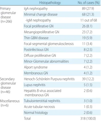
4. Histopathological diagnosis after renal biopsy The distributions of histopathological diagnoses are sum
marized in Table 2. Glomerular disease was identified in 98.1% (312/318) of cases, with a diagnosis of primary glo
merular disease in 85.2% (266/312) of these cases and of secondary glomerular disease in the remaining 14.7% (46/
312). Common causes of primary glomerulonephritis (GN) were IgA nephropathy, minimal change disease (MCD), and focal proliferative GN, with Henoch Schönlein pur
pura nephritis being the most common cause of secondary GN.
5. Histological classification of nephrotic syndrome The distribution of histological diagnoses with nephrotic syndrome is summarized in Table 3. Among the 102 cases diagnosed with nephrotic syndrome, 97 cases were histo
logically classified as a primary nephrotic syndrome (95.1
%) and 5 cases (4.9%) as a secondary nephrotic syndrome.
Among cases classified as a primary nephrotic syndrome, 68 cases (70.1%) presented with MCD, with IgM nephro
pathy accounting for 11 of these cases (16.2%). Focal seg
mental glomerulosclerosis (FSGS) was identified in 9 cases (9.3%), membranoproliferative GN (MPGN) in 7 cases (7.2
%), IgA nephropathy in 5 cases (5.2%), diffuse mesangial proliferative GN (DMPGN) in 4 cases (4.1%) and membra
nous glomerulopathy in 4 cases (4.1%).
Table 1. Clinical Diagnosis before Renal Biopsy
Clinical diagnosis No. of cases (%)
Asymptomatic urinary abnormalities 114 (35.9)
Isolated hematuria 44 (13.9)
Hematuria with proteinuria 70 (22.0)
Nephrotic syndrome 93 (29.3)
Acute glomerulonephritis 57 (18.0)
Henoch-Schönlein purpura nephritis 38 (11.9)
Lupus nephritis 4 (1.2)
Other 12 (3.7)
Total 318 (100.0)
Table 2. Histopathological Diagnosis after Renal Biopsy Histopathology No. of cases (%) Primary
glomerular disease (n=266)
IgA nephropathy 89 (27.9)
Minimal change disease 68 (21.3)
-IgM nephropathy 11 out of 68
Focal proliferative GN 26 (8.1) Mesangioproliferative GN 23 (7.2)
Thin GBM disease 19 (5.9)
Focal-segmental glomerulosclerosis 11 (3.4)
Postinfectious GN 8 (2.5)
Diffuse proliferative GN 7 (2.2) Minor-Glomerular abnormalities 7 (2.2)
Alport syndrome 4 (1.2)
Membranous GN 4 (1.2)
Secondary glomerular disease (n=46)
Henoch-Schönlein Purpura nephritis 39 (12.2)
Lupus nephritis 5 (1.5)
Hepatitis B virus associated
membranous GN 2 (0.6)
Miscellaneous
(n=6) Tubulointerstitial nephritis 3 (1.0)
Acute tubular necrosis 1 (0.3)
Normal histology 2 (0.6)
Total 318 (100.0)
Abbreviations: GN, Glomerulonephritis; Thin GBM disease, Thin basement membrane disease.
Recurrent macroscopic hematuria
Nephrotic syndrome associated with hematuria or hypertension Persistent microscopic hematuria with proteinuria
Steroid resistant, steroid dependent or frequent relapsed nephrotic syndome Evaluation of renal involvement in systemic disease
Persistent microscopic hematuria Others
35.8%
10.7%
10.1%
10.1%
8.5%
7.2%
17.6%
Fig1. Main indications for renal biopsy.
6. Clinical status of nephrotic syndrome as glomerular morphology
Among patients diagnosed with nephrotic syndrome, microscopic hematuria was identified in 48 patients (47.1
%), as well as in 21 of the 68 patients (30.8%) with MCD, 6 of the 7 patients (85.7%) with MPGN and 6 of the 9 patients (66.7%) with FSGS (Table 3). Microscopic hematuria was identified in all patients with membranous glomerulopathy, postinfectious glomerulonephropathy, Henoch Schönlein purpura nephritis, and nephropathy associated with he
patitis B viral infection. Hypertension was identified in 16 patients (15.7%), with decreased levels of serum comple
ment in 12 patients (11.8%), and decreased serum albumin concentration of <2.5 gm/dL in 61 patients (59.8%), 51 of
whom (83.6%) in the MCD group.
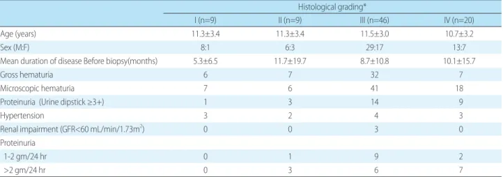
7. Clinical features according to histological grades in IgA nephropathy
Based on histological findings by Haas M
6), IgA nephro
pathy was classified as grade IV as in Table 4. In our study, 89 patients were diagnosed as having an IgA nephropathy following renal biopsy; a definite grade could not be esta
blished in 5 cases. The distribution of grades was as follows:
46 cases classified as grade III, 20 cases as grade IV, and 9 cases as grade I and II respectively. The maletofemale ratio of patients with IgA nephropathy was 2:1. Recurrent gross hematuria was identified in 52 cases (61.9%) with IgA nephropathy, with hypertension identified in 12 cases
Table 3. Clinical Status and Glomerular Morphology in Nephrotic Syndrome
Glomerular morphology No. of cases Microscopic
Hematuria Serum Albumin
<2.5 gm/dL Hypertension Complement 3 (C3) below normal
MCD 68 21 51 9 1
IgM Nephropathy 11 3 6 2 0
FSGS 9 6 4 3 1
MPGN 7 6 1 1 5
IgA Nephropathy 5 4 3 1 0
DMPGN 4 2 0 1 0
Membranous glomerulopathy 4 4 1 0 1
Postinfectious glomerulonephropathy 3 3 0 1 3
Henoch-Schönlein purpura nephritis 1 1 0 0 0
Hepatitis B Virus Associated 1 1 1 0 1
Total (%) 102 48 (47.1) 61 (59.8) 16 (15.7) 12 (11.8)
Abbreviations: MCD, Minimal change disease; IgM nephropathy, Immunoglobulin M nephropathy; FSGS, Focal segmental glomerulosclerosis; MPGN, Membranoproliferative glomerulonephritis DMPGN, Diffuse mesangial proliferative glomerulonephritis.
Table 4. Clinical Features According to Histological Grades in IgA Nephropathy
Histological grading*
I (n=9) II (n=9) III (n=46) IV (n=20)
Age (years) 11.3±3.4 11.3±3.4 11.5±3.0 10.7±3.2
Sex (M:F) 8:1 6:3 29:17 13:7
Mean duration of disease Before biopsy(months) 5.3±6.5 11.7±19.7 8.7±10.8 10.1±15.7
Gross hematuria 6 7 32 7
Microscopic hematuria 7 6 41 18
Proteinuria (Urine dipstick ≥3+) 1 3 14 9
Hypertension 3 2 4 3
Renal impairment (GFR<60 mL/min/1.73m
2) 0 0 3 0
Proteinuria
1-2 gm/24 hr 0 1 9 2
>2 gm/24 hr 0 3 6 7
*Haas M: Histologic subclassification of IgA nephropathy: A Clinicopathologic study of 244 cases, Am J of Kidney disease 28:829-42, 1997.
Abbreviation: GFR, Glomerular filtration rate.
(14.2%), proteinuria >1.0 gm/24 hr in 28 cases (33.3%), and renal impairment in 3 cases (3.6%).
Discussion
In this study, we examined the anthropometric charac
teristics of patients who underwent renal biopsy along with their indications for renal biopsy and common histopatho
logical findings. Our study revealed that the asymptomatic urinary abnormality is the most common clinical diagnosis and IgA nephropathy is the most common primary renal disease.
Renal biopsy in pediatric populations was introduced for diagnosing kidney disease in 1951 and has become a routine procedure in pediatric nephrology
7,8). Over a 27year period, 345 children underwent renal biopsy in our hospital and we identified an overall failure rate of renal biopsy of 1.2%
(4/345), which is comparable to previously published rates
9,10)
. Among our study sample, the maletofemale ratio was
1.8:1, a rate of male predominance that was comparable to previously reported sexspecific differences in prevalence
11)
. Printza et al.
12)reported that 48.0% of pediatric patients who underwent renal biopsy were 1114 years old, with 31.0
% between the ages of 610 years. We identified a similar age distribution, with 131 patients (41.2%) between the ages of 1115 years and 129 (40.5%) between the ages of 610 years.
Previous studies have identified idiopathic nephrotic syndrome to the most common indication for renal biopsy
1316)
. In contrast, 53.1% of patients in our study populations underwent renal biopsy to evaluate gross hematuria and/
or microscopic hematuria. Since 1998 in South Korea, more than 4 million students have participated in annual large
scale urine screening programs; additionally, many school
children with asymptomatic hematuria and/or proteinuria were detected and their diagnoses confirmed through renal biopsy
17). In current study, asymptomatic urinary abnor
malities were clinically diagnosed before renal biopsy in 114 patients (35.9%), with 89 patients (27.9%) diagnosed as having IgA nephropathy after renal biopsy.
Nephrotic syndrome can be associated with various types of glomerulonephritis and, therefore, an accurate histopa
thological identification of the glomerular lesion is essen
tial to establishing a correct diagnosis and providing the
appropriate treatment. Kim et al.
18)reported that among 52 pediatric patients with nephrotic syndrome, 90.4% were diagnosed with primary nephrotic syndrome and 9.6% with secondary nephrotic syndrome. In our study, primary ne
phrotic syndrome was identified in 95.1% of cases, with secondary nephrotic syndrome identified in 4.9% of cases.
According to Choi et al.
11), minimal change disease (MCD) was identified in 72.7% of all pediatric patients. A similar prevalence of MCD (70.1%) was found in our study. In ad
dition, Keeping in mind the controversy regarding the na
ture of these IgM deposits
1921), MCD with mesangial IgM deposit was identified in 16.2% in our study. Microscopic hematuria is present at diagnosis in 20% to 30% of children with MCD but rarely persists; macroscopic hematuria oc
curs in less than 1% of children with MCD
22). According to White et al.
23), microscopic hematuria was present in 13.0
% of cases with MCD. In our study, we identified hematuria in 30.8% of cases with MCD. Serum albumin levels usually fall below 2.0 g/dL and may be less than 1.0 g/dL in steroid sensitive nephrotic syndrome
24). Kim et al.
18)reported that the prevalence of low serum albumin levels <2.5 g/dL was 78.4% in children with nephrotic syndrome. In our study, we found that 59.8% of children with nephrotic syndrome and 75.0% of children with MCD showed low serum albu
min <2.5 g/dL. Elevated systolic and diastolic blood pressure are initially present in 520% of children with MCD, but hypertension usually does not persist
22). However, the fre
quency of hypertension occurring in other types of neph
rotic syndrome is as high as 80%
22). In this study, only 15.7
% of all cases of nephrotic syndrome presented hyperten
sion at diagnosis.
IgA nephropathy is the most prevalent form of glomeru
lonephritis worldwide. However, according to a large biopsy series, the prevalence of IgA nephropathy is influenced by significant race and ethnicityspecific variation
6). In their single center study, Choi et al.
11)reported a prevalence rate of IgA nephropathy of 18.2% in children, a prevalence rate similar to that reported in Japan
25). However, the prevalence rate of IgA nephropathy in current study was 27.9%, rela
tively high compared to other studies. The higher preva
lence of IgA nephropathy in our study reflects that many
school children with asymptomatic urinary abnormalities
have been detected through annual mass urinary screening,
which started in 1998 in South Korea
17). This screening pro
gram allows us to employ an active approach for evaluating children with persistent hematuria. Actually, the numbers of renal biopsy in our hospital increased annually com
pared to before 2000.
A majority of children with IgA nephropathy present with gross hematuria. In other patients, microscopic he
maturia and/ or proteinuria are the only signs at presenta
tion
26). Asymptomatic gross hematuria was identified in 61.9% of children with IgA nephropathy in our study and the prevalence of gross hematuria is similar to other studies
27,28)