Comparison of EMG Activity of the Posterior Oblique Sling Muscles and Pelvic Rotation During Prone Hip Extension With and Without
Lower Trapezius Pre-Activation
In-cheol Jeon1,2, MSc, PT, Sung-min Ha3, PhD, PT, Ui-jae Hwang1,2, BPT, PT, Sung-hoon Jung1,2, BPT, PT, Hyun-sook Kim4, PhD, PT, Oh-yun Kwon1,5, PhD, PT
1Kinetic Ergocise Based on Movement Analysis Laboratory
2Dept. of Physical Therapy, The Graduate School, Yonsei University
3Dept. of Physical Therapy, College of Health Science, Sangji University
4Dept. of Physical Therapy, Yeoju Institute of Technology
5Dept. of Physical Therapy, College of Health Science, Yonsei University
Abstract
1)Background: Prone hip extension (PHE) can be performed to measure the lumbopelvic motor patterns and motions. Imbalances in lumbopelvic muscle activity and muscle weakness can result in instability including pain in lumbopelvic region. The posterior oblique sling (POS) muscles contribute to dynamic lumbopelvic stability. In addition, POS are anatomically aligned with the trapezius muscle group according to shoulder positions.
Objects: This study compared the electromyography (EMG) activity of POS and pelvic compensations during PHE with and without pre-activation of lower trapezius muscle (lowT).
Methods: Sixteen healthy males were recruited. PHE was performed in randomized order: PHE with and without lowT pre-activation. Surface EMG signals were recorded for biceps femoris (BF), gluteus maximus (GM) (ipsilateral), lumbar multifidus (MF) (bilateral), and the lowT (contralateral). An electromagnetic tracking motion analysis was used to measure the angle of pelvic rotation and anterior tilting.
Results: The ipsilateral GM and bilateral MF EMG amplitudes were greater during PHE with lowT pre-activation compared to PHE without lowT pre-activation (p<.05). The BF amplitude during PHE without lowT pre-activation was significantly greater than that during PHE with lowT pre-activation (p<.05). The angles of pelvic rotation and anterior tilting during PHE with lowT pre-activation were significantly smaller compared to PHE without lowT pre-activation (p<.05).
Conclusion: PHE with lowT pre-activation, which is aligned with the POS, showed more increased MF and GM muscular activity with smaller lumbopelvic compensations in rotation and anterior tilting compared to PHE without lowT pre-activation.
Key Words: Gluteus maximus; Lumbopelvic instability; Posterior oblique sling; Prone hip extension.
Introduction
Prone hip extension (PHE) can be performed for the measurement of the lumbopelvic motor patterns (Tateuchi et al, 2012). The lumbopelvic region during PHE ideally remains neutral without lumbopelvic com- pensations in extension or rotation (Comerford and Mottram, 2012). Lumbopelvic instability during PHE
leads to limitations of controlling excessive compen- sations in lumbar extension and rotation and pelvic anterior tilt and rotation (Sahrmann, 2002).
Imbalances in lumbopelvic muscle activity and muscle weakness can result in lumbopelvic in- stability (Hodges and Moseley, 2003). Although the local and global muscles can contribute to lumbo- pelvic stability (Bergmark, 1989), the global muscles Corresponding author: Oh-yun Kwon [email protected]
are primarily involved with the spinal control and transfer loads directly from the spine to the legs during PHE (Danneels et al, 2001). Therefore, global muscle training can contribute to prevention and treatment of low-back pain (Comerford and Mottram, 2012).
A myofascial sling can contribute to the facili- tation of the force transfer through the trunk from the leg to the arm because these muscles are inter- connected anatomically (Kendall et al, 2005). The gluteus maximus (GM) is interconnected with the ipsilateral bicep femoris (BF) and the contralateral latissimus dorsi (LD) via the thoracolumbar fascia (Vleeming et al, 1995). Therefore, The GM acts as a load transfer through the hip. These muscles con- sist of the posterior oblique sling muscles con- tributing to dynamic lumbopelvic stability (Vleeming et al, 1995). Similarly, Myers (2009) described that three different major lines including superficial back line, superficial front line, and lateral line. In the superficial back line, according to shoulder positions, the posterior oblique sling muscles are anatomically aligned with the trapezius muscles (Myers, 2009).
The middle trapezius and lower trapezius (lowT) are especially important in the scapulothoracic joint as stabilizers (Myers, 2009).
The various studies have focused the LD, multi- fidus (MF), and GM muscle activation patterns dur- ing PHE exercise (Kim et al, 2013; Kim and Kim, 2015). However, to our knowledge, no study has assessed the posterior oblique sling muscles and pelvic compensations between PHE and PHE with and without lowT pre-activation. Therefore, this study compared the electromyography (EMG) activ- ity of the posterior oblique myofascial sling and the angle of lumbopelvic rotation and anterior tilting between PHE with and without lowT pre-activation.
We hypothesized that PHE with lowT pre-activa- tion would increase MF and GM muscular activity and decrease the angles of lumbopelvic rotation and anterior tilting compared to PHE without lowT pre-activation.
Methods
Subjects
Sixteen healthy male participants were recruited in this study (dominant leg: 16 right side). Their mean age (years) was 27.3±2.2 (mean±standard de- viation), their mean body mass (㎏) was 75.1±4.1, and their mean height (㎝) was 175.1±3.8. The ex- clusion criteria were as follows: 1) limited range of motion of the bilateral hip joint; 2) a history of lower back pain in the past 12 months; 3) lower extremity dysfunctions such as patellofemoral pain syndrome or anterior cruciate ligament sprains in the past 12 months; 4) pain in any joint of the body during PHE; 5) shoulder muscle weakness during all the PHE exercises. The experimental protocols were explained in detail to all of the par- ticipants, and an informed written consent was gathered. This study was approved by the Yonsei University Wonju Institutional Review Board (approval number: 1041849-201510-BM-075-01).
Experimental procedure
Sixteen participants were randomly performed PHE with and without lowT pre-activation. We used the metronome set at 60 beats per minute for subjects to perform each exercise in a standard manner (Nyland et al, 2004). Each participant was instructed to per- form PHE performance until hip extension angle (10°) reached the target bar. EMG data were col- lected for 5 sec during the isometric phase of exercise. The participants were instructed to maintain the initial position for 5 sec before they raise the dominant leg. Next, the hip extension was main- tained for 5 sec with the target bar. Each PHE per- formance was repetitive for three consecutive times with 5 min resting time between performances to minimize muscle fatigue (Sykes and Wong, 2003).
PHE without lowT pre-activation
The participant assumed a prone position on the table with the upper trunk, pelvis, and lower ex-
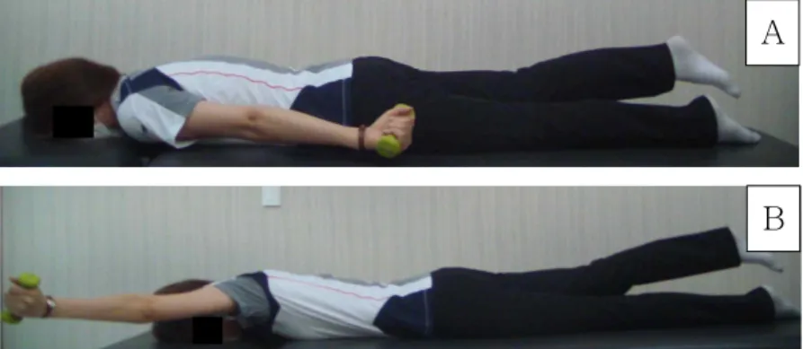
A
B
Figure 1. Prone hip extension without lower trapezius pre-activation (A) and Prone hip extension with lower trapezius pre-activation (B).
tremity in a straight line. Both arms were comfort- ably placed beside the trunk without pushing the ground with their hands. The hip joint was extended at 10° with knee extension touching the target bar (Figure 1A). This position was maintained for 5 sec, and then the participants slowly returned to the starting position. The electromagnetic motion sensor in sacral spine (S2) centrally located at the sacrum can monitor pelvic anterior tilting and rotation meas- urement during the exercises (Hungerford et al, 2007).
PHE with lowT pre-activation
The participant assumed a prone position on the table with the upper trunk, pelvis, and lower ex- tremity in a straight line. Both arms were comfort- ably placed beside the trunk without pushing the ground with their hands. The participants were asked to perform contralateral shoulder abduction at 125° angle and external rotation with 1 ㎏ load to activate lowT before PHE was performed until their ipsilateral leg touched the target bar (Kendall et al, 2005; Oyama et al, 2010) (Figure 1B). Also, the par- ticipants without limitation of motion were asked to perform the lowT activating motion by touching the target bar to avoid compensations such as trunk ex- tension and rotation. The hip joint was extended at 10° with knee extension touching the target bar. The position was maintained for 5 sec, and then the par- ticipant slowly returned to the starting position. The electromagnetic motion sensor in S2 can monitor for
pelvic anterior tilting and rotation measurement during the exercises.
Electromyography recording and data analysis The surface EMG-feedback with a wireless tele- metry system (TeleMyo 2400T, Noraxon, Scottsdale, AZ, USA) was used with the analyzing software.
Filtered movement artifacts were eliminated by a 20-450 ㎐ digital band-pass filter (Lancosh FIR). The sample rate was set to 1024 ㎐. Root mean square was used to process the EMG signals with a moving window of 50 ㎳. EMG signals were recorded for 5 sec (2 sec to 4 sec used for data analysis) while the dominant leg was maintained at the target bar during the isometric phase (Ayotte et al, 2007). The target regions were cleaned by cotton with isopropyl alcohol before electrodes were attached to minimize skin resistance. Disposable Ag/AgCl surface electrodes were attached on the target regions. Two electrodes were attached parallel to the proper muscle fiber along with the muscle fibers each on contralateral lowT (the inferior medial border of the scapula for the muscle mass and on an oblique angle, approximately 5 ㎝ down from the scapular spine at least a 90°
shoulder angle), bilateral MF (at a 2 finger-width distance lateral from the spinous process of L5), right GM (50% on the line extending between the sacrum and greater trochanter), right BF (70% on the line extending between the ischial tuberosity and lateral epicondyle) (Cram et al, 1998). MF, GM, BF,
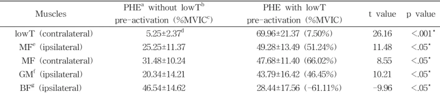
Muscles PHEa without lowTb pre-activation (%MVICc)
PHE with lowT
pre-activation (%MVIC) t value p value
lowT (contralateral) 5.25±2.37d 69.96±21.37 (7.50%) 26.16 <.001*
MFe (ipsilateral) 25.25±11.37 49.28±13.49 (51.24%) 11.48 <.05*
MF (contralateral) 31.48±10.24 47.68±11.40 (66.02%) 8.55 <.05*
GMf (ipsilateral) 20.34±14.21 43.79±16.42 (46.45%) 10.21 <.05*
BFg (ipsilateral) 46.54±14.62 28.44±17.56 (-61.11%) -9.96 <.05*
aprone hip extension, blower trapezius, cmaximal voluntary isometric contraction, dmean±standard deviation, emultifidus,
fgluteus maximus,gbiceps femoris, *significant difference between two conditions (p<.05).
Table 1. EMG amplitude of the various muscles and lowT muscles were performed for the manual muscle testing positions with the guideline recom- mended by Kendall et al (2005) to measure maximal voluntary isometric contraction normalization.
Kinematics measurements
The Polhemus Liberty™ (Polhemus, Colchester, VT, USA) was used to calculate pelvic rotation and anterior tilting at 120 ㎐. This electromagnetic track- ing device was accurate at .08 ㎝ for position and .15° for orientation (Mills et al, 2007). The electro- magnetic motion sensor was firmly attached to the skin on S2 with adhesive tape to diminish sensor to motion artifacts. The transmitter remained in the same position for all measurements during PHE performances.
The orientation of the electromagnetic tracker system was defined with +X parallel line to both anterior su- perior iliac crest, +Y parallel line to anterior-posterior axis, and +Z vertically upward line during PHE. In this study, S2 sensor in +X and +Y line was used to measure pelvic anterior tilting and rotation angle (in degrees). For kinematic angles, the differences between initial and final positions in the sagittal plane for pelvic anterior tilting and the transverse plane for pelvic rota- tion were measured during the performances.
Statistical analysis
The data are expressed as means±standard deviations.
One-sample Kolmogorov-Smirnov test was employed to ensure the normal distribution of the data. The sig- nificant difference in EMG muscular activities and pelvic
compensations between the two conditions (PHE without lowT pre-activation vs. PHE with lowT pre-activation) was assessed using paired t-test with the significance level set to .05. The SPSS ver. 18.0 (SPSS Inc., Chicago, IL, USA) was used for statistical analysis.
Results
EMG amplitude
The ipsilateral BF, GM, and bilateral MF EMG am- plitudes were significantly different between PHE with and without lowT pre-activation (p<.05). Bilateral MF and ipsilateral GM EMG amplitude were greatest in the PHE with lowT pre-activation compared to PHE without lowT pre-activation (Table 1). On the other hand, the BF amplitude in PHE with lowT pre-activation was significantly smaller than PHE without lowT pre-activation (Table 1).
Lumbopelvic kinematics
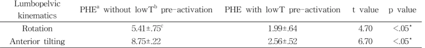
The angles of pelvic rotation and anterior tilting during PHE with lowT pre-activation were sig- nificantly smaller compared to PHE without lowT pre-activation (p<.05) as shown in Table 2.
Discussion
In this study, the EMG activities of the posterior oblique sling muscles and the angles of rotation and
Lumbopelvic
kinematics PHEa without lowTb pre-activation PHE with lowT pre-activation t value p value
Rotation 5.41±.75c 1.99±.64 4.70 <.05*
Anterior tilting 8.75±.22 2.56±.52 6.70 <.05*
aprone hip extension, blower trapezius, cmean±standard deviation, *significant difference between two conditions (p<.05).
Table 2. Lumbopelvic kinematics during two different exercises (Unit: °)
anterior tilting were assessed during PHE with and without lowT pre-activation. Our findings showed that the activity of the bilateral MF and ipsilateral GM was facilitated and the BF was inhibited during PHE with lowT pre-activation compared to PHE without lowT pre-activation. In the PHE with lowT pre-activation, the relative differences in right and left MF, ipsilateral GM, and BF muscle activities were 51.24%, 66.02%, 46.45%, and 61.11% compared to PHE without lowT pre-activation.
There are some possible mechanisms. First, should- er abduction at 125° and external rotation with 1 ㎏ load was performed before hip extension at 10° for the performance of PHE with lowT pre-activation.
This can contribute to increase the counterbalance forces compared to PHE without lowT pre-activation.
This counterbalance force would contribute to greater improvement of the bilateral MF and ipsilateral GM activity (Kim et al, 2013). In a previous study, dur- ing unilateral single-legged hold exercise on a round foam roll, the counterbalance force contributed to bi- lateral transverse abdominis and internal oblique muscle contractions to improve lumbar stability by enhancing the intra-abdominal pressure (Kim et al, 2011). In this study, the bilateral MF and ipsilateral GM activity contributed to this counterbalance force to stabilize the lumbopelvic region. In addition, ipsi- lateral GM was activated to perform hip extension with lumbopelvic stability. The BF muscle can be inhibited by the facilitated MF and GM as a muscle synergist during PHE with lowT pre-activation com- pared to without lowT pre-activation. Therefore, the relatively decreased GM muscular activity levels dur- ing PHE without lowT pre-activation may affect in- creased BF muscle activity to compensate for weak counterbalance force.
Second, the myofascial sling was anatomically inter- connected muscles as a chain (Myers, 2009). Muscle slings are considered for the facilitation of the load transfer from the lower to the upper body through the trunk (Page et al, 2010). The lowT muscle aligned with the elements of the posterior oblique sling can be also the muscle in the superficial back line (Myers, 2009). The lowT can act as a stabilizing mus- cle of the scapulothoracic joint. Therefore, the lowT pre-activation during PHE may improve greater my- ofascial sling co-activation stabilizing the thoracic and lumbar spine compared to PHE without lowT pre-ac- tivation because greater shoulder abduction and ex- ternal rotation angle were needed to activate the lowT (Kim et al, 2015).
In a previous study, the angle of anterior pelvic tilt was significantly increased during PHE without an abdominal drawing-in maneuver (ADIM) compared to PHE with ADIM (Oh et al, 2007). An ADIM with a pressure biofeedback unit during PHE was an effec- tive method for lumbopelvic stability (Oh et al, 2007).
This was consistent with the findings of our study.
We showed that pelvic rotation and anterior tilting during PHE without lowT pre-activation were sig- nificantly greater compared to PHE with lowT pre-activation (p<.05). These results imply that the different stabilizing strategies which were contributed from the facilitation of MF and GM and inhibition of BF muscles during PHE with lowT pre-activation might provide the counterbalance force. Accordingly, the different muscular activation strategies with this counterbalance force can improve lumbopelvic stabili- zation during PHE. In our study, PHE with lowT pre-activation can be recommended for individuals with GM weakness to effectively strengthen GM muscle without lumbopelvic compensations.
This study has several limitations. First, our result cannot be generalized to participants with chronic low back pain. Group differences in EMG amplitudes of posterior oblique sling muscles during exercises were needed for further study. Second, cross-talk between the lowT/MF and the BF/GM may be pos- sible because of surface EMG. Third, this study in- vestigated only posterior oblique sling muscle activ- ities during PHE exercises. A future study should investigate EMG onset time difference during differ- ent PHE exercises.
Conclusion
In this study, the purpose was to compare the EMG activity of the posterior oblique myofascial sling muscles and the angle of lumbopelvic rotation and anterior tilting between PHE with and without lowT pre-activation. Our findings suggest that PHE with lowT pre-activation contributes to increased MF and GM muscular activity, decreased BF muscular activ- ity, and decreased angles in lumbopelvic rotation and anterior tilting compared to PHE without lowT pre-activation. Therefore, the lowT pre-activation can be applied as a lumbopelvic stabilization technique during PHE to strengthen MF and GM muscles for minimizing lumbopelvic compensated motions.
References
Ayotte NW, Stetts DM, Keenan G, et al.
Electromyographical analysis of selected lower ex- tremity muscles during 5 unilateral weight-bearing exercises. J Orthop Sports Phys Ther. 2007;37(2):
48-55.
Bergmark A. Stability of the lumbar spine. A study in mechanical engineering. Acta Orthop Scand Suppl. 1989;230:1-54.
Comerford M, Mottram S. Kinetic Control: The man- agement of uncontrolled movement. 1st ed.
Chatswood, Churchill Livingstone Australia, 2012:
245-255.
Cram JR, Kasman GS, Holtz J. Introduction to Surface Electromyography. 1st ed. Gaithersburg, MD, Aspen Publishers, 1998:112-119.
Danneels LA, Vanderstraeten GG, Cambier DC, et al.
A functional subdivision of hip, abdominal, and back muscles during asymmetric lifting. Spine (Phila Pa 1976). 2001;26(6):E114-E121.
Hodges PW, Moseley GL. Pain and motor control of the lumbopelvic region: Effect and possible mechanisms. J Electromyogr Kinesiol. 2003;13(4):
361-370.
Hungerford BA, Gilleard W, Moran M, et al.
Evaluation of the ability of physical therapists to palpate intrapelvic motion with the Stork test on the support side. Phys Ther. 2007;87(7):
879-887.
Kendall FP, McCreary EK, Provance PG, et al.
Muscles: Testing and function with posture and pain. 5th ed. Baltimore, Williams & Wilkins, 2005:245-249.
Kim JS, Kang MH, Jang JH, et al. Comparison of selective electromyographic activity of the su- perficial lumbar multifidus between prone trunk extension and four-point kneeling arm and leg lift exercises. J Phys Ther Sci. 2015;27(4):1037-1039.
http://dx.doi.org/10.1589/jpts.27.1037
Kim JW, Han JY, Kang MH, et al. Comparison of posterior oblique sling activity during hip ex- tension in the prone position on the floor and on a round foam roll. J Phys Ther Sci. 2013;25(8):
977-979. http://dx.doi.org/10.1589/jpts.25.977 Kim SJ, Kwon OY, Yi CH, et al. Comparison of ab-
dominal muscle activity during a single-legged hold in the hook-lying position on the floor and on a round foam roll. J Athl Train. 2011;46(4):
403-408.
Kim TW, Kim YW. Effects of abdominal drawing-in during prone hip extension on the muscle activ- ities of the hamstring, gluteus maximus, and lumbar erector spinae in subjects with lumbar
This article was received November 25, 2015, was reviewed November 25, 2015, and was accepted January 28, 2016.
hyperlordosis. J Phys Ther Sci. 2015;27(2):383-386.
http://dx.doi.org/10.1589/jpts.27.383
Mills PM, Morrison S, Lloyd DG, et al. Repeatability of 3D gait kinematics obtained from an electro- magnetic tracking system during treadmill locomotion. J Biomech 2007;40(7):1504-1511.
Myers TW. Anatomy Trains: Myofascial meridians for manual and movement therapists. 2nd ed.
Edinburgh, Churchill Livingstone, 2009:238-251.
Nyland J, Kuzemchek S, Parks M, et al. Femoral an- teversion influences vastus medialis and gluteus medius EMG amplitude: Composite hip abductor EMG amplitude ratios during isometric combined hip abduction-external rotation. J Electromyogr Kinesiol. 2004;14(2):255-261.
Oh JS, Cynn HS, Won JH, et al. Effects of perform- ing an abdominal drawing-in maneuver during prone hip extension exercises on hip and back extensor muscle activity and amount of anterior pelvic tilt. J Orthop Sports Phys Ther. 2007;
37(6):320-324.
Oyama S, Myers JB, Wassinger CA, et al. Three-di- mensional scapular and clavicular kinematics and scapular muscle activity during retraction exercises. J Orthop Sports Phys Ther. 2010;40(3):
169-179. http://dx.doi.org/10.2519/jospt.2010.3018
Page P, Frank C, Lardner R. Assessment and Treatment of Muscle Imbalance: The Janda approach. 1st ed. Champaign, IL, Human Kinetics, 2010:159-165.
Sahrmann SA. Diagnosis and Treatment of Movement Impairment Syndromes. 1st ed. St Louis, MO, Mosby, 2002:345-349.
Sykes K, Wong YM. Electrical activity of vastus medialis oblique muscle in straight leg raise ex- ercise with different angles of hip rotation.
Physiotherapy. 2003;89(7):423-430.
Tateuchi H, Taniguchi M, Mori N, et al. Balance of hip and trunk muscle activity is associated with increased anterior pelvic tilt during prone hip extension. J Electromyogr Kinesiol.
2012;22(3):391-397. http://dx.doi.org/10.1016/j.jelekin.
2012.03.003
Vleeming A, Pool-Goudzwaard AL, Stoeckart R, et al.
The posterior layer of the thoracolumbar fascia.
Its function in load transfer from spine to legs.
Spine (Phila Pa 1976). 1995;20(7):753-758.