ABSTRACT
Objectives: Endosequence Bioceramic Root Repair Material (BC-RRM) is used in endodontic microsurgery. It is available as a paste and a putty. However, no studies to date have examined the sealing ability of these forms alone or in combination as root-end filling materials.
Hence, this study aimed to compare the sealing properties of these 2 forms of BC-RRM.
Materials and Methods: Forty-two extracted upper anterior teeth were divided into 3 experimental groups, a positive and negative control. After the root canal treatment, the root ends were resected, retroprepared and retrofilled with either putty, paste + putty or mineral trioxide aggregate (MTA). The teeth were mounted in tubes so the apical 3 mm was submerged in Brain Heart Infusion (BHI) broth. The coronal portions of the canals were inoculated with Enterococcus faecalis and BHI broth and incubated for 30 days. The broth in the tubes was analyzed for colony forming units to check for leakage of bacteria from the canal.
The teeth from the groups were sectioned and analyzed using scanning electron microscopy (SEM). The Kruskal-Wallis test and analysis of variance were used to analyze the data with a significance level p < 0.05.
Results: The BC-RRM and MTA groups showed similar sealing ability. The positive control showed leakage in all samples. The SEM imaging showed the presence of bacteria in all experimental groups at the material-tooth interface.
Conclusions: No significant differences were noted in the experimental groups, providing sufficient evidence that any combination could be effectively used during endodontic microsurgery.
Keywords: Bacteria; Bioceramic Paste; Bioceramic Putty; Mineral trioxide aggregate;
Sealability
INTRODUCTION
Endodontic microsurgical techniques have evolved over the past 2 decades and are considered an important therapeutic approach to deal with post-treatment apical periodontitis when orthograde treatment/retreatment fails or is contraindicated [1]. Endodontic microsurgical procedures have been shown to have high success rates due to the changes in technology, procedures and materials [2,3]. The main surgical steps involve curettage, root-end resection
Research Article
Received: Jan 26, 2021 Revised: Mar 3, 2021 Accepted: Mar 10, 2021
Rencher B, Chang AM, Fong H, Johnson JD, Paranjpe A
*Correspondence to
Avina Paranjpe, BDS, MS, MSD, PhD Professor, Department of Endodontics, School of Dentistry, University of Washington, 1959 NE Pacific Street, D-669 Health Science Center, Seattle, WA 98195, USA.
E-mail: avina@uw.edu
Copyright © 2021. The Korean Academy of Conservative Dentistry
This is an Open Access article distributed under the terms of the Creative Commons Attribution Non-Commercial License (https://
creativecommons.org/licenses/by-nc/4.0/) which permits unrestricted non-commercial use, distribution, and reproduction in any medium, provided the original work is properly cited.
Conflict of Interest
No potential conflict of interest relevant to this article was reported.
Author Contributions
Conceptualization: Paranjpe A; Data curation:
Rencher B, Chang AM; Formal analysis:
Rencher B; Investigation: Rencher B, Chang AM, Fong H; Methodology: Paranjpe A, Johnson JD; Project administration: Paranjpe A, Rencher B; Resources: Fong H, Chang AM; Supervision: Paranjpe A, Johnson JD;
Validation: Paranjpe A, Johnson JD, Fong H;
Visualization: Fong H, Chang AM; Writing - original draft: Rencher B; Writing - review &
editing: Paranjpe A, Johnson JD
Benjamin Rencher ,1 Ana M. Chang ,2 Hanson Fong ,3 James D. Johnson ,1 Avina Paranjpe 1*
1Department of Endodontics, School of Dentistry, University of Washington, Seattle, WA, USA
2Department of Periodontics, University of Washington, Seattle, WA, USA
3Department of Material Science and Engineering, University of Washington, Seattle, WA, USA
Comparison of the sealing ability
of various bioceramic materials for
endodontic surgery
ORCID iDs Benjamin Rencher
https://orcid.org/0000-0002-4729-7409 Ana M. Chang
https://orcid.org/0000-0003-2441-7911 Hanson Fong
https://orcid.org/0000-0003-1501-2621 James D. Johnson
https://orcid.org/0000-0002-2270-6229 Avina Paranjpe
https://orcid.org/0000-0002-8849-0141
and preparation, and root-end filling. Root-end resection removes the apical root third of the root, which is the region where persistent bacteria may be located [4]. After resection, the root end is prepared using specialized instruments to remove a portion of the gutta-percha and create space for the root-end filling [1].
Historically, several materials have been proposed for root-end fillings, including amalgam, reinforced zinc oxide and eugenol cements, gutta-percha, composite and epoxy resins, glass- ionomer and mineral trioxide aggregate (MTA), and more recently introduced bioceramic root repair material [3,5]. The ideal root-end filling material should be biocompatible, have an adequate apical seal, consistent handling properties, long-term clinical success, inhibit microorganism growth, and demonstrate dimensional stability [6,7]. MTA has been widely accepted as the gold standard due to its excellent chemical and biological properties and satisfactory surgical treatment outcomes [5,8]. Although MTA is the gold standard for root-end fillings, it has a few well-recognized drawbacks which include its long setting time, consistency, discoloration potential, and handling properties [9,10].
Endosequence Bioceramic Root Repair Material (BC-RRM, Brasseler, Savannah, GA, USA) has been introduced as a newer material for various applications including root-end filling during endodontic microsurgery [11]. The advantages of this material include improved handling characteristics and shorter setting times with all other properties being similar to those of MTA [11-14]. BC-RRM comes in a ready-to-use, premixed paste or a putty consistency. Several aspects of this material have been studied including its biological properties, antibacterial and antifungal properties, biocompatibility, and outcomes [14-16].
The manufacturer outlines many different methods of use of this material during endodontic microsurgery. Based on these recommendations, some clinicians use only the paste or the putty whereas others prefer to use the combination of the paste first followed by the putty to seal the top. However, there have been no studies that have demonstrated whether using the paste and putty in combination or separately affects or enhances the sealing ability of this material as a root-end filling.
Hence, the purpose of this in vitro study was to compare the sealing ability of bioceramic putty, alone and in combination with bioceramic paste when used as a root-end filling material. The null hypothesis was that there is no difference in the sealing abilities of the various materials when used for root-end fillings during microsurgical endodontic procedures.
MATERIALS AND METHODS
Tooth selection and preparation
Forty-two maxillary incisors with a single canal were selected for this study. Teeth were selected after the radiographs were analyzed for the presence of a single canal. Tooth lengths were standardized to 16 mm by decoronation at or below cemento-enamel junction. Root canals were accessed, and patency was established with a size #15 K-file. ProTaper SX (Dentsply Sirona, Tulsa, OK, USA) was used to coronally flare the canal. Instrumentation was completed with Vortex Blue (Dentsply Sirona) to size #45/0.04 to working length (15 mm) under 2 mL of saline positive pressure irrigation. The canals were dried with paper points; the teeth were wrapped in moist 2 × 2 gauze and sterilized using an autoclave at 116°C at 20 psi for 40 minutes. The canals were filled with Brain Heart Infusion (BHI) broth (Millipore Sigma, St. Louis, MO, USA) and sampled with 3 coarse paper points. These paper points were plated
on BHI agar plates to ensure that the samples were sterile prior to the experiment. Canals were obturated using warm vertical compaction with Vortex Blue gutta-percha (Dentsply Sirona) and a zinc-oxide -eugenol based sealer, Tubli- Seal (Kerr Corporation, Brea, CA, USA) to about 8 mm. Approximately 7 mm of space was left unobturated in the coronal portion of the root for the bacterial specimens (see the section on bacterial leakage). The samples were placed in an incubator at 37°C and 5% CO2 for 7 days to allow the sealer to be completely set [17]. After 7 days, 3 mm of the root-end was resected using a Lindemann bur under water cooling at a zero-degree bevel [18]. Root-end preparation was done using diamond-coated ultrasonic KiS tips (Kerr Corporation) except the positive control group. The tips were used with sterile water to a depth of 3 mm into the canal space. The root ends were retrofilled with a root-end filling material (explained in the next section). The outer root surfaces were covered with 2 layers of nail varnish to prevent any leakage from possible lateral or accessory canals, leaving only the resected surface uncovered. The methodology to analyze the sealing ability of the different groups in this study is similar to a previous study by Antunes et al. [19].
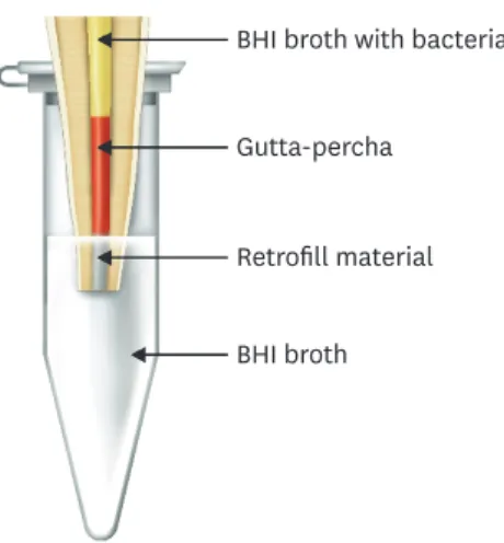
After the material was set (see next section), the teeth were mounted in sterile Eppendorf tubes containing BHI broth so the apical 3 mm of the root end was submerged in the BHI broth (Figure 1). The teeth were stabilized and sealed onto the top of the Eppendorf tubes with cyanoacrylate, and Parafilm® M (Millipore Sigma) was used to secure the lids of the tubes to ensure there was no evaporation or contamination of the broth (Figure 1).
Root-end filling procedure
After obturating and resecting the root ends, the root-end preparations were irrigated with sterile saline and dried with a Stropko Irrigator (J Bar B Co., Carefree, AZ, USA) syringe attachment. The teeth were randomly divided into 3 experimental groups with 12 teeth each and 2 control groups with 3 teeth each. All materials were mixed and used according to the manufacturer's directions.
Group 1: Bioceramic putty (Brasseler).
Group 2: Bioceramic paste + putty (Brasseler).
Group 3: ProRoot Grey MTA (Dentsply Sirona).
Group 4: Gutta-percha only (positive control).
Group 5: Sealed apex (negative control).
BHI broth with bacteria Gutta-percha
Retrofill material BHI broth
Figure 1. Schematic representation of the methodology used in this study.
BHI, Brain Heart Infusion.
For group 1 the putty BC putty was rolled into small, 2- to 3-mm cones and delivered into the root-end preparation in increments by using the back end of a plastic filling instrument, was placed into the root-end preparations. For group 2 the paste was first inserted into the root-end preparation with the BC putty tip provided by the manufacturer followed by the putty as mentioned before. For group 3, the MTA was mixed with sterile water according to the manufacturer's instructions and placed on a wipe-on Lee block (Lee Endo Bloc, San Francisco, CA, USA) in order to deliver 2–3 mm blocks of MTA to the root-end. All the materials were compacted into the root-end preparation with the help of a microplugger.
The excess materials from the above groups were cleaned from the root-end with the help of microbrushes. In the positive control group, the root-end were resected and left unfilled, no retropreparation or retrofilling was done. For the negative control group, utility wax was utilized to seal the resected surface and the entire root surface along with the wax filling were covered with 2 layers of nail varnish. Radiographs were taken of the teeth from each of the experimental groups to confirm that the retrofilling material was adequately placed. All teeth were wrapped in moist gauze, placed in an incubator at 37°C and 5% CO2 for 48 hours to allow the material to set. The material setting was verified with an endodontic explorer. Once the setting was verified the teeth were mounted in sterile Eppendorf tubes as described in the previous section.
Bacterial preparation and inoculation.
Sterile BHI broth was added to the Eppendorf tubes of each sample, enough to submerge the apical 3 mm of the tooth root. Pure isolated 24-hour colonies of Enterococcus faecalis (E. faecalis) (ATCC 19433) from BHI agar plates were suspended into 3 ml of sterile BHI broth and grown aerobically at 37°C overnight. The following day, 120 μL of this overnight bacterial culture was sub-cultured into 20 ml of fresh BHI broth until log phase was achieved (about 4 hours and an optical density of 600 nm [OD600] of 1). Finally, log phase cultures were adjusted to 1.5 × 10⁸ colony-forming units (CFUs)/mL (equivalent to 0.5 McFarland standard) and 20 μL of this bacterial inoculum was added to the coronal 7 mm of the teeth. All experiments were performed under aerobic conditions at 37°C for 30 days to ensure adequate penetration of bacteria into the canal space and replenished with fresh bacterial inoculum/BHI mixture every 2–3 days. Bacteria were inoculated into all experimental and positive control samples.
Negative controls were inoculated with BHI broth alone without bacteria.
Sample evaluation
After 30 days of inoculation with E. faecalis within tooth canals, the suspension broth within Eppendorf tubes was visually examined for turbidity and analyzed at an OD600. Teeth were then removed and prepared for scanning electron microscopy (SEM). The 100 μL of suspension broth from each tube was plated onto blood agar plates and incubated aerobically at 37°C overnight. CFUs were counted, and the purity was confirmed by gram staining and colony morphology.
Scanning electron microscopy examination
After the teeth were removed from the Eppendorf tubes, 3 teeth from each group, except negative control, were longitudinally grooved with a diamond disk and split with a custom wedge-splitting device. These samples were rinsed in phosphate-buffered saline and fixed with 2.5% glutaraldehyde (Sigma Aldrich, St. Louis, MO, USA) for 1 hour, post fixed in 1%
osmium tetroxide (OsO4) (Sigma Aldrich) for 30 minutes and then rinsed with deionized water. Samples were then dehydrated, mounted on SEM discs, and sputter-coated with 5 nm of gold-palladium for conductivity. The samples were scanned using JEOL JSM 7000F
(Jeol USA Inc, Peabody, MA, USA) at 10 kV acceleration voltage. Images were taken at various sections of the tooth and at various magnifications (20×, 250×, 2,500×, and 5,000×) to visualize bacteria within the canal and interface between material and canal wall.
Statistical analysis
SigmaPlot 11.0 (Systat Software, Inc. San Jose, CA, USA) was used for all the statistical testing and analysis. The required sample size was calculated to be 12 samples for the experimental groups. This gave the study at least 95% power to detect a difference between group means using 1-way analysis of variance (ANOVA) with a significance level of 0.05. The power was set to 95% to allow for possible loss of samples. With the loss of one sample per group, a sample size per group of 11 would yield at least 90% power and a loss of 2 samples per group would still yield at least 85% power. Viable counts/positive cultures were calculated. The outcome measures were CFU counts, which indicates a positive culture. The Kruskal-Wallis test was used to test for differences in CFUs across groups. ANOVA with a post hoc Tukey test was used to test for differences in the OD readings between the different groups. The significance level was set at p < 0.05 for all tests.
RESULTS
Bacterial counts
After 30 days of incubation, samples that were visibly turbid exhibited an OD600 reading of > 1, whereas samples that were not visibly turbid exhibited an OD600 reading of < 1.
Group 1: BC putty: none of the samples appeared turbid after 30 days, and no colonies were visible on the blood agar plates. All OD600 readings were < 1, which suggests no leakage of bacteria occurred from the tooth into the suspension broth.
Group 2: BC putty + paste: none of the samples appeared turbid after 30 days, and no colonies were visible on the blood agar plates. The OD600 readings were all < 1.
Group 3: Grey MTA: One sample turned turbid during the 30-day incubation. The turbid sample showed multiple colonies on the blood agar plate; however, the other samples within this group were not turbid and did not have any colonies on the plates. The OD600 reading for the turbid sample in Group 3 was > 1, and the remaining samples were < 1.
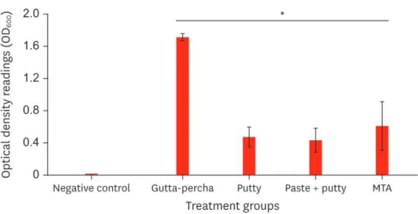
Group 4: Gutta-percha only (positive control): All samples of the positive control group were turbid (Figure 2) during the 30-day incubation. The CFU counts for all samples in the positive control group were > 50 and the OD600 readings were > 1 for all teeth.
Group 5: Sealed apex (negative control): No samples appeared turbid, and no CFUs could be counted on the agar plates, which suggests no contamination during the experimental procedures. All OD600 readings for the negative control were < 1.
There were no statistically significant differences between the 3 experimental groups.
However, there were statistically significant differences between the experimental and positive control groups (Figures 2 and 3).
Scanning electron microscopy imaging
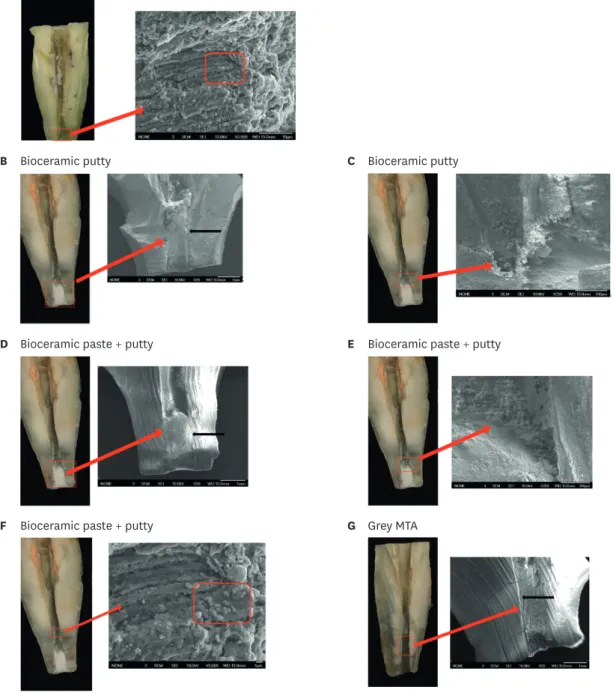
The SEM imaging revealed detectable bacteria in the canal space of all the samples (Figure 4A-I).
The gutta-percha from the canals of all teeth was displaced/lost during sample preparation. The SEM images demonstrated adequate marginal adaptation of all the experimental materials to
the dentin walls (Figure 4B-I). No significant visible differences were noted between putty, paste + putty, and MTA groups. The gutta-percha only (positive control group) showed the presence of bacteria throughout the length of the canal and at the apical portion where the root resection was completed (Figure 4A). The experimental groups showed the presence of bacteria within the middle 1/3rds of canals, but not within suspension broths demonstrating that bioceramic and MTA groups provided sufficient sealability to prevent leakage of bacteria into the broth. Figure 4F and I showed the presence of bacteria in the apical 3 mm.
DISCUSSION
Calcium silicate based root-end filling materials have gained tremendous popularity over the past decade and are routinely used in endodontics for various purposes including root- end fillings during surgical procedures. Previous studies have evaluated various physical properties of these materials including biocompatibility, sealing ability, and marginal
0 0.8 0.4 1.2 1.6 2.0
Negative control Gutta-percha
Treatment groups
*
Optical density readings (OD600)
Putty Paste + putty MTA
Figure 2. Bacterial counts were determined with the optical density of 600 nm (OD600) readings. There were statistically significant differences between the gutta-percha (positive control) and experimental groups. Analysis of variance with a post hoc Tukey test was used to test for differences in the OD readings between the different groups.
MTA, mineral trioxide aggregate.
*The significance level was set at p < 0.05.
0 100 250 200 150
50 300 350
Gutta-percha
Treatment groups Bacterial counts
*
CFUs
Putty Paste + putty MTA
Figure 3. Colony forming units (CFUs) were calculated for all the groups. CFU data corroborated optical density of 600 nm readings. Gutta-percha (positive control) had statistically higher numbers as compared to the other experimental groups. The negative control had no colonies. The Kruskal-Wallis test was used to test for differences in CFUs across groups.
MTA, mineral trioxide aggregate.
*The significance level was set at p < 0.05.
adaptability [19-21]. BC-RRM is available in 2 forms, the paste and the putty. There is currently no evidence to support or refute the concurrent use of both the paste and the putty formulation of BC-RRM together as a root-end filling. The paste and putty formulations have A Gutta-percha (positive control)
B Bioceramic putty C Bioceramic putty
D Bioceramic paste + putty E Bioceramic paste + putty
F Bioceramic paste + putty G Grey MTA
H Grey MTA I Grey MTA
Figure 4. Scanning electron microscopic (SEM) images demonstrated the surface canal interface for the positive control and the experimental groups. The gutta-percha from the canals for all groups was displaced during specimen preparation. (A) Bacteria present in the canal (red box); the resected root surface in the gutta-percha (positive control) group (red arrow). (B) Root-end filling material in the bioceramic putty (group 1) group (20×). The marginal adaptation of the material to the root surface (black arrow). (C) Material and canal interface in the bioceramic putty group (group 1). This SEM image further demonstrates the marginal adaptation of the material to the root surface (250×). (D) Root-end filling material in the bioceramic paste + putty group (group 2) (20×). The marginal adaptation of the material to the root surface (black arrow). (E) Material and canal interface in the bioceramic paste + putty group (group 2) (250×). This SEM image demonstrates the marginal adaptation of the material to the root surface. (F) Presence of bacteria (red box) in the tooth canal space in group 2 (5,000×).
(G) Root-end filling material in the Grey mineral trioxide aggregate (MTA) (group 3) (20×). The marginal adaptation of the material to the root surface (black arrow). (H) Material and canal interface in the Grey MTA (group 3) (250×). This SEM image demonstrates the marginal adaptation of the material to the root surface. (I) Presence of bacteria (red box) in the canal space in the tooth in group 3 (5,000×).
SEI, scanning electron imaging; WD, working distance. (continued to the next page)
similar properties but differ in the quantity of thickening agents [22]. It is hypothesized that the paste may have better flow and adaptation properties. However, studies that compare similarities or differences in sealability, adaptability, or other properties for these formulations are lacking. Hence, this study evaluated the sealing ability of BC-RRM in the putty and paste formulation when used as a root-end filling material. The sealing properties were compared to those of MTA in this study along with appropriate positive and negative controls.
MTA has been widely used for various procedures in Endodontics. The efficacy of MTA as a retro-filling material has been previously well established and various studies have demonstrated tissue healing after endodontic surgery [23-26]. It is considered the gold standard and has been used in many previous studies when comparing various bioceramic materials like BC-RRM. Chen et al. [11] found BC-RRM had a favorable histological healing response in dogs when comparing periradicular tissue healing after surgery using MTA and BC-RRM as root-end filling materials. Another study demonstrated that BC-RRM was also associated with high healing rates [16]. Amongst other factors, the good clinical results reported for these materials are potentially related to their sealing ability as shown in this study. A more recent randomized control trial compared the outcomes of endodontic microsurgery using MTA and RRM. The data from this study demonstrated that both materials demonstrated similar success rates [3].
This study used E. faecalis as this has been the bacteria of choice in similar in vitro studies and is commonly found in teeth with post-treatment disease [14,15,27,28]. After 30 days of inoculation with E. faecalis, 1 out of 12 samples from the MTA group was contaminated.
This could be due to several reasons such as incomplete material set, root degradation, user contamination during sampling or incubation, or material washout.
This study evaluated root-end filling materials in single canal anterior teeth because these could be placed in the apparatus (Figure 1) with ease without the introduction of additional variables. Posterior teeth such as molars possess more complicated anatomy and technical challenges. Some studies have found that tooth type had no significant impact on the outcome of root-end surgery [16]. In contrast, other studies have reported that molar root- Sealing ability of bioceramic materials
B Bioceramic putty C Bioceramic putty
D Bioceramic paste + putty E Bioceramic paste + putty
F Bioceramic paste + putty G Grey MTA
H Grey MTA I Grey MTA
Figure 4. (Continued) Scanning electron microscopic (SEM) images demonstrated the surface canal interface for the positive control and the experimental groups. The gutta-percha from the canals for all groups was displaced during specimen preparation. (A) Bacteria present in the canal (red box); the resected root surface in the gutta-percha (positive control) group (red arrow). (B) Root-end filling material in the bioceramic putty (group 1) group (20×). The marginal adaptation of the material to the root surface (black arrow). (C) Material and canal interface in the bioceramic putty group (group 1). This SEM image further demonstrates the marginal adaptation of the material to the root surface (250×). (D) Root-end filling material in the bioceramic paste + putty group (group 2) (20×). The marginal adaptation of the material to the root surface (black arrow). (E) Material and canal interface in the bioceramic paste + putty group (group 2) (250×). This SEM image demonstrates the marginal adaptation of the material to the root surface. (F) Presence of bacteria (red box) in the tooth canal space in group 2 (5,000×). (G) Root-end filling material in the Grey mineral trioxide aggregate (MTA) (group 3) (20×). The marginal adaptation of the material to the root surface (black arrow). (H) Material and canal interface in the Grey MTA (group 3) (250×). This SEM image demonstrates the marginal adaptation of the material to the root surface. (I) Presence of bacteria (red box) in the canal space in the tooth in group 3 (5,000×).
SEI, scanning electron imaging; WD, working distance.
end surgery had a lower healing rate than single canal anterior teeth and attributed this difference to the difficulty of access and the complexity of the root anatomy [29]. The outer surfaces of the teeth were sealed with varnish in order to ensure that bacterial leakage of bacteria can only originate from the apical portion of the tooth and not other areas into the suspension broth.
Bacteria and bacterial leakage have been suggested to be the primary cause of apical
periodontitis and post-treatment disease [30,31]. This bacterial leakage model has been used in a previous study and tries to simulate the clinical conditions [19]. BHI broth was used as it is the preferred media and the ideal source of nutrients for E. faecalis. Replenishing nutrients within this system is important to keep the bacteria viable during the 30-day experimental period.
The results of this in vitro study may differ from a clinical situation since other variables may influence the results, including the presence of blood and tissue fluids during the placement of the retro-filling materials, presence of various bacterial species versus 1 bacteria the bacterial counts in the canal, presence of a periapical lesion, and anatomic variations. Other factors, which may influence differences between studies or outcomes include case selection criteria, experimental design and operator technique. However, the study was designed to verify the sealing ability of the different materials and the data demonstrated that there were no statistically significant differences between BC putty, BC putty and paste, and MTA.
Therefore, any of them could be successfully used during endodontic microsurgery.
The clinical implications of this study relate to the importance of a root-end filling material during endodontic microsurgery. Furthermore, all the experimental groups sealed the canal space which implies that either of the root-end fillings materials would be acceptable during Endodontic microsurgical techniques. Based on the data from this study we can accept the null hypothesis.
CONCLUSIONS
This study underscores the significance and the importance of the placement of a root- end filling material after root-end resection since all the positive control samples showed bacterial leakage. No significant differences were noted when using the paste and the putty in combination in this in vitro system, providing sufficient evidence that this combination could be effectively and efficiently used during endodontic microsurgical procedures. Furthermore, the ease of placement of the paste and putty may provide an additional benefit. This data also reiterated the findings from previous studies where the BC-RRM groups had similar sealing properties to those of MTA.
ACKNOWLEDGEMENTS
The authors would like to acknowledge Dr. Richard P. Darveau for his support with the microbiological aspect of this study.
REFERENCES
1. Kim S, Kratchman S. Modern endodontic surgery concepts and practice: a review. J Endod 2006;32:601-623.
PUBMED | CROSSREF
2. Huang S, Chen NN, Yu VSH, Lim HA, Lui JN. Long-term success and survival of endodontic microsurgery.
J Endod 2020;46:149-157.e4.
PUBMED | CROSSREF
3. Safi C, Kohli MR, Kratchman SI, Setzer FC, Karabucak B. Outcome of endodontic microsurgery using mineral trioxide aggregate or root repair material as root-end filling material: a randomized controlled trial with cone-beam computed tomographic evaluation. J Endod 2019;45:831-839.
PUBMED | CROSSREF
4. Siqueira JF Jr, Rôças IN, Ricucci D, Hülsmann M. Causes and management of post-treatment apical periodontitis. Br Dent J 2014;216:305-312.
PUBMED | CROSSREF
5. Torabinejad M, Pitt Ford TR. Root end filling materials: a review. Endod Dent Traumatol 1996;12:161-178.
PUBMED | CROSSREF
6. Chong BS, Pitt Ford TR, Watson TF, Wilson RF. Sealing ability of potential retrograde root filling materials. Endod Dent Traumatol 1995;11:264-269.
PUBMED | CROSSREF
7. Saxena P, Gupta SK, Newaskar V. Biocompatibility of root-end filling materials: recent update. Restor Dent Endod 2013;38:119-127.
PUBMED | CROSSREF
8. Torabinejad M, Parirokh M. Mineral trioxide aggregate: a comprehensive literature review--part II: leakage and biocompatibility investigations. J Endod 2010;36:190-202.
PUBMED | CROSSREF
9. Parirokh M, Torabinejad M. Mineral trioxide aggregate: a comprehensive literature review--part III:
clinical applications, drawbacks, and mechanism of action. J Endod 2010;36:400-413.
PUBMED | CROSSREF
10. Butt N, Talwar S, Chaudhry S, Nawal RR, Yadav S, Bali A. Comparison of physical and mechanical properties of mineral trioxide aggregate and Biodentine. Indian J Dent Res 2014;25:692-697.
PUBMED | CROSSREF
11. Chen I, Karabucak B, Wang C, Wang HG, Koyama E, Kohli MR, Nah HD, Kim S. Healing after root-end microsurgery by using mineral trioxide aggregate and a new calcium silicate-based bioceramic material as root-end filling materials in dogs. J Endod 2015;41:389-399.
PUBMED | CROSSREF
12. Abusrewil SM, McLean W, Scott JA. The use of Bioceramics as root-end filling materials in periradicular surgery: a literature review. Saudi Dent J 2018;30:273-282.
PUBMED | CROSSREF
13. Mahgoub N, Alqadasi B, Aldhorae K, Assiry A, Altawili ZM, Tao Hong . Comparison between iroot bp plus (endosequence root repair material) and mineral trioxide aggregate as pulp-capping agents: a systematic review. J Int Soc Prev Community Dent 2019;9:542-552.
PUBMED
14. Lovato KF, Sedgley CM. Antibacterial activity of endosequence root repair material and proroot MTA against clinical isolates of Enterococcus faecalis. J Endod 2011;37:1542-1546.
PUBMED | CROSSREF
15. Alanezi AZ, Jiang J, Safavi KE, Spangberg LS, Zhu Q. Cytotoxicity evaluation of endosequence root repair material. Oral Surg Oral Med Oral Pathol Oral Radiol Endod 2010;109:e122-e125.
PUBMED | CROSSREF
16. Shinbori N, Grama AM, Patel Y, Woodmansey K, He J. Clinical outcome of endodontic microsurgery that uses EndoSequence BC root repair material as the root-end filling material. J Endod 2015;41:607-612.
PUBMED | CROSSREF
17. Allan NA, Walton RC, Schaeffer MA. Setting times for endodontic sealers under clinical usage and in vitro conditions. J Endod 2001;27:421-423.
PUBMED | CROSSREF
18. Gilheany PA, Figdor D, Tyas MJ. Apical dentin permeability and microleakage associated with root end resection and retrograde filling. J Endod 1994;20:22-26.
PUBMED | CROSSREF
19. Antunes HS, Gominho LF, Andrade-Junior CV, Dessaune-Neto N, Alves FR, Rôças IN, Siqueira JF Jr. Sealing ability of two root-end filling materials in a bacterial nutrient leakage model. Int Endod J 2016;49:960-965.
PUBMED | CROSSREF
20. Toia CC, Teixeira FB, Cucco C, Valera MC, Cavalcanti BN. Filling ability of three bioceramic root-end filling materials: a micro-computed tomography analysis. Aust Endod J 2020;46:424-431.
PUBMED | CROSSREF
21. Tran D, He J, Glickman GN, Woodmansey KF. Comparative analysis of calcium silicate-based root filling materials using an open apex model. J Endod 2016;42:654-658.
PUBMED | CROSSREF
22. Zamparini F, Siboni F, Prati C, Taddei P, Gandolfi MG. Properties of calcium silicate-monobasic calcium phosphate materials for endodontics containing tantalum pentoxide and zirconium oxide. Clin Oral Investig 2019;23:445-457.
PUBMED | CROSSREF
23. Chong BS, Pitt Ford TR, Hudson MB. A prospective clinical study of mineral trioxide aggregate and IRM when used as root-end filling materials in endodontic surgery. Int Endod J 2003;36:520-526.
PUBMED | CROSSREF
24. Christiansen R, Kirkevang LL, Hørsted-Bindslev P, Wenzel A. Randomized clinical trial of root-end resection followed by root-end filling with mineral trioxide aggregate or smoothing of the orthograde gutta-percha root filling--1-year follow-up. Int Endod J 2009;42:105-114.
PUBMED | CROSSREF
25. Kruse C, Spin-Neto R, Christiansen R, Wenzel A, Kirkevang LL. Periapical bone healing after apicectomy with and without retrograde root filling with mineral trioxide aggregate: a 6-year follow-up of a
randomized controlled trial. J Endod 2016;42:533-537.
PUBMED | CROSSREF
26. Lindeboom JA, Frenken JW, Kroon FH, van den Akker HP. A comparative prospective randomized clinical study of MTA and IRM as root-end filling materials in single-rooted teeth in endodontic surgery. Oral Surg Oral Med Oral Pathol Oral Radiol Endod 2005;100:495-500.
PUBMED | CROSSREF
27. Nair U, Ghattas S, Saber M, Natera M, Walker C, Pileggi R. A comparative evaluation of the sealing ability of 2 root-end filling materials: an in vitro leakage study using Enterococcus faecalis. Oral Surg Oral Med Oral Pathol Oral Radiol Endod 2011;112:e74-e77.
PUBMED | CROSSREF
28. Zhang C, Du J, Peng Z. Correlation between Enterococcus faecalis and persistent intraradicular infection compared with primary intraradicular infection: a systematic review. J Endod 2015;41:1207-1213.
PUBMED | CROSSREF
29. Song M, Jung IY, Lee SJ, Lee CY, Kim E. Prognostic factors for clinical outcomes in endodontic microsurgery: a retrospective study. J Endod 2011;37:927-933.
PUBMED | CROSSREF
30. Kakehashi S, Stanley HR, Fitzgerald RJ. The effects of surgical exposures of dental pulps in germ-free and conventional laboratory rats. Oral Surg Oral Med Oral Pathol 1965;20:340-349.
PUBMED | CROSSREF
31. Vieira AR, Siqueira JF Jr, Ricucci D, Lopes WS. Dentinal tubule infection as the cause of recurrent disease and late endodontic treatment failure: a case report. J Endod 2012;38:250-254.
PUBMED | CROSSREF