INTRODUCTION
The differentiation between acute total occlusion (ATO) of coronary artery and chronic total occlusion (CTO) is important because the therapeutic strategy of these two entities differ (1, 2). Acutely occluded coronary artery in patients with acute cor- onary syndrome (ACS) must be treated immediately, using re- perfusion therapy, whereas, many patients with CTO are man- aged conservatively (3-5). Although the diagnosis of coronary artery disease is made comprehensively, using cardiac biomark-
ers, patient symptoms, electrocardiography (ECG), and imaging findings, it is not always possible to determine whether the pa- tient has a culprit lesion or not (6-9). Furthermore, when com- plete interruption of enhanced coronary arterial lumen is en- countered, in coronary computed tomography angiography (CCTA), it is difficult to decide whether it is a culprit lesion be- cause the presenting symptom and the laboratory results may sometimes not be helpful in properly triaging the patient.
With advances in multi-detector computed tomography (MDCT) technology, many studies have demonstrated that
J Korean Soc Radiol 2012;67(2):93-99
Received May 17, 2012; Accepted July 24, 2012 Corresponding author: Hyon Joo Kwag, MD Department of Radiology, Kangbuk Samsung Hospital, Sungkyunkwan University School of Medicine, 29 Saemunan-ro, Jongno-gu, Seoul 110-746, Korea.
Tel. 82-2-2001-1031 Fax. 82-2-2001-1030 E-mail: [email protected]
Copyrights © 2012 The Korean Society of Radiology
Purpose: To compare the features of coronary computed tomography angiography (CCTA) imaging of the patients with acute total occlusion (ATO) of coronary artery with those of chronic total occlusion (CTO).
Materials and Methods: CCTA of 26 patients with complete interruption of the coronary artery in CCTA and occlusion in conventional coronary angiography, were retrospectively analyzed. Discrimination between the ATO group (n = 11, patients with non ST-elevation myocardial infarction or unstable angina) and the CTO group (n = 15, patients with stable angina or nonspecific symptom) was arbitrarily deter- mined by clinical diagnosis. Lesion length, remodeling index (RI), plaque density measured by Hounsfield units (HU), plaque composition, percentage attenuation drop across the lesion, and presence of myocardial thinning were evaluated.
Results: Comparisons between the ATO and CTO groups revealed significantly shorter lesion length in the ATO group (0.40 cm vs. 1.87 cm, respectively; p = 0.001), and significantly higher RI (1.56 vs. 1.10, respectively; p = 0.004). Plaque density of the ATO group was lower (37.0 HU vs. 104.7 HU, respectively; p < 0.001) and non- calcified plaque was frequently seen in the ATO group (72.7% vs. 26.7%, respective- ly; p = 0.02). Percentage attenuation drop across the lesion was lower for the ATO group (10.92% vs. 25.44%, respectively; p = 0.005). Myocardial thinning was exclu- sively observed in the CTO group (seven of 15 patients, p = 0.01).
Conclusion: CCTA shows various statistically significant differences between the ATO and CTO groups.
Index terms Cardiac Imaging
Tomography, X-ray Computed Coronary Artery Disease Chronic Total Occlusion Acute Coronary Syndrome
Differentiation of Acute Total Occlusion of Coronary Artery from Chronic Total Occlusion in Coronary Computed Tomography Angiography
관상동맥 CT 혈관촬영술을 이용한 관상동맥의 급성완전폐쇄와 만성완전폐쇄의 감별
Hyon Joo Kwag, MD
Department of Radiology, Kangbuk Samsung Hospital, Sungkyunkwan University School of Medicine, Seoul, Korea
complete interruption of enhanced coronary arterial lumen in CCTA with stable angina or nonspecific symptoms. Diagnoses of NSTEMI, unstable angina, and stable angina were made on the basis of the American College of Cardiology/American Heart Association definition (8).
CCTA was performed before CAG in all patients. The medi- an interval between CCTA and CAG was not significantly dif- ferent between the ATO group (2.0 days; range, 0-7 days) and the CTO group (5.0 days; range, 1-17 days) (p = 0.26).
Institutional review board approval was obtained for this ret- rospective study, and informed consent was waived.
MDCT Protocols
Patients with a heart rate exceeding 70 beats/min received 10-30 mg intravenous esmolol (Jeil Pharm., Seoul, Korea), be- fore MDCT imaging. CCTA was performed with Philips Bril- liance 64 scanner (Philips Medical Systems, Best, The Nether- lands). A standard scanning protocol was applied, with 64 × 0.625 mm section collimation, 420-ms rotation time, 120-kV tube voltage, and 800-mA tube current. All scans were per- formed with ECG-gated dose modulation. A bolus of 80 mL Iomeprol 400 (Bracco, Milan, Italy) was intravenously injected (4 mL/s), followed by 50 mL of saline. A region of interest was placed in the descending thoracic aorta, and image acquisition was automatically initiated, once a selected threshold [150 Houn- sfield units (HU)] had been reached with bolus tracking. The ECG of each patient was simultaneously recorded to allow for the retrospective segmental data reconstruction. Images were initially reconstructed at a mid-diastolic phase (75% of the R-R interval) of the cardiac cycle for coronary artery evaluation, and end-diastolic phase (90-100% of the R-R interval) for measure- ment of the left ventricular myocardial thickness. Additional reconstructions were performed if the motion artifacts were present.
Image Analysis
All scans were analyzed with a dedicated workstation (Bril- liance; Philips Medical Systems, Cleveland, OH, USA). Each le- sion was assessed with multiplanar reconstruction (MPR), maxi- mum intensity projections, and three-dimensional volume rendered reconstruction, as well as image data sets with 1-mm reconstructed slice thickness.
CCTA in patients with acute chest pain is safe, feasible, and fa- cilitates early triage of acute chest pain in low-risk patients, which primarily relies on their negative MDCT result (10-14).
Several clinical studies have suggested that CCTA can predict culprit coronary lesions in patients with ACS (15, 16). Other studies demonstrated CCTA differences in plaque morphology and composition between ACS and stable angina (9, 17).
When we encounter a complete interruption of contrast-en- hancement of coronary artery in CCTA, the distinction be- tween acute and chronic occlusion has particular importance for the management of patients presented with acute chest pain. To date, however, there is a paucity of information for pa- rameters to differentiate coronary artery ATO from CTO, by using CCTA (18).
The purpose of this study was to compare the CCTA imaging features of patients with ATO of coronary artery with those of CTO.
MATERIALS AND METHODS
Study Population
Among the 164 patients, who showed complete interruption of coronary artery in CCTA, between January 2006 and June 2009, 26 patients with total occlusion in conventional coronary angiography (CAG) were selected. Patients who underwent CAG before CCTA were excluded due to an intervention pro- cedure ahead of CCTA can change the lesion characteristics.
Patients with previous bypass surgery, as well as lesions with stents and patients who did not show 100% stenosis in their CAG, were excluded.
These patients were divided into two groups, arbitrarily (ATO and CTO), according to the clinical diagnosis. The ATO group (n = 11, nine men and two women; age range, 48-86 years;
mean age, 63.5 years) was comprised of patients who showed complete interruption of enhanced coronary arterial lumen in CCTA with non ST-elevation myocardial infarction (NSTE- MI), or unstable angina. The author considered that an acutely occluded coronary artery will lead to ACS, including STEMI, NSTEMI, and unstable angina. However, the STEMI patients who underwent CAG, prior to CCTA, were excluded. The CTO group (n = 15, 11 men and four women; age range, 47-84 years;
mean age, 61.9 years) was comprised of patients who showed
RESULTS
Several statistically significant differences were evident be- tween the ATO group and that of the CTO group, concerning the complete interruption of enhanced coronary artery (Figs. 1, 2, Table 1). Demographic findings between the two groups are summarized in Table 2.
In the ATO group, lesion length was shorter than the CTO group [median (25th-75th percentiles); 0.40 (0.38-0.60) cm vs.
1.87 (0.89-3.00) cm, respectively; p = 0.001] and RI was higher in the ATO group than in the CTO group [1.56 (1.10-2.20) vs.
1.10 (0.95-1.34), respectively; p = 0.004]. Plaque density of the ATO group was lower than the CTO group [37.0 (28.0-45.0) HU vs. 104.7 (70.0-142.0) HU, respectively; p < 0.001], and the non-calcified plaque was seen more frequently in the ATO group than in the CTO group (72.7% vs. 26.7%, respectively; p = 0.02).
Percentage attenuation drop across the lesion was lower for the ATO group than the CTO group [10.92 (6.75-17.32)% vs. 25.44 (17.50-39.10)%, respectively; p = 0.005]. Myocardial thinning was exclusively observed in the CTO group (seven of 15 pa- tients, p = 0.01).
DISCUSSION
In this study, various CCTA imaging characteristics were demonstrated that can be used to help differentiate between ATO of coronary artery in patients with ACS and CTO. Short The following parameters were evaluated, retrospectively, by
one radiologist with 4 years of CCTA experience: coronary ar- tery assessment including lesion length, remodeling index (RI), plaque density, plaque composition, as well as the attenuation drop across the lesion, and myocardial thinning. Results of CAG were reviewed by experienced cardiologists in all patients.
Lesion length was measured on curved MPR images, rendered exactly in the plane of the lesion. The length of the vessel seg- ment, with complete absence of detectable luminal enhance- ment, was measured. RI was defined as the cross-sectional ves- sel area within the lesion, divided by the reference vessel (non- diseased vessel immediately proximal to the lesion) cross- sectional area (19). Plaque densities of corresponding artery, at least five regions of interests (each region area = 1 mm2) posi- tioned in non-calcified area, were measured; the minimum density value was decided as the plaque density (20). The plaques were reported as non-calcified, calcified, or mixed plaques.
Based on prior data (17), plaque was defined as a non-calcified plaque when the HU was < 130. Calcification was recognized as the plaque with the density of > 130 HU on curved MPR im- ages. Mixed plaque was defined if the plaque area consisted of
> 50% of non-calcified plaque. The percentage attenuation drop across the lesion was defined as the difference in CT attenua- tion, proximal and distal, to the lesion divided by the attenua- tion value of proximal to the lesion and multiplied by 100 (19).
The presence of myocardial thinning was defined as the mea- sured myocardial thickness < 6 mm on the short axis image of the end-diastolic phase. The myocardium was divided in 17 standardized segments, using the American Heart Association/
American College of Cardiology classification (21).
Statistical Analyses
Statistical analyses were performed using SPSS 16.0 (SPSS, Chi- cago, IL, USA). To test for differences in the lesion length, RI, plaque density, and percentage attenuation drop across the lesion between the ATO and CTO groups of patients, the Mann-Whit- ney U test was used due to the distribution of variables, which did not show normal distribution. Chi-square test and Fisher’s exact test were used for the comparison of plaque composition and presence of myocardial thinning between the two groups. Quan- titative variables are presented as the median (25th-75th percen- tiles). A p-value < 0.05 was considered to indicate significance.
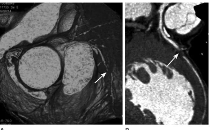
A B
Fig. 1. A 48-year-old male with acute chest pain. Volume rendering image (A) reveals complete interruption of coronary artery at mid left anterior descending artery (arrow). Curved multiplanar image (B) re- veals discrete total occlusion with non-calcified plaque and severe positive arterial remodeling as a culprit lesion in the same area (ar- row). Final diagnosis is non ST-elevation myocardial infarction.
non-calcified plaques contribute to a greater degree to the total plaque burden than those in a stable angina.
In a study in which the drop in attenuation over the lesion was compared in total occlusion and high-grade stenosis by us- ing CCTA, the attenuation drop across the lesion was higher in total occlusion than high-grade stenosis. However, it was not correlated with the lesion length (19). In the present study, the percentage attenuation drop across the lesion was lower in the ATO group, compared with that of the CTO group. This was an unexpected result, since the average attenuation of the distal portion of the occluded coronary artery was expected to be higher in the CTO group, due to more prominent collateral cir- culation, compared with the ATO group. It is difficult to sug- gest the possible reasons because of the small sample number lesion length, high RI, low plaque density, non-calcified plaque,
low percentage attenuation drop across the lesion, and no evi- dence of myocardial thinning indicated acute complete inter- ruption of the coronary artery in CCTA.
The present results echo those of the other studies that com- pared the culprit lesions in ACS with those of the stable angina in CCTA, with respect to the frequency of non-calcified plaque and positive remodeling. Motoyama et al. (22) found that presence of non-calcified plaque, positive arterial remodeling, and spotty cal- cification was associated with a high positive predictive value for the culprit plaque associated with ACS. Hoffmann et al. (9) re- ported greater plaque area, higher RI, and higher prevalence of non-calcified plaque, associated with culprit plaques in patients with ACS. Leber et al. (17) noted that, in myocardial infarction,
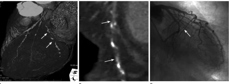
A B C
Fig. 2. A 71-year-old male with acute chest pain. Volume rendering image (A) and curved multiplanar image (B) reveals diffuse occlusion of left circumflex coronary artery with multifocal calcified plaques (arrows), which is confirmed by conventional angiography (C) (arrow). Final diagno- sis is unstable angina.
Table 1. CT Findings in Complete Interruption of Enhanced Coronary Artery between Patients in the Acute Occlusion Group and Chronic Oc- clusion Group
Acute Occlusion Group Chronic Occlusion Group p-Value
Lesion length (cm) 0.40 1.87 0.001
Remodeling index 1.56 1.10 0.004
Plaque density (HU) 37.0 104.7 < 0.001
Plaque composition 0.02
Noncalcified 8/14 (72.7%) 4/15 (26.7%)
Calcified 0/14 (0%) 3/15 (20.0%)
Mixed 3/14 (27.3%) 8/15 (53.3%)
Attenuation drop (%) 10.92 25.44 0.005
Presence of myocardial thinning 0.01
No 11/11 (100%) 8/15 (53.3%)
Yes 0/14 (0%) 7/15 (46.7%)
Note.-HU = Hounsfield unit
of patients with acutely occluded coronary artery.
In conclusion, CCTA shows various statistically significant differences between the ATO and CTO groups.
REFERENCES
1. Boden WE. Management of chronic coronary disease: is the pendulum returning to equipoise? Am J Cardiol 2008;
101:69D-74D
2. Stone GW, Kandzari DE, Mehran R, Colombo A, Schwartz RS, Bailey S, et al. Percutaneous recanalization of chroni- cally occluded coronary arteries: a consensus document:
part I. Circulation 2005;112:2364-2372
3. Hoe J. CT coronary angiography of chronic total occlu- sions of the coronary arteries: how to recognize and eval- uate and usefulness for planning percutaneous coronary interventions. Int J Cardiovasc Imaging 2009;25 Suppl 1:43-54
4. Werner GS, Gitt AK, Zeymer U, Juenger C, Towae F, Wien- bergen H, et al. Chronic total coronary occlusions in pa- tients with stable angina pectoris: impact on therapy and outcome in present day clinical practice. Clin Res Cardiol 2009;98:435-441
5. Fefer P, Knudtson ML, Cheema AN, Galbraith PD, Osherov AB, Yalonetsky S, et al. Current perspectives on coronary chronic total occlusions: the Canadian Multicenter Chron- ic Total Occlusions Registry. J Am Coll Cardiol 2012;59:
991-997
6. Braunwald E, Antman EM, Beasley JW, Califf RM, Cheitlin MD, Hochman JS, et al. ACC/AHA guideline update for the management of patients with unstable angina and non- ST-segment elevation myocardial infarction--2002: sum- mary article: a report of the American College of Cardiol- ogy/American Heart Association Task Force on Practice Guidelines (Committee on the Management of Patients With Unstable Angina). Circulation 2002;106:1893-1900 7. Gibler WB, Cannon CP, Blomkalns AL, Char DM, Drew BJ,
Hollander JE, et al. Practical implementation of the guide- lines for unstable angina/non-ST-segment elevation myo- cardial infarction in the emergency department: a scientific statement from the American Heart Association Council on Clinical Cardiology (Subcommittee on Acute Cardiac Care), in the present study. Further studies are necessary.
The findings of case series, in which acute coronary occlu- sion was compared with chronic occlusion (18), suggested that myocardial thinning indicates chronicity of coronary occlu- sion. The authors showed typical CCTA appearance of chronic coronary occlusion, including extensive calcification, negative arterial remodeling, presence of collateral circulation, and im- paired left ventricular function. The present study demonstrat- ed characteristic findings of acute coronary occlusion, com- pared to that of chronic occlusion. Also, myocardial thinning was frequently observed in the CTO group. These findings are consistent with the view that stable occlusive coronary lesions, in patients with stable angina/non-specific symptoms, are most likely related to previous myocardial infarction. These findings may be useful in guiding a decision to perform a revasculariza- tion of a total coronary occlusion, which is a technically demand- ing, potentially harmful, and is often an unsuccessful procedure in patients with CTO.
This study had several limitations. The small sample size and retrospective analysis are its major limitations. There was a lack of a gold standard by using a clinical diagnosis, considering pa- tient’s symptom, ECG, and biomarkers as the reference stan- dard for the acute and chronic occlusion groups. The exclusion of cases of STEMI because of CAG ahead of CCTA might have induced a patient selection bias and influenced the proportion Table 2. Characteristics of the Twenty Six Patients according to Group
Acute Occlusion
Group Chronic Occlusion Group
No. of patients 11 15
Sex
Male 9 11
Female 2 4
Age (years) 63.5 61.9
Related-artery
LAD 6 5
LCX 5 4
RCA 0 6
Clinical diagnosis
NSTEMI 6 0
Unstable angina 5 0
Stable angina 0 5
Nonspecific symptom 0 10
Note.-LAD = left anterior descending artery, LCX = left circumflex artery, NSTEMI = non-ST-elevation myocardial infarction, RCA = right coronary artery
14. Goldstein JA, Gallagher MJ, O’Neill WW, Ross MA, O’Neil BJ, Raff GL. A randomized controlled trial of multi-slice coronary computed tomography for evaluation of acute chest pain. J Am Coll Cardiol 2007;49:863-871
15. Huang WC, Wu MT, Chiou KR, Mar GY, Hsiao SH, Lin SK, et al. Assessing culprit lesions and active complex lesions in patients with early acute myocardial infarction by multide- tector computed tomography. Circ J 2008;72:1806-1813 16. Huang WC, Chiou KR, Liu CP, Lin SK, Huang YL, Mar GY, et
al. Multidetector row computed tomography can identify and characterize the occlusive culprit lesions in patients early (within 24 hours) after acute myocardial infarction.
Am Heart J 2007;154:914-922
17. Leber AW, Knez A, White CW, Becker A, von Ziegler F, Muehling O, et al. Composition of coronary atherosclerotic plaques in patients with acute myocardial infarction and stable angina pectoris determined by contrast-enhanced multislice computed tomography. Am J Cardiol 2003;91:
714-718
18. Lehman SJ, Schlett CL, Bamberg F, Nieman K, Abbara S, Hoffmann U. Appearance of acute and chronic coronary occlusions in contrast-enhanced cardiac computed to- mography. JACC Cardiovasc Imaging 2008;1:809-811 19. von Erffa J, Ropers D, Pflederer T, Schmid M, Marwan M,
Daniel WG, et al. Differentiation of total occlusion and high-grade stenosis in coronary CT angiography. Eur Radi- ol 2008;18:2770-2775
20. Kitagawa T, Yamamoto H, Ohhashi N, Okimoto T, Horiguchi J, Hirai N, et al. Comprehensive evaluation of noncalcified coronary plaque characteristics detected using 64-slice computed tomography in patients with proven or suspected coronary artery disease. Am Heart J 2007;154:1191-1198 21. Cerqueira MD, Weissman NJ, Dilsizian V, Jacobs AK, Kaul S,
Laskey WK, et al. Standardized myocardial segmentation and nomenclature for tomographic imaging of the heart: a state- ment for healthcare professionals from the Cardiac Imaging Committee of the Council on Clinical Cardiology of the American Heart Association. Circulation 2002;105:539-542 22. Motoyama S, Kondo T, Sarai M, Sugiura A, Harigaya H,
Sato T, et al. Multislice computed tomographic character- istics of coronary lesions in acute coronary syndromes. J Am Coll Cardiol 2007;50:319-326
Council on Cardiovascular Nursing, and Quality of Care and Outcomes Research Interdisciplinary Working Group, in Collaboration With the Society of Chest Pain Centers.
Circulation 2005;111:2699-2710
8. Anderson JL, Adams CD, Antman EM, Bridges CR, Califf RM, Casey DE Jr, et al. ACC/AHA 2007 guidelines for the man- agement of patients with unstable angina/non ST-elevation myocardial infarction: a report of the American College of Cardiology/American Heart Association Task Force on Prac- tice Guidelines (Writing Committee to Revise the 2002 Guidelines for the Management of Patients With Unstable Angina/Non ST-Elevation Myocardial Infarction): developed in collaboration with the American College of Emergency Physicians, the Society for Cardiovascular Angiography and Interventions, and the Society of Thoracic Surgeons: en- dorsed by the American Association of Cardiovascular and Pulmonary Rehabilitation and the Society for Academic Emergency Medicine. Circulation 2007;116:e148-e304 9. Hoffmann U, Moselewski F, Nieman K, Jang IK, Ferencik M,
Rahman AM, et al. Noninvasive assessment of plaque morphology and composition in culprit and stable lesions in acute coronary syndrome and stable lesions in stable angina by multidetector computed tomography. J Am Coll Cardiol 2006;47:1655-1662
10. White CS, Kuo D, Kelemen M, Jain V, Musk A, Zaidi E, et al.
Chest pain evaluation in the emergency department: can MDCT provide a comprehensive evaluation? AJR Am J Roentgenol 2005;185:533-540
11. Rubinshtein R, Halon DA, Gaspar T, Jaffe R, Karkabi B, Flugelman MY, et al. Usefulness of 64-slice cardiac comput- ed tomographic angiography for diagnosing acute coronary syndromes and predicting clinical outcome in emergency department patients with chest pain of uncertain origin.
Circulation 2007;115:1762-1768
12. Chang SA, Choi SI, Choi EK, Kim HK, Jung JW, Chun EJ, et al. Usefulness of 64-slice multidetector computed tomog- raphy as an initial diagnostic approach in patients with acute chest pain. Am Heart J 2008;156:375-383
13. Hoffmann U, Nagurney JT, Moselewski F, Pena A, Ferencik M, Chae CU, et al. Coronary multidetector computed to- mography in the assessment of patients with acute chest pain. Circulation 2006;114:2251-2260
관상동맥 CT 혈관촬영술을 이용한 관상동맥의 급성완전폐쇄와 만성완전폐쇄의 감별
곽현주
목적: 관상동맥 급성완전폐쇄와 만성완전폐쇄의 관상동맥 CT 혈관촬영술 영상소견을 비교하고자 하였다.
대상과 방법: 관상동맥 CT 혈관촬영술에서 관상동맥 조영증강이 완전히 중단되고 고식적 관상동맥혈관촬영술에서 관 상동맥 완전폐쇄가 있던 26명의 환자를 후향적으로 분석하였다. 급성완전폐쇄군(11명, 비ST절상승 심근경색 또는 불안 정협심증 환자)과 만성완전폐쇄군(15명, 안정협심증 또는 비특이적 증상을 보이는 환자)의 구분은 임상진단을 통해 임의 로 결정하였다. 병변의 길이, 혈관재형성 지수, 죽상판 음영, 죽상판 유형, 병변 전후 감쇄 정도와 심근 얇아짐 유무에 대 하여 분석하였다.
결과: 급성완전폐쇄군에서 병변의 길이가 짧았고(0.40 cm vs. 1.87 cm, p = 0.001) 혈관재형성 지수가 높았다(1.56 vs. 1.10, p = 0.004). 급성완전폐쇄군의 죽상판 음영이 낮았고[37.0 Hounsfield unit (이하 HU) vs. 104.7 HU, p < 0.001] 비석회화 죽상판의 빈도가 높았다(72.7% vs. 26.7%, p = 0.02). 병변 전후 감쇄 정도는 급성완전폐쇄군에서 낮았다(10.92% vs. 25.44%, p = 0.005). 심근 얇아짐은 만성완전폐쇄군에서만 보였다(15명 중 7명, p = 0.01).
결론: 관상동맥의 급성완전폐쇄군과 만성완전폐쇄군 사이에서 통계적으로 유의한 차이를 보이는 다양한 관상동맥 CT 혈관촬영술 영상소견을 보인다.
성균관대학교 의과대학 강북삼성병원 영상의학과