INTRODUCTION
The intrapulmonary bronchogenic cyst appears a sharply circumscribed, round or oval nodule or mass, usually in the medial third of the lungs. The lesion do not communicate with the tracheobronchial tree until they become infected, a complication that occurs in about 75% of cases recognized clinically (1, 2). Bronchial polyps are rare findings and clas- sified as multiple papillomas, solitary papillomas, and inflam- matory polyps (3). Bronchial polyp occurs spontaneously or after airway injury such as chronic infection, foreign bodies, asthma, and inhalation injury. We report a case of polypoid intrapulmonary lung cyst, which was the first case among Koreans, has been removed by bronchoscopy.
CASE REPORT
A 68-yr-old woman came to our hospital with a month history of discomfort and pain in the right upper chest. She denied a history of fever, upper respiratory infection, or aller- gies. She had never smoked. On admission her blood pres- sure was 120/80 mmHg, pulse rate 84/min, respiratory rate 24/min, and body temperature 36.6℃. Cardiac examination showed normal findings. The lungs on auscultation showed
a decrease in vesicular sounds in the right upper lung field.
Laboratory tests revealed normal findings. On spirometry, her forced vital capacity and forced expiratory volume in 1 sec were 2,480 mL (120% of the predicted value) and 2,000 mL (139% of the predicted value), respectively. Chest radio- graphy revealed cystic lesion in the right upper lung (Fig. 1A).
Computed tomography revealed a 4×5 cm sized large cyst before polypectomy (Fig. 2). Neither enlarged mediastinal lymph nodes nor extrabronchial involvements were observed.
Flexible bronchoscopy revealed a peduncular polyp about 2 cm in length originating from the anterior segment of the right upper lung (Fig. 3). The surface of the polyp was smooth.
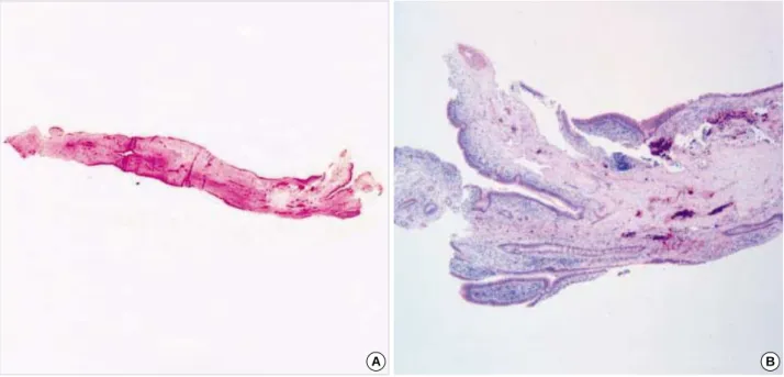
After bronchoscopic removal with biopsy forceps of the polyp, cystic lesion of the right upper lung disappeared (Fig. 1B) and the discomfort and pain in the right upper chest of patient improved. Gross appearance showed polyp with stalk and light-microscopical examination showed a lymphocyte infil- tration with epithelial lining cells that was consistent with the diagnosis of a bronchial inflammatory polyp (Fig. 4).
DISCUSSION
This is a rare case of polypoid intrapulmonary bronchogenic cyst, which was disappeared after bronchoscopic removal. There
Jung-Hoon Kim, An-Soo Jang, Jong-Sook Park, June-Hyuk Lee, Sung-Woo Park, Eun-Suk Koh*, Jai-Soung Park�, Choon-Sik Park
Department of Internal Medicine, Pathology*, Radiology�, Soonchunhyang University Hospital, Bucheon, Korea
Address for correspondence Choon-Sik Park, M.D.
Division of Allergy and Respiratory Medicine, Department of Internal Medicine, Soonchunhyang University Bucheon Hospital, 1174 Jung-dong, Wonmi-gu, Bucheon 420-021, Korea Tel : +82.32-621-5105, Fax : +82.32-621-5016 E-mail : [email protected]
*This work supported by a grant of the Korea Health 21 R4D Project, Ministry of Health & Welfare, Republic of Korea (01-PJ3-PG6-01GN04-003).
892 J Korean Med Sci 2005; 20: 892-4
ISSN 1011-8934
Copyright � The Korean Academy of Medical Sciences
Polypoid Endobronchial Lung Cyst with Bronchoscopic Removal : A Case Report
Pulmonary bronchogenic cyst in adults is rare and the typical appearance is a sharply circumscribed, round or oval nodule or mass, usually in the medial third of the lungs. Bronchial polyps are rare histopathologically distinct nonneoplastic endobronchial lesions and are classified as multiple papillomas, solitary papillo- mas, and inflammatory polyps. We herein report a patient with polypoid endo- bronchial lung cyst. A 68-yr-old woman presented with a discomfort and pain in the right upper chest of four weeks’ duration. Chest radiography revealed a cystic lesion in the right upper lung. Computed tomography revealed a 4××5 cm sized large cyst. Neither enlarged mediastinal lymph nodes nor extrabronchial involve- ments were observed. Flexible bronchoscopy revealed a peduncular polyp about 2 cm in length originating from the anterior segment of right upper lung. After bron- choscopic removal of polyp, cystic lesion of the right upper lung disappeared.
Key Words : Bronchial Cyst; Lung Disease; Polyp; Bronchus; Bronchoscopy
Received : 7 July 2004 Accepted : 22 September 2004
Polypoid Endobronchial Lung Cyst 893
were possible diagnoses for the initial radiographic chest find- ings of the cystic lesion in the right upper lung, including infected cyst, infected large bullae, pulmonary sequestration.
After bronchoscopic removal, the right upper cystic lesion disappeared immediately. Bronchogenic cysts are usually soli- tary, thin walled, unilocular, and roughly spherical in shape.
They are filled with either mucoid or serous fluid and do not communicate with the tracheobronchial tree unless they become infected, in which case the cyst fluid may be pus or
by pus and air (4). In this case cystic fluid is not aspirated during bronchoscopy. The cyst wall often contains cartilage and respiratory epithelium. Most are discovered incidentally and cause no symptoms. However they may communicate with the tracheobronchial tree and become infected and some enlarge to cause airway obstruction. Bronchial polyps are clas- sified as multiple papillomas, solitary papillomas, and inflam- matory polyps (3). Inflammatory polyps of the airways are now regarded as histopathologically distinct nonneoplastic
Fig. 1.Chest radiography shows a very large cyst in right upper lung (A) and a large cyst disappears after removal of endobronchial polyp but remains cystic wall (B).
A B
Fig. 2.Chest computed tomography reveals a 4×5 cm sized large cyst before polypectomy.
Fig. 3.Bronchoscopic examination shows a polyp at anterior seg- ment of right upper lung.
894 J.-H. Kim, A.-S. Jang, J.-S. Park, et al.
endobronchial lesions, which in adults are associated with a variety of chronic inflammatory insults such as chronic infec- tion, foreign bodies, asthma, or inhalation injury (5-7). Gener- ally, benign bronchogenic cysts need not be resected unless they cause symptoms. In treatment of bronchial polyp there are several methods such as bronchoscopic excision and lobec- tomy. The developments in bronchoscopic techniques have made it possible to remove superficial tumors by bronchoscopy.
Removal of the foreign body or inhalation of corticosteroid may resolve bronchial polyp (5). In the present case, bronchial polyp was removed by bronchoscopy and then lung cyst dis- appeared suggesting that intrapulmonary cyst communicate with bronchus. Histologically the bronchogenic cyst wall is lined by a pseudostratified, ciliated epithelium and contains cartilage and strands of smooth muscle important for diag- nosis. In the present case, polyp composed of epithelial lin- ing cells without cartilage in their wall may represent intra- pulmonary cyst as a result of ingrowth from communicat- ing airway epithelium.
REFERENCES
1. Rogers LF, Osmer JC. Bronchogenic cyst: A review of 46 cases. Am J Roentgenol 1964; 91: 273-90.
2. Ribet ME, Copin MC, Gosselin B. Bronchogenic cysts of the medi- astinum. J Thorac Cardiovasc Surg 1995; 109: 1003-10.
3. Dream JM, Douglas AC. Solitary papilloma of a bronchus. J Clin Pathol 1965; 18: 401-2.
4. Beecham JE. Fine needle aspiration biopsy of peripheral congenital bronchial cyst. Acta Cytol 1987; 32: 663-6.
5. Niimi A, Ikeda AT, Kubo Y, Tanaka E, Kuze F. Inflammatory bronchial polyps associated with asthma: resolution with inhaled corticosteroid.
Eur Respir J 1995; 8: 1237-9.
6. Roberts C, Devenny AM, Brooker R, Cockburn JS, Kerr KM. Inflam- matory endobronchial polyposis with bronchiectasis in cystic fibro- sis. Eur Respir J 2001; 18: 612-5.
7. Yong SJ, Won PS, Min KH, Soo KJ. A case of intratracheal polyp simulating asthma. Pediatr Allergy Respir Dis 2002; 12: 231-5.
Fig. 4.Gross appearance of bronchial polyp (A) and bronchial polyp composed of lymphocytes and epithelial lining cells (B). (Hema- toxylin-Eosin stain ×100).
A B