L-Aspartic Acid의 무질서하게 분포된 매질에서 파동전개와 변동에 관한 연구
김기준✝․이주엽
대진대학교 공과대학 화학공학과
(2012년 2월 28일 접수 ; 2012년 3월 22일 수정 ; 2012년 3월 25일 채택)
Studies on the Wave Propagation and Fluctuation in Randomly Ditribution Media of L-Aspartic Acid
Ki-Jun Kim✝․Joo-Youb Lee
Department of Chemical Engineering, Daejin University, Pochun 487-711, Korea (Received February 28, 2012 ; Revised March 22, 2012 ; Accepted March 25, 2012)
요약 : L-arpartic acid의 산란혼탁매질에서 형광, 산란과 응집의 영향은 파장과 산란된 형광세기 로 나타내는데, laser induced fluorescence(LIF) 분광학에 의한 분자특성으로 나타난다. 산란매질에 서 광학적 효과는 광학적 파라미터들((
μ
s,μ
a,μ
t)에 의해 표현되고 응집은 고-액상 분리공정에서 중 요하게 활용되고 있다. 따라서 입자가 서로 접근될 때 콜로이드 입자들의 상호작용을 LIF와 응집효 과로 분석하였다. 레이저 광원에서 검출기까지 농도의 함수에 의해 농도가 묽어짐에 따라 산란세기 가 기하급수적으로 감소함을 알 수 있다. 이는 유지화학, 생의학 생성물, 레이저 의학, 의공학 분야 적용에 LIF와 입자이동 현상은 아주 적합한 모델 연구에 큰 도움이 될 것이다.Abstract : The influences of fluorescence, scattering, and flocculation in turbid material by light scattering of L-aspartic acid, preservative and emulsifier were interpreted for the scattered fluorescence intensity and wavelength. They have been studied the molecular properties by the spectroscopy of laser induced fluorescence (LIF) and flocculation. The effects of optical properties in scattering media have been found by the optical parameters(
μ
s,μ
a,μ
t). Flocculation is an important step in many solid-liquid separation processes and is widely used. When two particles approach each other, interactions of several colloid particles can come into play which may have major effect on the flocculation and LIF process. The values of scattering coefficientμ
s are large by means of the increasing scattering of scatterer, The values have been found that the slope decays exponentially as a function of concentration from laser source to detector by our experimental result. It may also aid in designing the best model for oil chemistry, bio-pharmaceutical products, laser medicine and application of medical engineering on LIF and coagulation in particle transport mode.Keywords : L-aspartic acid, light scattering, laser induced fluorescence, optical parameters, flocculation, biopharmaceutical products.
✝
주저자 (E-mail : [email protected])
1. 서 론
In-situ 분석에서 활용되는 시료에 산란, 흡수 계수의 물리적 의미가 불확실하였으나, 기기분 석의 크나큰 발전에 의해 분석에 사용되는 산 란 횡단면과 흡수 횡단면[1-2]에 의해 광학적 매개변수[3]의 해석이 가능하게 되었다. 오늘 날 외과적인 수술시 종양조직을 완전히 제거하 지 못할 뿐만 아니라 방사선 치료와 약물 요법 에서 항암제의 대부분이 높은 독성을 가지고 있기 때문에 종양세포 뿐만 아니라 정상세포에 작용하여 부작용을 줄 수 있다[4-5]. 이러한 문 제점을 해결하기 위하여 항암제를 암조직에 선 택적으로 수송하여 치료함은 물론, 정상조직에 는 영향을 미치지 않게 하는 연구들이 많이 진 행되고 있다[6]. Dougherty 등이 광역학적 치 료(photodynamic therapy : PDT)에서 인체에 발생하는 많은 종양에 대한 새로운 치료 모델 링[7]을 제한하였으나, 지금까지 이 분야에 대 한 연구는 아직 초기 단계에 있다. 본 연구는 이 분야가 급격히 발전하게 될 LIF spectroscopy의 생의학 응용에서 종양의 진단 및 치료에 효과적으로 이용할 수 있도록 모델 링을 세우는 데 있다. 산란 매질에서 빛 에너지 전파가 의학의 진단학적 분광 분석의 발전과 치료적 레이저광 전달에 따른 방사량에서 PDT 의 임상실험과 광생물학 등의 여러 분야에 응 용함에 있어서 살아 있는 세포조직에서 산란 및 뭉침의 측정은 광학적 특성을 결정하는 데 가장 중요하다[8].
본 연구에 적합한 L-aspartic acid는 인체구 성뿐만 아니라 효소와 호르몬 등을 구성하는 가장 중요한 단백질요소로서 분자내에 카르복 실기와 아미노기를 가지는 화합물로 생물이 생 체내에서 합성하는 것이 L-아미노산으로 광학 활성을 가지고 있다.
산란과 응집에 의한 시료에 빛의 정확한 량 이 조사될 경우에 효과적일 수도 있으나, 비효 과적, 치명적인 손상을 입히게 될 수도 있다[9].
살아있는 세포의 분광분석에서 Beer의 법칙이 적용되기 어려운 "광학적으로 시료농도가 진한"
상태인 문제점들을 극복하고자 레이저, 검출기, 광섬유 및 단색화장치 등을 연결한 LIF 분광법 은 실시간 분석(real time analysis)과 현장분석 (in situ analysis)을 가능하게 하였다[10]. 또한 본 연구는 혼탁매질에서 L-Aspartic acid인 아
미노산(L-aspartic acid), 보존제(preservative) 와 유화제(emulsifier)의 시료에 대한 광원에서 검출기까지의 빛이 산란매질에서 확산과 이동 으로 변화하는 광학적 매개변수와 입자크기를 측정하였다.
2. 실 험
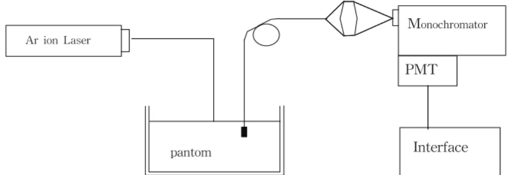
본 실험에서 아미노산인 L-aspartic acid(Acros Co., USA)를 사용하였으며, 단일파 장의 모드의 argon ion laser(Coherent Co., model No. Innova70, USA)를 이용하여 520 부 터 900 nm 사이의 형광을 측정하였다. Laser beam을 L-Aspartic acid, 보존제인 파라옥시안 식향산이소부틸(isobutyl-p-hydroxy benzoate, Sigma Co. USA), 유화제(emusifier)는 솔비탄 지방산 에스테르(sorbitan ester of fatty acid, Stamfords Co., Cytec, USA)을 사용하였다. 광 섬유(diameter : 4 mm)로 측정된 빛을 모아서 광전 증배관(photomultiplier tube)에서 검출하 며, 광자 계수기로 광자세기를 측정하였다. 실 험장치는 Fig. 1과 같다.
또한 연속파(continuous wave, CW)인 argon laser를 사용하여, Dye laser(Coherent Co., model No CR-599, USA)를 동기식 펌핑하였으 며, 형광염료는 RA-6G를 사용하였다. 산란된 빛은 광섬유를 통하여 원하는 빛만 검출기에 도달할 수 있는 중성밀도 필터(Esco Co., density 1.97, USA)를 지나 단색화 장치 (Monochromator, SPEX Co., model No 1870, 50 cm focal length, 1800 groove/mm)에 입사 하는데, Monochromator의 파장은 측미계인 Minidrive(SPEX Co., model No. 1872, USA)로 작동하며, Monochromator에 연결된 광전증배 관(Products for Research Co., model No RCA 31034A, USA)의 열적 노이즈를 줄이기 위해 수냉식과 DC 전원 공급장치(Power supply, Product for Research Co., model No.
TE104RF)를 이용하여, -20 ℃에서 실험하였다.
또한 디지털 광도계 (SPEX Co., model No.
DPC-2, USA), single photon amplifier discriminator(SPEX Co., model No. 453, USA), photon counter(Stanford Research Co., model No. SR400, USA)를 이용하여 실험하였 다. 데이터 처리 보드(Data Aquisition Board,
Ar ion Laser
pantom
M
onochromator
PMT
Interface
Fig. 1. Diagram of experimental equipment.
National Instrument Lad-PC) 등을연결하여 컴 퓨터 계간시스템을 구성하여, 300 rpm, 3 분동 안 교반하여 만든 L-aspartic acid, isobutyl-p-hydroxybenzoate인 보존제와 유화 제의 시료 농도를 변화시켜면서 매질의 산란세 기를 실험하였다. 시료를 담는 통은 표면반사, 투광도의 영향을 없애기 위해 크기 200 X 200 X 150 mm인 검정색 통을 사용하였으며, 시료 의 정밀 측정기기인 XYZ stainless steel multi axis stage(Newport Co., model No.
M-TSX-1A, USA)를 이용하여 입자파동을 측 정하였다. 본 실험에서 흡광계수(
μ
s), 산란계수 (μ
s), 감쇠계수(attenuation coefficient,μ
t =μ
s+
μ
a)를 측정하였다. 시료의 입자크기 분포 (particle size distribution)는 Hollow fiber module(Needham, A/G Technology Co., USA) 와 레이저 산란의 Coulter(Miami FL Co., model No. LS230, USA)로 측정하며, Hollow fiber module은 6개로 구성되어 있고, 길이 10.9 cm, 막표면은 20 ㎠이며, 공극크기는 0.2 ㎛인 정밀 여과막을 사용하였다.3. 결과 및 고찰
L-aspartic acid인 아미노산의 산란계수를 결 정하기 위해서 광원과 검출기까지 파동변화에 따라 흡수와 산란된 빛의 양을 측정하였으며, Added Absorber Technique를 사용하여 감쇠 계수(
μ
t =ρσ
t)는 산란 입자만 존재하기 때문에 산란 계수(μ
s)에 일치한다[12]. 전체 감쇠 계수 측정은 단일입자 산란만이 일어나도록 하기 위 해 묽은 농도에서 실험하였고, 산란되지 않고나오는 광자를 검출하기 위해서 아이리스를 사 용하였다. Fig. 2는 광원에서부터 검출기까지 L-aspartic acid와 isobutyl-p-hydroxybenzoate 에 대한 LIF.변화에 의해 측정되어지는 scattering spectra를 나타내었다. 이 값들은 L-aspartic acid의 산란세기가 isobutyl-p- hydroxybenzoate보다 증폭되어 크게 측정된 값 들이다.
Fig. 2. Scattering spectra of (a) L-aspartic acid, (b) isobutyl-p-hydroxybenzoate measured by means of LIF.
산란체인 아미노산에 유화제인 sorbitan ester
of fatty acid 1.0 wt%을 혼합하여 초음파 처리 한 시료의 농도 대 광자세기(I/I0)의 기울기로 부터 얻은
μ
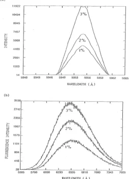
s값은 Table 1과 같다. 이는 광원에 서 검출기까지의 산란체의 농도에 따라 산란이 많이 일어날수록 투과가 많이 일어나고, 반면에 광원에서 검출기까지의 산란체의 농도가 묽을 수록 산란에 의해 축상 광자세기가 크게 작아 짐을 알 수 있다. 이는 L-aspartic acid 농도가 진해짐에 따라 검출기에 닿는 비율이 적어짐을 의미하는 데, 매질 층이 두꺼워서 산란이 증가 함을 예상할 수 있다. 이 실험은 광자가 시료 속에 입사될 때부터 검출기에 도달할 때까지의 농도에 대하여 레이저를 이용한 광원과 광섬유 간의 깊이를 1.0 cm에 고정시키고, 농도가 각각 1.0 %, 2.0 %, 3.0 %, 4.0 % 따라 측정한 형광 세기를 도시하였다. 위 결과로부터 광자의 분포 를 조사하면 매질에서 농도가 진할수록 산란세 기가 증가함을 알 수 있었다.Fig. 3. The detected scattering and fluorescence lineshapes with different concentrations of (a): L-aspartic acid 3.0 wt%, (a): L-aspartic acid 3.0 wt%, 2.0 wt%, 1.0 wt%, (b): sorbitan ester of fatty acid 3.0 wt%, 2.0 wt%, 1.0 wt%.
Table 1. Measured Mean Values versus Optical Parameters of L-aspartic acid 3.0 wt%, 2.0 wt%, 1.0 wt% + isobutyl-p-hydroxybenzoate 1.0 wt%
I
μ
t(mm-1) Concentration(wt%) 8,19312,168 18,363 22,971
09,974 12,785 19,034 25,911
1.0 2.0 3.0 4.0
Fig. 3은 유화제인 sorbitan ester of fatty acid를 상온에서 광원에서 검출기까지의 농도별 로 흡수되지 않고 투과한 빛의 세기를 측정한 결과를 도시한 것이다. L-aspartic acid가 유화 제인 sorbitan ester of fatty acid보다 빛의 세 기가 매우 크게 측정되었다. (a)와 (b)에서도 농 도가 진함에 따라 세기가 크게 나타났다. Table 2에서는 L-aspartic acid 3.0 wt%, 2.0 wt%, 1.0 wt% + sorbitan ester of fatty a챠d 1.0 wt%인 시료에 대한 산란강도 및 감쇠계수 값 을 측정하여 나타낸 값들이다. 이것은 광자가 효소의 시료 속에 입사될 때부터 검출기에 도 달할 때까지의 농도에 대하여 레이저를 이용한 광원과 광섬유간의 깊이를 1.0 cm에 고정시키 고, 아미노산의 농도 차이에 따라 측정한 빛의 세기로 측정한 값들이다. 광자의 분포를 조사하 면 매질에서 농도가 진할수록 산란세기가 증가 하였으나, Fig. 3에서 L-aspartic acid의 시료보 다 작은 유화제의 영향에 따른 세기를 나타낸 것은 산란이 적게 발생한 것으로 사료된다.
Table 2. Measured Mean Values versus Optical Parameters of L-aspartic acid 3.0 wt%, 2.0 wt%, 1.0 wt% + sorbitan ester of fatty aicd 1.0 wt%
I
μ
t(mm-1) Concentration(wt%) 5,0112,953 839
22,195 6,028 1,279
1.0 2.0 3.0
Table 3. Experimental Values of Optical Parameters on Scattering Media at 595nm Wavelength
species(medium) I
μ
t(cm)δ
t concentration(wt%) AA + SEF 1.0wt% 5023 21.563 0.148 1.0AA + SEF 2.0wt% 4917 13.425 0.293 2.0 AA + SEF 3.0wt% 3702 9.167 0.334 3.0 AA + SEF 4.0wt% 2956 7.378 0.591 4.0 AA + IBHB 1.0wt% 4023 11.563 0.343 1.0 AA + IBHB 2.0wt% 5271 23.415 0.139 2.0 AA + IBHB 3.0wt% 6767 34.167 0.118 3.0 AA + IBHB 4.0wt% 7439 47,382 0.086 4.0 AA: L-aspartic acid, SEF: sorbitan ester of fatty acid,
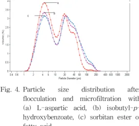
IBHB: isobutyl-p-hydroxybenzoate Fig. 4. Particle size distribution after
flocculation and microfiltration with (a) L-aspartic acid, (b) isobutyl-p- hydroxybenzoate, (c) sorbitan ester of fatty acid.
아미노산의 농도가 증가함에 따라 산란세기 가 증가하고, 뭉침에 의해 빛의 흡수가 강해진 다. 이는 투과된 빛의 세기가 약해지는 현상이 다. 이 현상은 광자가 상호 작용하여 매질의 농 도가 클수록 입자간의 거리[13], 즉 MFP(mean free path)가 작아지며 축방향 투광도 역시 작 아짐을 확인하였다.
또한 광역학적 작용이란 어떤 물질이 빛을 흡수하면 산화될 수 있는 조건(dye + light + oxygen)하에서 생물학적 분자계를 변화시키거 나 파괴시키는 작용을 의미하며, 일중항 산소에 의해 PDT결과를 얻어 낼 수가 있다[14].
Fig. 4는 300 rpm, 3분동안 교반한 다음, Hollow fiber module에 의한 정밀여과를 통과 한 시료의 입자크기에 대한 응집된 부피를 측 정하였다. (a)는 L-aspartic acid 입자크기를 나 타내었다. 이는 글루코스의 산란세기가 증가함 에 따라 부피(%)는 증가하였으나, 입자크기의 직경은 점점 감소한 것으로 나타났다. (b)는 isobutyl-p-hydroxybenzoate, (c) sorbitan ester of fatty acid의 입자크기 분배를 측정한 그래 프이다. 이것은 산란세기가 감소함에 따라 부피 는 감소하였으나, 입자크기의 직경은 점점 증가 하였음을 알 수 있다. 이 결과는 혼탁매질 속에 서 위치함수로 산란이 증가함에 따라 MFP는 짧아지며, 충돌에 의해 에너지 흐름이 많아져서 감쇠계수가 증가한 현상으로 사료된다.
Table 3은 L-aspartic acid(AA) + sorbitan ester of fatty acid(SEF 1.0 wt%, 2.0 wt%, 3.0 wt%) 와 AA + isobutyl-p- hydroxybenzoate (IBHB 1.0 wt%, 2.0 wt%, 3.0 wt%)에 대한 I,
μ
t 와δ
t 등을 측정하여 나타낸 값들이다.여기에서 SEF와 IBHB 농도가 진함에 따라 I 와
μ
t는 감소하나δ
t값은 증가하였다. 광증감 제로 사용되는 isobutyl-p-hydroxybenzoate를 첨가한 L-aspartic acid와 보존제에 각각 광학적 매개변수들을 측정하여 나타내었다.
L-aspartic acid의 경우가 보존제의 경우보다 농도가 진할수록 산란세기와 감쇠계수가 크게
나타났으며, 침투깊이(
δ
= 1 /μ
t)는 감소함을 알 수 있었다.SEF이 IBHB보다 I와
μ
t의 측정치보다 크 게 나타났으나,δ
t 측정치는 작게 나타났다. 이 는 산란이 증가되어 나타낸 결과로 생각이 된 다. 유화된 량은 유화물질의 흡착매(absorbent) 의 단위면적, 단위질량 당에 흡착분자의 물질량 이 수소결합을 형성하고 정전기적 힘이 흡착분 자, 분자표면하고 접촉으로 분산력이 발생하고 전자의 이동으로 결합된 . B접촉r법칙에서 벗어 난 결과로서 L-Aspartic acid의 입자가 많을수 록 광자세기가 증가하였으나, 유화가 많이 발생 할수록 광자세기가 작아진 것은 역 반사의 영 향이라고 사료된다.5. 결 론
본 연구에서 LIF에 의한 L-aspartic acid, isobutyl-p-hydroxybenzoate, sorbitan ester of fatty acid에 의한 광학적 파라미터를 측정한 결과 다음과 같은 결론을 얻었다.
1. 산란매질에 레이저 광이 입사됨에 따라 광자 를 검출하는 광섬유까지의 L-aspartic acid의 농도가 진할수록 산란세기가 증가하였으며, L-aspartic acid의 농도가 묽을수록 산란세기 가 작아짐을 알 수 있었다.
2. 무질서한 산란매질에서 L-aspartic acid 시료 가 isobutyl-p-hydroxybenzoate시료보다 산 란계수기가 더 증가하였으며, L-aspartic acid의 농도가 묽을수록 산란계수가 작아짐 을 알 수 있었다.
3. 광자 축적에 따른 공간적 침투에 대한 광자 의 발견확률, 침투거리 (
δ
)는 L-aspartic acid + isobutyl-p-hydroxybenzoate의 농도가 묽 음에 따라 L-aspartic acid + sorbitan ester of fatty acid의 경우보다 더 크게 나타났으 나, I 와μ
t는 작게 측정되었다.4. 빛의 파장에 의해 광자의 확산과 열적반응을 뭉침과 정밀여과 공정을 거친 후에 입자크기 분포에서 L-aspartic acid, isobutyl-p- hydroxybenzoate, sorbitan ester of fatty acid의 시료순으로 입자크기가 나타났다.
참고문헌
1. K. J. Kim and K. C. Sung, A Study on Spectra of Laser Induced Fluorscence in Phantom, J. of Korean Oil Chemist' Soc,, 16(4), 329 (1999).
2. K. J. Kim and K. C. Sung, Monte Carlo Simulation on Light Distribution in Turbid Material, J. of Korean Oil Chemists Soc., 15(4), 11 (1998).
3. K. J. Kim and K. C. Sung, Studies on Measurements of Optical Parameters in Turbid Material by Light Scattering, J. of
Korean Oil Chemists Soc., 12(2), 151
(1995).4. S. L. Jacques and S. A. Prahl, Modeling Octical and Thermal Distribution in Tissue During Laser Irradiation, Laser in
Surgery and Medicine 6, 494 (1987).
5. B. C. Wilson, Y. D. Park, Y. Hefetz, M.
S. Patterson, S. J. Madsen and S. L.
Jacqu es, The Potential of Timeresolved Reflectance Measurements for the Noninvasive Determination of Tissue Optical Properties, in Thermal and Optical Interactions with Biological and Related Composite Materials, M. J. Berry and G.
M. Harpole, eds., Proc. S. P. I. E., p. 97, (1989).
6. S. L. Jacques, C. A. Alter, and S. A.
Prahl, Angular Dependence of HeNe Laser Light Scattering by Human Dermis, Laser
in the Life Science, 1(4), 309 (1987).
7. T. J. Dougherty, C. J. Gomer, and K. R.
Weishaupt, Energetics and Effeciency of Photoinactivation of Murine Tumor Cells Containing Hematoporphyrin,
Cancer Research 36, 2330 (1976).
8. R. R. Alfano, G. C. Tang, A. Pradhan, W.
Ran, S. J. Daniel, ,and E Opher, Fluor- escence Spectro from Cancerone and Normal Human Breast and Lung Tissues,
IEEE J. Quan. Elec. QE, 23(10), 1806
(1987).9. S. R. Wickramasinghe, Y. Wu, and B.
Han, Enhanced Microfiltration of sorbitan ester of fatty acid by Flocculation,
Desalination 147, 25, (2002).
10. L. O. Poicard, C. J. Gomer, and A. E.
Profio, Laser-Induced Hyperthermia of Ocular Tumors, Appl. Opt., 28(12), 2318 (1989).
11. M. Winkelmann, B. Chance, and B. C.
Wilson, Time Resolved Reflectance and Transmittanance for the Noninvasive Measurement of Tissue Optical Properties, Appl. Opt., 28(12), 2331 (1989).
12. K. Lee, I. J. Forbes, and W. H. Betts, Oxygen Dependency of Photocytotoxicity with Hematoporphyrin Derivative,
Photochem. Photobiol 39(5), 631 (1984).
13. R. Hilf, P. B. Leakey, S. J. Solltt, and S.
L. Gibson, Photodynamic Inactivation of R3230AC Mammary Carcinoma in vitro with isobutyl-p-hydroxybenzoate: Effects of Dose, Time and Serum on Uptake and Phototoxicity,
Photochem. Photobiol.
37(6), 633 (1983).
14. H. Schnecknburger, M. Frenz, Y.
Tsnchiya, U.. Denzer, and L. Schleinkofer, Picosecond Fluorescence Microscopy for Measuring Chlorophyll and Porphyrin Components in Conifers and Cultured Cells, Lasers in the Life Science 1(4), 299 (1987).
15. S. R. Wickramasinghe, Y. Wu, and B.
Han, Enhanced Microfiltration of sorbitan ester of fatty acid Flocculation,