박봉욱∙하영술
1∙김진현
1∙조희영
1∙정명희
1∙김덕룡
2∙김욱규
3∙김종렬
4∙장중희∙변준호
경상대학교 의학전문대학원 구강악안면외과학교실, 경상대학교 건강과학연구원, 의생명과학사업단(BK21),
1
경상대학교병원 임상의학연구소,
2경상대학교 의학전문대학원 생화학교실, 경상대학교 건강과학연구원, 의생명과학사업단(BK21),
3
부산대학교 치의학전문대학원 구강악안면외과학교실,
4온 종합병원 턱얼굴센터
혈관내피세포 채취의 원천으로 인간 지방조직의 활용
Use of Human Adipose Tissue as a Source of Endothelial Cells
Bong-Wook Park, Young-Sool Hah
1, Jin-Hyun Kim
1, Hee-Young Cho
1, Myeong Hee Jung
1, Deok Ryong Kim
2, Uk-Kyu Kim
3, Jong-Ryoul Kim
4, Jung-Hui Jang, June-Ho Byun
Department of Oral and Maxillofacial Surgery, Gyeongsang National University School of Medicine and Institute of Health Sciences, Biomedical Center (BK21),
1Clinical Research Institute, Gyeongsang National University Hospital,
2
Department of Biochemistry, Gyeongsang National University School of Medicine and Institute of Health Sciences, Biomedical Center (BK21), Jinju, Korea,
3Department of Oral and Maxillofacial Surgery, School of Dentistry, Pusan
National University, Yangsan, Korea,
4Maxillofacial Center, Onhospital, Pusan, Korea
Purpose: Adipose tissue is located beneath the skin, around internal organs, and in the bone marrow in humans. Its main role is to store energy in the form of fat, although it also cushions and insulates the body. Adipose tissue also has the ability to dynamically expand and shrink throughout the life of an adult.
Recently, it has been shown that adipose tissue contains a population of adult multipotent mesenchymal stem cells and endothelial progenitor cells that, in cell culture conditions, have extensive proliferative capacity and are able to differentiate into several lineages, including, osteogenic, chondrogenic, endothelial cells, and myogenic lineages.
Materials and Methods: This study focused on endothelial cell culture from the adipose tissue. Adipose tis- sues were harvested from buccal fat pad during bilateral sagittal split ramus osteotomy for surgical correc- tion of mandibular prognathism. The tissues were treated with 0.075% type I collagenase. The samples were neutralized with DMEM/and centrifuged for 10 min at 2,400 rpm. The pellet was treated with 3 vol- ume of RBC lysis buffer and filtered through a 100 μm nylon cell strainer. The filtered cells were cen- trifuged for 10 min at 2,400 rpm. The cells were further cultured in the endothelial cell culture medium (EGM-2, Cambrex, Walkersville, Md., USA) supplemented with 10% fetal bovine serum, human EGF, human VEGF, human insulin-like growth factor-1, human FGF-β, heparin, ascorbic acid and hydrocorti- sone at a density of 1 × 10
5cells/well in a 24-well plate. Low positivity of endothelial cell markers, such as CD31 and CD146, was observed during early passage of cells.
Results: Increase of CD146 positivity was observed in passage 5 to 7 adipose tissue-derived cells. However, CD44, representative mesenchymal stem cell marker, was also strongly expressed. CD146 sorted adipose tissue-derived cells was cultured using immuno-magnetic beads. Magnetic labeling with 100 μl microbeads per 10
8cells was performed for 30 minutes at 4℃ a using CD146 direct cell isolation kit. Magnetic separa- tion was carried out and a separator under a biological hood. Aliquous of CD146+ sorted cells were evalu- ated for purity by flow cytometry. Sorted cells were 96.04% positivity for CD146. And then tube formation
Abstract
서 론
줄기세포와 관련하여 가장 많이 알려진 원천세포는 골수 기원줄기세포(bone marrow-derived mesenchymal stem cells)이다. 주로 흡입(aspiration) 방법을 통하여 채 취되는 골수기원줄기세포가 다능성 세포로서 조골세포, 연 골세포 및 지방세포등으로 분화할 수 있는 능력을 가지고 있다는 것은 이제 보편적인 사실이나 술후 동통의 증대, 감 염 가능성 및 채취시 세포수의 부족함과 같은 단점과 함께, 특히 임상적으로 치과분야에서 이를 채취, 이용시 번거로움 과 같은 것으로 인하여 줄기세포의 다른 원천들에 대한 연 구가 많이 진행되고 있다.
골수와 마찬가지로 지방조직도 중배엽 기원 조직으로 풍 부한 지방세포 뿐 아니라, 대식세포 및 평활근 세포등을 포 함하여 다양한 세포들을 함유하고 있으며 줄기세포 획득의 원천으로 최근에 많이 보고되고 있다.
1-4)지방조직에는 특이 적으로 지방세포를 둘러싸고 있고 기질-혈관 분획 세포 (stromal-vascular fraction cells)라 불리우는 비특정 세 포군(non-characterized cells population)이 존재하는데 이 기질-혈관 분획 세포부분에는 자가 증식 능력이 있고 특 정세포들로 분화할 수 있는 줄기세포 혹은 전구세포라 불리 우는 세포들이 존재하여 특정 조건하에서 지방세포 뿐 아니 라 조골세포, 연골세포 및 근육세포등으로 분화될 수 있다 고 보고되고 있다. 특히 지방조직 기원 줄기세포(adipose tissue-derived stem cells)로부터 조골세포로의 분화에 대해서는 이미 많은 연구가 진행된 상태라 할 수 있다.
5-7)뿐 만 아니라 Zuk 등
8)에 의하면 각종 표지자(markers)들을 이용한 연구에서 지방조직기원 줄기세포가 신경세포 표현 형도 나타내는 것을 관찰하여 중배엽 뿐 아니라 외배엽 조 직으로의 분화도 보고되고 있다.
골 조직은 복잡하면서도 고도로 혈관화 되어있는 조직이 므로 골의 재형성이나 골절 치유등과 관련하여 가장 중요하 게 고려하여야 할 요소중의 하나는 활성화된 혈류공급이라 할 수 있다. 혈관계(vasculature)는 산소, 영양물 및 각종 인자들을 신체 조직에 운반하며 성숙화된 혈관 구조의 발달 이 기관형성(organogenesis)의 가장 초기에 일어나는 현상
이라 할 수 있다. 일반적으로 골 손상이 있을 경우, 관련 부 위 혈관손상으로 산소부족에 따른 저산소 상태가 초래되어 이에 대한 보상 반응으로 혈류회복을 통한 산소증가를 위하 여 혈관신생 기전이 발전하게 된다. 그러므로 사실 골의 재 형성이 일어나기 전, 혈류의 회복은 골의 재형성에 가장 중 요한 요소중의 하나가 되는 것이다. 그러므로 적절한 혈류 공급이 골 조직에 제공되지 못할 경우 골격성 병변을 야기 할 수 있다.
9-12)이를 고려하여 골 조직공학과 관련된 최신 경향은 다공성의 담체에 미리 혈관형성을 이루어 혈관화 담 체 형성물(vascularized scaffold construct)을 제작하고 이에 골 전구세포를 적용하여 골재생(osteogenesis)시 미 리 형성된 혈관재생(angiogenesis)을 통하여 골형성이 더 더욱 잘 일어날 수 있게 하는 것이라 할 수 있다. 최근 일부 의 연구에서 골수기원줄기세포를 혈관내피세포(endothe- lial cells)와 함께 합동배양(coculture)시 골수기원줄기세 포가 조골세포로 더 잘 분화됨이 보고되고 있다. 이러한 것 들을 고려할 때, 어떤 원천이 되는 조직으로부터 혈관내피 세포를 쉽게 추출, 배양, 증식시켜 적절한 방법으로 다공성 담체에 적용하여 혈관화된 담체를 형성한다면 골 조직공학 의 본연의 목표를 더 잘 이루리라 사료된다.
13,14)이에 본 연구에서는 골 조직공학을 위하여 적용될 수 있는 담체에 혈관내피세포를 적용하기 위한 전 실험으로 지방조 직으로부터 혈관내피세포를 분화시키고자 한다. 일반적으 로 혈관내피세포에 대한 세포배양이 쉽지 않은 상황에서, 쉽게 접근할 수 있는 원천으로 혈관내피세포를 추출하고 활 용한다면 조직공학의 부가적인 장점을 제공하는 것이라 할 수 있다. 구강내에서 간단한 시술을 통하여 협부 지방체 (buccal fat pad)는 쉽게 얻을 수 있다. 치과적으로 국소마 취하에 매복치 발치 등을 포함한 일반적인 시술을 통하여 쉽게 채취할 수 있는 골막에서 골막기원세포를 얻어 조골세 포로 분화시켜 활용하는 것과 마찬가지로 구강내 간단한 시 술을 통하여 협부 지방체를 채취하여 그 내부에 존재하는 혈관내피세포를 추출할 수 있다면, 그 세포를 활용할 수 있 는 어떤 조건에서는 조직공학적으로 큰 잇점을 제공하는 것 이라 할 수 있다. 이에 본 연구에서는 협부 지방체로부터 혈 관내피세포의 추출과 관련된 현상을 관찰하고자 하였다.
was examined. These CD146 sorted adipose tissue-derived cells formed tube-like structures on Matrigel.
Conclusion: These results suggest that adipose tissue-derived cells are endothelial cells. With the fabrica- tion of the vascularized scaffold construct, novel approaches could be developed to enhance the engineered scaffold by the addition of adipose tissue-derived endothelial cells and periosteal-derived osteoblastic cells to promote bone growth.
Key words: Adipose tissue, Endothelial cells, Adipose tissue-derived endothelial cells
연구 재료 및 방법
1. 협부 지방체로부터 혈관내피세포의 채취
협부 지방체의 채취는 하악골 시상 분할술 과정동안 채취 하였다. 채취한 지방 조직을 인산염 식염수에 세척한 후, 1 시간정도 0.075% 제1형 콜라겐분해효소(Sigma-Aldrich, St. Louis, USA) 처리하였다. 동일량의 Dulbecco's mod- ified Eagle's medium/10% fetal bovine serum (DMEM/10% FBS)을 통하여 콜라겐 분해효소를 비활성 화 시킨 후, 10분간 2,400 rpm에서 원심분리를 실시하였 다. 세포층을 DMEM/10% FBS로 부유시키고 100-μm 나 일론 세포여과채에서 여과시켰다. 여과된 세포들을 다시 10분간 2,400 rpm에서 원심분리를 시행하였다. 인산염 식 염수에 세척한 후, 세포들을 1 × 10
5cells/well의 밀도로 2 μg/cm
2로 fibronectin이 코팅되어 있는 24-well plate에 주입하고 FBS, human epidermal growth factor, human vascular endothelial growth factor, human insulin-like growth factor-1, human fibroblast growth factor-B, heparin, ascorbic acid 및 hydrocortisone 등 이 포함된 혈관내피세포 유도배지인 혈관내피세포 유도배 지-2 (EGM-2, Cambrex, Walkersville, Md., USA)에 서 배양하여 혈관내피세포를 추출하였다.
2. 세포 표면 표지자 분석
배양 후, 관련 세포들을 트립신 처리하고 1 × 10
5개 정도 의 세포들을 30분간 얼음에 일차항체와 배양시켜 조혈세포 표지자에 대한 세포 표면 표지자 분석(Cell Surface Markers Analysis)을 시행하였다. 조혈세포 표지자로는 CD31과 CD146을 이용하였으며 간엽 줄기세포 표지자로 는 CD44, CD90, 그리고 CD166를 이용하였다. 이들에 대 한 Fluorescein isothiocyanate (FITC)-conjugated 항체 를 이용하고 mouse IgG-FITC를 음성 대조군으로 이용하 였다.
3. CD146 양성 세포의 분리
지방조직은 간엽조직으로 세포배양 과정을 거치더라고 조 직에 포함된 모든 세포들이 완전히 혈관내피세포등의 조혈 세포만 발현되는 것은 아니므로 조혈세포의 특이성을 다수 가지고 있는 세포만을 추출하기 위하여 자기 세포분리 (magnetic cell sorting)를 이용하여 CD146에 양성인 세 포들을 추출하였다. CD146 MicroBead Kit (Miltenyi Biotec, CA, USA)를 사용하여 1 × 10
8개의 세포당 100 μ l microbeads로 labeling을 시행하고 column과 분리기를
이용하여 자기 세포분리를 시행하였다.
4. CD146 양성 세포의 matrigel에 의한 혈관내피세 포 세포망 형성
자기 세포분리를 통해 획득한 CD146 양성 세포들이 혈 관내피세포라 표현할 수 있을지, 관모형(tube formation) 형성정도로 평가하였다. 일반적으로 혈관내피세포는 이의 기질역할을 하는 matrigel을 적용할 경우, 3차원적으로 혈 관 형태인 관모형을 형성하는 것으로 알려져 있다. 그러므 로 CD146 양성 세포들이 matrigel에 적용되었을 경우, 일 반적인 혈관내피세포처럼 관모형을 형성하는 지를 관찰하 였다. Chamber slides를 matrigel (BD bioscience, CA, USA)로 입히고 1시간 정도 지난 후, chamber slides에 well당 2 × 10
4세포들을 주입하였다. 48시간 후, 세포망 형성정도를 관찰하였다. HUVEC 세포를 양성 대조군으로 사용하였고 음성 대조군으로는 대장암 세포주를 이용하였 다.
연구 결과
1. 세포 표면 표지자 분석
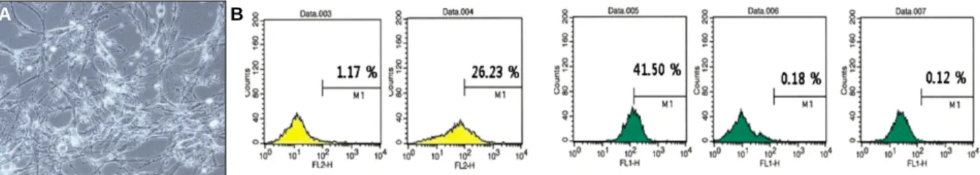
지방조직에서 추출한 세포들은 계대배양이 지속되면서 조 혈세포 표지자중 CD146의 발현은 증가를 관찰하였다. 대 체적으로 passage 5내지 7단계에서 세포들의 CD146 발현 이 증가되어 나타남을 관찰하였다. 계대배양을 지속하면서 조혈세포 표현형의 발현이 강하게 나타나는 passage 5내지 7단계에서 간엽 줄기세포 표현형의 발현도 일부 유지되는 것을 관찰하였다(Figs. 1-5).
2. CD146 양성 세포의 분리
계대배양을 지속하면서 조혈세포 표현형의 발현이 증가되
는 시기인 passage 5내지 7단계에서 간엽 줄기세포 표현형
의 발현도 일부 유지되는 것은 추출한 세포들이 혈관내피세
포의 특성을 나타내지만 원천적으로 간엽조직이므로 간엽
줄기세포 표현형의 발현이 완전히 없어지지는 않음을 알 수
있었다. 이러한 점을 고려하고 passage 5내지 7단계에서
조혈세포 표지자중 발현이 뚜렷한 CD146에 대한 양성 세
포를 자기 세포분리 원리를 이용하여 추출하였다. 자기 세
포분리한 CD146 양성 세포들은 CD146에 96% 이상의 양
성을 나타내었다(Fig. 6). 이를 통하여 자기 세포분리한
CD146 양성 세포들은 혈관내피세포의 특성을 상당히 가진
다고 할 수 있다.
Fig. 2. Representative photograph of passage 3 adipose tissue-derived cells. (A) Cellular morphology. (B) FACS analysis of cul- tured human adipose tissue-derived cells. Low positivity of endothelial cell markers was observed in passage 3 adipose tissue- derived cells.
A B
Fig. 3. Representative photograph of passage 5 adipose tissue-derived cells. (A) Cellular morphology. (B) FACS analysis of cul- tured human adipose tissue-derived cells. Increase of positivity for endothelial cell markers was observed in passage 5 adipose tissue-derived cells. However, CD44, representative mesenchymal stem cell marker, was also strongly expressed.
A B
Fig. 4. Representative photograph of passage 7 adipose tissue-derived cells. (A) Cellular morphology. (B) FACS analysis of cul- tured human adipose tissue-derived cells. Positivity for endothelial cell markers was moderately observed in passage 7 adipose tissue-derived cells. However, expression of CD44 was not clearly decreased.
A B
Fig. 5. Representative photograph of passage 9 adipose tissue-derived cells. (A) Cellular morphology. (B) FACS analysis of cul- tured human adipose tissue-derived cells. Expression of both of endothelial cell markers and mesenchymal stem cell markers was decreased in passage 9 adipose tissue-derived cells.
A B
Fig. 1. Representative photograph of passage 1 adipose tissue-derived cells. (A) cellular morphology. (B) FACS analysis of cul- tured human adipose tissue-derived cells. Low positivity of endothelial cell markers was observed in passage 1 adipose tissue- derived cells.
A B
3. CD146 양성 세포의 matrigel에 의한 혈관내피세 포 세포망 형성
자기 세포분리된 CD146 양성 세포들이 진정한 혈관내피 세포라 표현할 수 있을지, 관모형 형성정도로 평가하였다.
HUVEC 세포를 양성 대조군으로 사용하였고 음성 대조군 으로는 대장암 세포주인 SW620 세포를 이용하였다. 관찰 결과, 자기 세포분리된 CD146 양성 세포들도 HUVEC 세 포와 마찬가지로 matrigel을 적용할 경우, 일반적인 혈관형 태인 관모형을 형성하는 것으로 관찰되었으며 음성 대조군 인 SW620 세포는 matrigel이 적용되더라도 관모형을 형 성하지 못하였다(Fig. 7).
고 찰
골신생과 혈관신생사이의 밀접한 상호작용이 골의 재형성 이나 골절의 치유과정에서 중요한 역할을 하는 것은 주지의 사실로 여러 동물 모델에서 이에 대한 연구가 지속적으로 이루어지고 있다. Gerber 등
15)에 의하면 대표적인 혈관신 생인자인 혈관내피세포성장인자의 내인성 발현이 억제될 경우 골형성의 장애가 유도된다고 하였으며 Maes 등
16)은
혈관내피세포성장인자 isoforms이 부족한 마우스의 모델에 서 혈관계 및 골형성/연골형성 프로그램의 손상이 나타났다 고 보고하였다.
골 조직공학의 궁극적인 목표는 정상적인 골조직과 유사 한 조직을 형성하는 것이므로 이를 위해서는 적절한 담체가 필요하다. 이상적인 담체는 적용된 후, 주위조직과의 생물 학적 반응동안 생체적합성(biocompatibility)과 서서히 체 내에서 분해되며 자가조직으로 치환되는 생분해성 (biodegradability)을 가지고 있는 것이라 할 수 있으며 이 에 덧붙여 조직공학적으로는 배양된 세포와의 상호작용을 통하여 관련 세포의 표현형(phenotype)의 발현에 긍정적 인 효과를 제공하여야 한다. 또한 배양된 세포가 담체로 유 입되어 활성을 나타내기 위하여 적절한 다공성 구조 및 적 절한 다공 크기를 가지고 있어야 한다.
17-19)최근에는 골 조 직공학에서 혈관계의 활용을 이용한 혈관화 담체 형성물을 제작하고 이를 기초로 하여 골 전구세포에서 분화시킨 조골 세포를 적용하여 골재생시 미리 형성된 혈관재생을 통하여 골형성을 촉진하는 것이 보고되고 있다. 본 연구에서는 이 러한 점을 고려하여 혈관내피세포 채취의 원천으로 지방조 직을 이용하였다.
조골세포로의 분화를 포함하여 최근 조직공학의 여러분야
Fig. 6. CD146 sorted-adipose tissue-derived endothelial cells.
A B
CD146 sorted cells HUVEC cells SW620 cells
Fig. 7. Tube formation of adipose tissue-derived endothelial cells.
에서 지방조직의 활용이 부각되고 있다. 인간의 지방조직에 는 지방세포를 둘러싸고 있으며 기질-혈관 분획 세포라 불 리우는 비특정 세포군이 있어 특정 조건하에서 조골세포, 연골세포 및 근육세포등으로 분화될 수 있다고 보고되고 있 다. 조골세포 획득의 원천으로 지방조직의 이용에 대한 보 고가 증가되면서 골 조직공학에서 지방조직의 활용에 대한 보고가 증가하고 있다.
1-3)최근에는 지방조직의 기질-혈관 분획 세포가 간엽 세포군 뿐 아니라, 조혈 세포 채취의 원천 으로 가능함이 알려지고 있다.
5-7)또 다른 조혈 세포 채취의 원천으로 인간 제정맥 혈관내피 세포(human umbilical vein endothelial cells, HUVEC)의 이용이 주목받고 있다. 인간 제정맥 혈관내피 세포가 신체의 비자가세포에 대한 거부반응에서 중요한 역 할을 하는 MHC (major histocompatibility complex) 항 원에 대하여 극히 낮은 수준을 나타내어 다방면의 치료 영 역에서 활용가치가 높다고 알려져 있고 상업적으로도 쉽게 구할 수 있어 조직공학적 연구 뿐 아니라 치료영역에서도 많이 이용되고 있다. 그러나 실지 이는 태아와 관련된 파생 조직이라 할 수 있으므로 신체의 일반적인 내피계 (endothelial system)와는 다소 다른 비전형적 산물이다.
내피(endothelium)는 상당할 정도로 이질성을 가지고 있 으며 대혈관계 및 미세혈관계(macrovascular and microvascular system) 혈관내피세포는 다소 다른 조직학 적 특성을 나타낸다고 알려져 있다. 그러나 사실 인간의 모 든 혈관내피세포의 95%는 미세혈관 혈관내피세포 (microvascular endothelial cells)이므로 혈관내피세포를 응용하여 의학과 관련된 생리학적/병리학적 상태를 연구하 는 모델에서는 미세 혈관내피세포를 이용하는 것이 유리하 다고 할 수 있다. 최근 이와 관련하여 지방조직에 존재하는 미세혈관 혈관내피세포가 혈관내피세포를 응용하는 여러 분야에서 좋은 성과를 제공해 주고 있다. 지방조직은 상대 적으로 고도로 혈관화 되어 있으며 지방세포를 제외하고 간 질 부분이 비교적 적어 혈관내피세포를 추출하기에 용이함 을 제공한다고 알려져 있다.
20-23)본 연구에서는 협부 지방체에서 혈관내피세포를 추출하였 다. 일반적으로 혈관내피세포에 대한 세포배양이 쉽지 않은 상황에서, 쉽게 접근할 수 있는 원천으로 혈관내피세포를 추출하고 활용한다면 조직공학의 부가적인 장점을 제공하 는 것이라 할 수 있을 것이다. 본 연구에서 지방조직을 추출 하여 혈관내피세포 배양을 위한 배지에서 배양할 경우 어렵 지 않게 혈관내피세포를 추출할 수 있을 것으로 생각되었으 나 조혈세포 표지자에 양성이고 간엽 줄기세포 표지자에 특 이적인 반응을 나타내지 않는 세포군을 획득하는 것은 쉽지 않았다. 이는 지방조직이 본질적으로 간엽조직이므로 채취 된 세포들도 어느 정도 간엽세포 특성을 함유하고 있다는 것을 의미한다고 할 수 있다. 그리하여 본 연구에서는 자기
세포분리 방법을 이용하여 혈관내피세포 표현형으로 알려 진 CD146 양성 세포들을 추출하였고 혈관내피세포에 대한 특성을 증명하기 위하여 혈관망 형성 정도를 평가하여 추출 한 세포들이 혈관내피세포에 근접한 것을 관찰하였다.
24-26)이를 토대로 향후 적절한 다공성의 담체에 본 연구를 통하 여 추출한 지방조직기원 혈관내피세포를 적용하여 혈관형 성이 동반된 혈관화 담체 형성물을 제작할 수 있다면 골 조 직공학 분야에서 좀 더 진보된 목표를 이룰 수 있으리라 여 겨진다.
결 론
경상대학교 병원의 윤리위원회를 따르고 환자 동의하에 매복된 하악지 시상 분할술 과정에서 채취한 지방조직에서 지방조직기원 혈관내피세포를 추출하는 과정에서 다음과 같은 현상을 발견하였다.
1. 지방조직에서 추출한 세포들을 혈관내피세포 유도배지 에서 배양하여 계대배양이 지속되면서 조혈세포 표지 자중 CD146의 발현은 증가를 관찰하였다. 대체적으 로 passage 5내지 7단계에서 세포들의 CD146 발현 이 증가되었다. Passage 5내지 7단계에서 간엽 줄기 세포 표현형의 발현도 일부 유지되었다.
2. Passage 5내지 7단계에서 조혈세포 표지자중 발현이 뚜렷한 CD146에 대한 양성 세포를 자기 세포분리 원 리를 이용하여 추출하였으며 자기 세포분리된 CD146 양성 세포들은 CD146에 96% 이상의 양성을 나타내 었다.
3. 자기 세포분리된 CD146 양성 세포들을 혈관내피세포 기질역할을 하는 matrigel에 적용할 경우, 일반적인 혈관형태인 관모형을 형성하는 것으로 관찰되었다.
References