서 론
이상적인 자가골 이식재를 대체하기 위한 연구들이 오래 전 부터 시행되었고 다양한 골대체 재료들(동종골, 이종골, 합성 골 등)이 임상에서 사용되고 있지만 만족스런 결과들을 얻지 못하는 경우가 많다.
Recombinant human bone morphogenetic protein-2와 anorganic bovine bone 혼합이식 후
치유과정에 관한 실험적 연구
안교진1ㆍ박주철2,3ㆍ김영균1,3,*
1분당서울대학교병원 치과 구강악안면외과, 2서울대학교 치의학대학원 구강조직학교실, 3서울대학교 치의학연구소
Experimental study on healing procedure after combined grafting of recombinant human bone morphogenetic
protein-2 and anorganic bovine bone
Kyo-Jin Ahn1, Ju-Cheol Park2,3, Young-Kyun Kim1,3,*
1
Department of Oral and Maxillofacial Surgery, Section of Dentistry, Seoul National University Bundang Hospital, Seongnam,
2
Department of Oral Histology, School of Dentistry, Seoul National University, Seoul,
3
Dental Research Institute, Seoul National University, Seoul, Korea
ABSTRACT
Purpose: The purpose of this study was to evaluate a bone healing procedure using anorganic bovine bone and recombinant
human bone morphogenetic protein-2 (rhBMP-2).Materials and Methods: Anorganic bovine bone (Bio-Oss®; Geistlich Pharma AG, Wolhausen, Switzerland) was grafted at two
preformed alveolar bone defects in beagle dogs. One was filled only with Bio-Oss® while the other was filled with Bio-Oss®mixed with rhBMP-2. Both animals were sacrificed at 2, 4, and 8 weeks after the experiment. Block specimens, including grafted bone and surrounding bone, were harvested and histological healing procedure evaluated.
Results: Increased strength and radiopacity were observed in both groups in a time-dependent manner. However, when using
rhBMP-2 together, more new and matured bone was formed histologically.Conclusion: In this study, we showed that rhBMP-2 can promote bone formation effectively, and anorganic bovine bone can be
used as a scaffold.Key Words: Bio-Oss, Recombinant human bone morphogenetic protein-2, Scaffold
Received Jun 18, 2014; Revised version received Jul 31, 2014 Accepted Aug 25, 2014
Corresponding author: Young-Kyun Kim
Department of Oral and Maxillofacial Surgery, Section of Dentistry, Seoul National University Bundang Hospital, 82 Gumi- ro 173beon-gil, Bundang-gu, Seongnam 463-707, Korea
Tel: 82-31-787-7541, Fax: 82-31-787-4068
E-mail: [email protected]
1965년 Urist [1]가 처음 bone morphogenetic protein (BMP) 을 발견하여 보고한 이래 많은 학자들에 의해 관련 연구들이 진행되어 왔다. BMP는 골 기질, 골육종 조직, 상아질 기질 등 에 존재하며 중간엽 줄기 세포를 연골과 골로 분화시키는 것 으로 알려져 있다[2-5]. 많은 학자들이 소, 토끼, 쥐 등의 동 물 치아로부터 BMP를 추출하여 실험한 결과 골 유도성 효 과를 관찰하였다[6-13]. 그러나 치아로부터 BMP 추출은 양 적으로 매우 부족하기 때문에 임상적으로 적용하는 데 한계 가 있다. 따라서 최근에는 다양한 recombinant human bone morphogenetic protein (rhBMP)이 포유동물이나 대장균의 세 포에서 유전자 재조합에 의해 제조되고 있다[14-19].
치과 영역에서 성장인자는 임프란트를 이용한 수복 시 치 주, 재건, 보철 전의 수술에서 치조골 재생의 목적으로 연구되 어 왔다. 현재의 연구는 성장인자의 방출과 활동력을 조절하 고 국소화하는 것을 가능하게 하는 효과적인 운반 체계의 개 발에 집중하고 있다. 실제로 성장인자와 관련된 주요 쟁점 중 의 하나는 시간이 경과하면서도 성장인자의 방출을 조절하는 방법과 골치유의 여러 단계에 있어 이들의 효과적인 활동을 보장할 수 있느냐는 것이다. 최근 국내에서 골 유도 능력을 개 선시키기 위한 조직공학적 연구를 통해 rhBMP-2를 이용한 골 이식재들이 상용화되었다. 그러나 rhBMP-2를 수술 부위에 잘 전달할 수 있는 운반체가 미비한 실정이다.
본 연구는 동물실험을 통해 새로 개발된 rhBMP-2와 이종골 의 복합 적용이 골치유 과정을 촉진시키고 양질의 골재생 효 과를 얻을 수 있는지 평가하고자 하였다. 이를 통해 두개악안 면부 경조직 결손 재건을 위한 rhBMP-2의 효과를 검증하고 성장인자를 적절히 운반할 수 있는 운반체 효과를 검증하고 자 하였다.
재료 및 방법
본 동물실험연구는 분당서울대학교병원 Institutional Animal Care and Use Committee (IACUC)의 승인하(BA1304-
126/028-01)에 시행되었다.
전신마취 및 발치
동물은 임상적으로 건강한 평균 체중 10 kg의 비글 견을 암 수 구별 없이 사용하였다. 각각의 케이지에 사육하며, 상품 화된 고형사료(Dog Chow GoldPet, #35520; Cargillagripurina Inc., Pyeongtaek, Korea)를 급여하고, 실험 전 12시간 절식 시켰다. 전마취제로 Atropine 0.005 mg/kg (아트로핀황산 염, 0.5 mg/mL; Daihan Pharm., Ansan, Korea)을 피하주사하 고, 약 15분 후에 Xylazine 0.2 mg/kg (Rompun, Bayer Korea, Ansan, Korea)과 Zoletil 5 mg/kg (Zoletil 50; Virbac S.A, Carros, France)을 근육 주사하였다. 전신마취 유도 후 6.5 size endotracheal tube를 기관 내 삽관하여 마취기(Datex-Ohmeda GE, Helsinki, Finland)에 연결하고 Enflurane 2.2% (Gerolan;
Jw-Pharma, Hwaseong, Korea)와 oxygen 3.0 L/min을 사용하 여 마취를 유지하였다. 기관삽관 및 마취유도 후 1:200,000 Epinephrine이 함유된 2% lidocaine 1 mL를 편도주변에 국 소 침윤하고, 수술 시작 전 항생제로서 Cefazolin 30 mg/kg (Cefazolin; Chong Kun Dang Pham., Cheonan, Korea)을 근육 주사하였다. 수술 부위를 소독한 후 멸균 방포로 격리시키고 출혈 최소화 목적으로 2% Lidocain HCl (Huons, Seongnam, Korea) (1:100,000 에피네프린 함유) 침윤마취 하에서 비글견 상악 우측 제2, 3 소구치들을 치과용 발치겸자 및 기자와 외과 용 드릴을 이용하여 발치하였다.
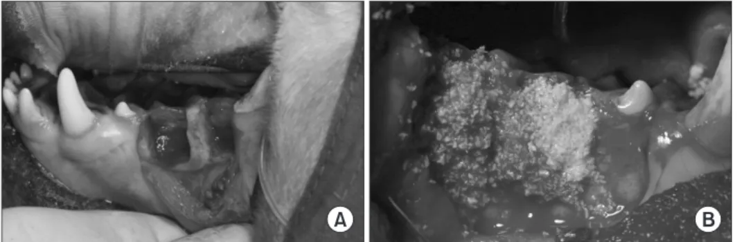
골이식 실험(Fig. 1)
발치창 주변의 점막골막피판을 거상하고 외과용 드릴을 사 용하여 발치창 협측벽을 근첨부까지 삭제함으로써 인위적으 로 결손부를 형성하였다.
결손부에 다음과 같이 골이식을 시행한 후 창상을 일차봉합 하였다.
- Group 1: 상악 우측 제2 소구치, Bio-Oss® (Geistlich Pharma AG, Wolhausen, Switzerland) 단독이식
Fig. 1. (A) Intentional bone defects
were formed. (B) Bone graft materials
were filled.
- Group 2: 상악 우측 제3 소구치, Bio-Oss®+NOVOSIS (Bioalpha Inc., Seongnam, Korea) rhBMP-2 혼합 이식
실험 후 항생제 및 진통제를 일정기간 투여하고 사육실에 서 희생 전까지 건강하게 사육하였다. 수술 후 항생제로는 Cefazolin 25 mg/kg을 3일간 1일 1회 주사하였다.
치유과정 평가
동물희생
골이식 수술 시와 같은 방법으로 전신마취 유도 후 외부경 정맥삽관술(external jugular vein cannulation)과 총경동맥삽 관술(common carotid artery cannulation)을 시행하였다. 삽 관이 끝난 후 마취상태를 확인하여 심마취 상태임을 확인하 고 염화칼륨 5 mL (Potassium Chloride Inj.; Jw-Pharma)을 정 맥 주입하였다. 청진하여 심정지를 확인 후 총경동맥 혈관 내 로 10% 포름알데히드를 주입함으로써 관류고정(perfusion fixation)을 시행하였다.
실험 2, 4, 8주 후에 각각 2마리씩 동물을 희생시킨 후 주변 골조직과 이식골을 포함한 블록시편을 채취하였다. 채취한 시편을 이동식 방사선 촬영기(DXR-1; Exaro, Seoul, Korea)와 디지털 센서(X-vision; DigiMed, Seoul, Kora)를 이용하여 촬 영한 후(관전압 60 kvp, 관전류 2 mA, 노출시간 0.12초) 10%
Formalin 용액(Sigma Aldrich Co. LLC., St. Louis, MO, USA)에 고정하였다. 고정한 시편은 검사기관으로 발송하여 시편제작 을 의뢰하였다.
조직시편 제작 및 평가
고정된 조직시편들은 물로 세척한 후 tissue processor (Sha- don Citadel 2000; Thermo Fisher Scientific Inc., Kalamazoo, MI, USA) 로 탈회하고 파라핀 왁스로 포매하였다. Microtome (Shadon Finesse 325; Thermo Fisher Scientific Inc.)를 사용하 여 3.0 µm 두께로 절편을 제작한 후 hematoxylin and eosin (H&E) 염색을 시행하였다.
조직학적 관찰 및 조직형태계측학적 평가
2, 4, 8주 후 치유과정을 광학현미경으로 관찰하였다. 이후 analysis LS starter program (OLYMPUS Soft Imaging Solution, Müster, Germany)을 이용하여 광화 골조직의 양을 측정하였 다. 총 관찰면적당 광화 골조직의 비율을 측정하였으며 평균 과 표준편차를 계산하였다(mean±standard deviation). 2, 4, 8 주에서 광화 골조직 형성 정도에 대한 Group 1, 2 간의 차이에 대해 분석하였다. IBM SPSS Statistics version 20.0 (IBM Co., Armonk, NY, USA)의 non-parametric Mann-Whitney test를 이용하여 통계적 유의성을 분석하였다(p<0.05).
결 과
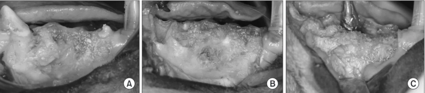
육안적 소견
수술 부위의 창상열개, 감염 등의 합병증은 발생하지 않았 다. 2, 4, 8주 후에 채취한 시편의 골 결손부는 모두 골이식재가 잔존하며 골이식재들이 연조직 및 신생 골조직과 잘 섞여있 는 양상을 관찰할 수 있었다. 4주부터는 이식재가 육안적으로 더 안정적으로 치유되는 양상을 관찰할 수 있었고 8주 후의 시 편은 더욱 우수한 골치유 양상을 보였다(Fig. 2).
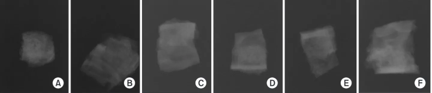
방사선학적 소견
두 Group 모두에서 시간이 경과하며 방사선 불투과성이 증 가하는 양상을 보였으나 Group 간의 뚜렷한 방사선학적 차이 를 관찰할 수는 없었다(Fig. 3).
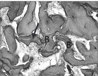
조직학적 소견 2주 소견(Figs. 4, 5)
Group 1에서는 골이식재 주위에 소량의 미성숙 골형성 (woven bone formation)이 관찰되었고(Fig. 4), Group 2에서는 골이식재 주위의 신생골이 woven-lamellar bone 형태로 잘 형 성되어 있는 것이 관찰되었다. 형성된 골의 양도 Group 1에 비 하여 2배 가량 많이 관찰되었다(Fig. 5).
Fig. 2. (A) Two weeks after bone graft. (B) Four weeks after bone graft. (C) Eight weeks after bone graft.
4주 소견(Figs. 6, 7)
Group 1에서는 골이식재 주위에 소량의 woven-lamella bone 형성이 관찰되었고(Fig. 6), Group 2에서는 신생골과 기존 골이 구분되지 않을 정도의 성숙도를 보이는 mature (lamellar) bone이 잘 형성되어 있었다. 형성된 골의 양도 Group 1에 비하여 50% 이상 증가된 소견을 보였다(Fig. 7)
8주 소견(Figs. 8, 9)
Group 1에서는 결손부의 대부분이 새로 형성된 woven- lamellar bone으로 채워져 있는 것이 관찰되고(Fig. 8), Group 2에서는 거의 모든 결손부가 mature (lamellar) bone으로 수복 되어 있는 것이 관찰되었다. 형성된 골의 양은 Group 1에 비하 여 70% 이상 증가된 소견을 보였다(Fig. 9).
조직형태계측학적 평가(Table 1, Fig. 10)
형성된 광화 골조직의 비율을 분석하였다. 2, 4, 8주 모두에
Fig. 3. (A) Radiographic view. Bio-Oss
®only, 2 weeks after bone graft. (B) Bio-Oss
®with recombinant human bone morphogenetic protein-2 (rhBMP-2), 2 weeks after bone graft. (C) Bio-Oss
®only, 4 weeks after bone graft. (D) Bio-Oss
®with rhBMP-2, 4 weeks after bone graft. (E) Bio- Oss
®only, 8 weeks after bone graft. (F) Bio-Oss
®with rhBMP-2, 8 weeks after bone graft.
Fig. 4. Histologic finding 2 weeks of Group 1. Small woven bone for- mation (arrows) was observed. B: Bio-Oss
®(H&E, ×100).
Fig. 5. Histologic finding 2 weeks of Group 2. The woven-lamellar bone (arrows) was formed around the Bio-Oss
®(B) particles (H&E,
×100).
Fig. 6. Histologic finding 4 weeks of Group 1. Small amount of
woven-lamellar bone (arrow) was formed around the Bio-Oss
®(B)
particles (H&E, ×100).
서 Group 2에서 높은 비율로 형성된 것으로 평가되었고, 2주 와 8주에서는 통계적으로 유의한 차이가 있었다.
고 찰
오래 전부터 가장 많이 연구된 성장인자는 BMPs이다. BMP 는 Urist [1]가 뼈를 탈회, 동결건조하여 얻은 단백질 혼합물을 토끼의 근육 조직에 이식한 후 이것이 골 형성에 관여했다는 것을 발견하였다.
BMP는 transforming growth factor-β superfamily에 속하는 다기능 cytokine이다. 다양한 기능을 가진 20개 이상의 BMP가 인간에서 확인되었다. BMP는 embryogenesis와 성인에서 많
은 골격과 비골격 조직의 유지 및 회복에 중요한 역할을 한다 [20].
Urist의 실험 후, BMP는 토끼, 소, 인간 등 다양한 종의 뼈에 서 분리되어 얻어졌다. 요즘은 DNA 재조합 기술과 포유류 세 포와 박테리아의 두 개의 expression 시스템을 사용하여 BMP 를 제작하고 정제한다[21]. rhBMP-2와 rhBMP-7은 현재 사람 에게 임상적인 목적으로 사용되고 있으며 미 식품 의약국(The
Fig. 7. Histologic finding 4 weeks of Group 2. Mature lamellar bone
(arrow) was formed well around the Bio-Oss
®(B) particles (H&E,
×100).
Fig. 8. Histologic finding 8 weeks of Group 1. Woven-lamellar bone (arrow) was observed around the Bio-Oss
®particles (H&E, ×100).
Fig. 9. Histologic finding 8 weeks of Group 2. Most bone defect was filled with mature lamellar bone (H&E, ×100).
Table 1. Mineralized Tissue Formation
Group 1 (%) Group 2 (%) p-value 2 Weeks
4 Weeks 8 Weeks
23.97±3.42 31.12±10.01
31.38±5.65
51.86±4.36 45.17±12.64
52.32±0.14
<0.05 -
<0.05 Values are presented as mean±standard deviation.
Fig. 10. Mineralized tissue formation in vivo. BMP: bone morphoge-
netic protein.
US Food and Drug Administration)에서 승인한 유일한 BMP 이다[22].
Jung 등[23]은 국소적인 치조능 증대술을 위해 BMP-2, BMP-7, GDF-5, platelet-derived growth factor (PDGF), and parathyroid hormone (PTH)를 사용한 후 임상적, 조직학적, 방사선학적 결과에 대해 체계적인 문헌 고찰을 시행하였다.
모두 74개의 연구가 포함되었고 이 중 6개는 사람에서 치조 능 증대술을 위해 사용한 BMP-2의 결과였고 나머지는 BMP- 2, BMP-7, GDF-5, PDGF, PTH가 포함된 전임상 연구였다.
BMP-2를 사용한 동물연구의 대부분에서(45개 중 43개) 긍정 적인 효과를 보였다. 8개의 연구 중 6개는 BMP-7의 긍정적인 효과를 보고했다. 1개의 GDF-5에 대한 동물 실험에서는 통계 적으로 유의하게 골 면적 비율이 증가하였다고 하였다. PDGF 를 사용한 10개의 연구 중 5개에서도 통계적으로 유의한 골 면 적 비율 증가가 보고되었다. 4개의 동물 연구에서 PTH를 사용 한 경우 대조군에서 보다 유의하게 더 많은 골 재생을 확인하 였다. BMP-2는 골 면적 비율에 있어 투여 의존적인 증가로 국 소적인 골의 증강에 긍정적인 영향을 주었다. 전달된 BMP-2 는 높은 투여량에서는 더 많은 국소적 골재생을 보이고, 발치 와에서 골 높이의 감소를 줄여주는 치료 효과를 보였다[24- 27]. 사람에 대한 BMP-2의 6개의 연구 중 5개에서는 운반체로 absorbable collagen sponge (ACS)를 사용하였으나 Jung 등[28]
은 demineralized bovine bone matrix을 사용하였다.
Boyne 등[29]은 2-stage의 상악동 증대술에서 ACS로 전달 되는 BMP-2 이식(rhBMP/ACS)의 안전성과 효과에 대한 연구 를 했다. rhBMP-2의 투여량은 환자당 1.77 mg에서 3.40 mg이 었다. Computed tomography 분석에서 모든 환자에 있어 평균 8.51 mm 높이의 유의한 골 성장이 관찰되었다. 임프란트 식립 시 채취한 core bone에 대한 조직검사에서 rhBMP/ACS에 의 해 유도된 양질의 골이 확인되었다.
BMP-2와 BMP-7은 골개조 동안 골과 연골의 성장과 항상성 유지에 뚜렷한 효과를 지니고 있다[30]. 하지만 이것의 한계 중 하나는 인체 내에서 조직 재생 결과에 대해 예측할 수 없다 는 점이다. rhBMP의 임상적 효과는 이식한 부위에 적절한 단 백질 농도가 효과적으로 전달되는 시스템에 의존한다고 알려 져 왔다[31]. BMP는 수용성 단백질이고 완충용액에서 운반되 며 빠른 분해가 일어나기 때문에 불충분한 생체이용률을 가 지고 있다. 단백질 경쟁, 효소활성, 온도, pH, 염분농도와 같은 다른 요소들도 BMP의 작용에 영향을 미친다[32]. 따라서 단 독으로 사용할 경우 즉시 용해되면서 골 유도성 효과를 거의 발휘하지 못한다[33-35]. 그러므로 BMP, 성장인자, 줄기세포 를 임상에 적용하기 위해서는 운반체 역할을 수행할 수 있는 이상적인 scaffold가 필요하다. 이상적인 scaffold는 성장인자
나 세포의 방출을 조절하면서 분해와 비활성화에 대해 보호 하는 역할을 수행할 수 있어야 한다[36]. 또한 생체적합성, 예 측 가능한 생체분해성, 적절한 염증반응의 유도 능력과 같은 일반적인 요구 조건을 충족시켜야 한다[37]. Scaffold로 사용 될 수 있는 재료들은 human collagen, atelocollagen, albumin, osteonectin, osteocalcin, bone matrix, blood clot, polylactic polyglycolic acid polymers, calcium sulfate, hydroxyapatite, β -tricalcium phosphate (TCP), titanium oxide이며 임상에서 많 이 적용되고 있는 것은 collagen과 TCP이다[38]. 그 밖에도 성 장인자들을 여러 종류의 운반체를 이용하여 임상에 적용하는 연구가 많이 진행되어 왔다[26,28,39-48].
Anorganic bovine bone을 성장인자의 운반체로 이용하여 운반체로서의 효과와 골유도 효과가 있다는 것을 증명한 연 구가 있었고[49], Bio-Oss® 단독으로 사용 시에도 여러 골 형 성 세포의 물리적인 scaffold 역할을 하여 효과적인 골 형성을 할 수 있었다는 연구 결과도 있었다[50].
이번 연구에서는 anorganic bovine bone인 Bio-Oss®를 scaffold로 이용하였는데 이는 효과적인 골이식재로의 역할도 수행하면서 rhBMP 첨가 시 성장인자를 효과적으로 전달하여 골 형성에 좋은 영향을 미칠 수 있었다.
현재 임상적으로 사용되는 BMP는 BMP-2와 BMP-7이다.
여러 회사에서 다양한 제품이 나오고 있는데 BMP-2나 BMP-7 을 활용한 미국 Medtronic사(Minneapolis, MN, USA)의 Infuse 나 Stryker사(Kalamazoo, MI, USA)의 OP-1이라는 제품이 있 다. 해당 제품들은 동물 진핵세포로부터 제조되기 때문에 수 율(원자재에 화학적 과정을 가해 원하는 물질을 얻을 때, 실제 로 얻어진 양과 이론적으로 기대했던 양을 백분율로 나타낸 비율)이 낮고 생산성이 떨어져 가격이 매우 고가이다. 고농도 로 사용할 경우엔 악성종양 등을 유발할 위험성이 있으며 치 과 분야의 소규모 결손부에 적용하기 위해서는 적절한 운반 체가 있어야 가능하다. 따라서 치과분야에 적용하기에는 아 직 무리가 있으며 특히 국내 상용화는 현 시점에서는 불가능 하다고 판단된다. 이번 연구에서 사용한 성장인자는 국내에 서 제작된 rhBMP-2인 NOVOSIS (Bioalpha Inc.)로 우수한 골 형성 증가를 보였고 회사측에서는 다른 제품에 비해 높은 순 도와 낮은 엔도톡신을 가지고 있다고 소개하고 있다.
본 연구에서 동물을 대상으로 실험한 결과 Bio-Oss® 단독으 로도 2, 4, 8주에 있어 신생골 형성이 관찰되었으나 rhBMP-2 인 NOVOSIS와 혼합 사용 시에 더 빠른 시간에 골이 성숙되었 고, 같은 기간에 있어 더 많은 양의 신생골이 형성되는 것을 관 찰할 수 있었다. 모든 관찰 기간에 있어 NOVOSIS 혼합군에 서 더 많은 양의 신생골이 형성되었고, 2주와 8주에서는 통계 적으로 유의한 차이가 있었다. 이 논문은 Bio-Oss®에 BMP를
복합 사용하는 것이 골 형성에 어떤 영향을 미치는가를 연구 한 것이므로 이식재와 연조직의 양보다는 새로 만들어진 경 조직의 양이 중요한 것으로 생각되어 조직계측학적 분석으로 골성조직만 분석하였다. 향후 잔존이식재, 신생골, 연조직 및 성숙골과 미성숙골의 비율 등을 좀 더 자세히 분석하는 추가 연구가 필요할 것으로 생각된다. 본 연구에서 woven-lamellar bone이라는 용어를 사용하였는데 조직 소견에서 염색의 차이 를 보이는 두 가지 종류의 골조직이 관찰되어 이와 같은 용어 를 사용하였다. 조직 시편의 개수가 적고 염색 과정에서의 오 류 가능성이 있기 때문에 역시 추가 연구에서는 이에 대한 평 가를 다시 할 필요가 있다.
이번 연구에서 rhBMP-2는 우수한 골유도성 치유능력을 지 니고 있다는 것을 관찰할 수 있었고, anorganic bovine bone은 자체가 골 이식재로서의 역할을 수행하면서 동시에 rhBMP-2 의 운반을 위한 효과적인 scaffold로 유용하게 사용될 수 있다 고 판단된다.
Acknowledgments
This study was supported by graft no 02-2013-008 from the SNUBH Research Fund.
References