서론
최근에 골 결손부위의 임플란트 성공률을 높이기 위해 임플 란트 표면에 생체분자들을 부착하는 생체모방적인 표면처리 기법에 관한 연구들이 활발히 진행되고 있다.1골 유착과 치유
를 매개하는 생체분자로 Arg-Gly-Asp (RGD) 펩타이드,2세포외 기질 단백질(collagen),3성장인자4등이 이용되고 있으며, 그 중에 서도 중간엽 줄기세포로부터 골모세포로의 분화와 증식을 촉 진하고 골기질의 생합성을 도와 골 형성을 증진시키는 역할을 하는 골형성단백질(bone morphogenetic protein, BMP)과 같은 성장
양극산화 임플란트 표면에 적용된 헤파린과 골형성단백질(rhBMP-2)이 치조골 증대에 미치는 효과: 방사선학적 평가
이소현∙조재영∙윤미정∙전영찬∙허중보∙정창모*
부산대학교 치의학전문대학원 치과보철학교실, 중개치의학연구소
Effect of immobilization of the recombinant human bone morphogenetic protein 2 (rhBMP-2) on anodized implants coated with heparin for improving alveolar ridge augmentation
in beagle dogs: Radiographic observations
So-Hyoun Lee, DDS, Jae-Young Jo, DDS, MSD, Mi-Jung Yun, DDS, MSD, PhD,
Young-Chan Jeon, DDS, MSD, PhD, Jung-Bo Huh, DDS, MSD, PhD, Chang-Mo Jeong*, DDS, MSD, PhD Department of Prosthodontics, School of Dentistry, Institute of Translational Dental Sciences, Pusan National University, Yangsan, Korea
Purpose: The aim of this study was to evaluate the effect of immobilization of the recombinant human bone morphogenetic protein 2 (rhBMP-2) on anodized titaum implants coated with heparin to enhance the vertical alveolar ridge augmentation in the supraalveolar peri-implant defect region. Materials and methods: 18 pure titanium implants (7.0 mm in length, 3.5 mm in diameter) were manufactured for this study. All implants were anodized and designed insertion reference line marked with laser at the apical 2.5 mm from the fixture platform. Implantation of 6 noncoated anodized implants (Control group), 6 anodized implants physically adsorbed with rhBMP-2 by dip and dry method (BMP group) and 6 anodized implants chemically immobilized 3,4-dihydroxyphenylalanine (DOPA)-heparin/ rhBMP-2 (Hep-BMP group) was performed in the both mandibular of three male adult beagle dogs using split-mouth design. Radiologic examinations were performed immediately after implant placement and 4 and 8 weeks after implant place- ment. The amount of mesio-distal bone augmentation was evaluated by measuring the vertical distance from the platform to the marginal bone. Statistical analysis was performed using one-way analysis of variance (SPSS version 18.0) and multiple comparison analysis of The Kruskal-Wallis test and the Mann-Whitney U test. Statistical significance was established at the 5% significant level. Results: At the 4 weeks vertical alveolar ridge augmentation of Control group, BMP group and Hep-BMP group is 0.09 ± 0.22 mm, 1.02 ± 0.72 mm, and 1.29 ± 0.51 mm, At the 8 weeks 0.11 ± 1.26 mm, 1.11 ± 0.58 mm, 1.59 ± 0.79 mm according to radiographic observations. The two experimental groups showed a significantly increasing in vertical bone height compared with the control group (P<.05). However, there is no significant difference between the BMP group and Hep-BMP group (P>.05). Conclusion: The rhBMP-2 coated implants were enhanced the vertical bone growth in the supraalveolar peri-implant defect area. However, there is no significant difference between chemically and physically coating method. (J Korean Acad Prosthodont 2013;51:307-14)
Key words: Anodized implants; Dopamine; Heparin; Bone morphogenetic protein
c cc
2013 The Korean Academy of Prosthodontics
This is an Open Access article distributed under the terms of the Creative Commons Attribution Non-Commercial License (http://creativecommons.org/licens- es/by-nc/3.0) which permits unrestricted non-commercial use, distribution, and reproduction in any medium, provided the original work is properly cited.
*Corresponding Author: Chang-Mo Jeong
Department of Prosthodontics, School of Dentistry, Pusan National University, Beomeo-ri, Mulgeum-eup, Yangsan, Gyeongnam, 626-870, Korea
+82 55 360 5130: e-mail, [email protected]
Article history: Received September 16, 2013 / Last Revision October 2, 2013 / Accepted October 8, 2013
*This research was supported by Basic Science Research Program through the National Research Foundation of Korea (NRF) funded by the Ministry of Education, Science and Technology (2012R1A1A1005122).
인자의 적용이 큰 관심을 받고 있다.5,6BMP-2는 BMP 중 가장 높 은 골유도성을 지닌 것으로 보고되고 있으며7상악동 거상술,8 치조골 보존술,9골 증대술,10치주적 회복술11등을 포함하는 다 양한 연구를 통해 골재생에 관한 잠재성이 입증되었다. 최근 에는 티타늄 임플란트에 코팅된 recombinant human (rh)BMP-2가 임플란트 주변골의 재생과 유착에 미치는 효과에 관한 연구가 활발히 진행되고 있다. Huh 등12은 Escherichia coli-rhBMP-2가 코팅된 양극산화 임플란트를 비글견에 이식한 후 방사선학적 분석과 Implant stability quotient (ISQ) values측정을 시행해 본 결과 수직적인 골 증대와 임플란트의 안정성이 향상되었음을 보고 하였다. 그러나 rhBMP-2의 적용이 골형성에 별다른 기여를 하 지 못했다는 보고도 있으며, 이는 rhBMP-2의 빠른 반감기로 인 한 급격한 분해, 표준화되지 못한 최적 농도, 생체 내 다른 성장 인자와의 상호작용 없이 단독 사용된 것 등이 원인으로 추정되 고 있다.13-15이중 빠른 반감기를 극복하여, BMP-2의 지속적이고 국소적인 방출을 유도할 수 있는 collagen gels,16hyaluronic acid,17 fibrin gel18등을 이용한 BMP-2 전달 시스템이 연구되어 왔다. 본 연구에서는 BMP-2의 방출을 조절하기 위해 성장인자와 결합 친화성을 가진 헤파린19을 사용하였다. 헤파린을 결합시킨 생 체 물질 시스템이 성장 인자들의 방출을 조절할 수 있다는 이 점이 많은 연구를 통해 보고되고 있으며,20,21본 실험의 이전 연 구22에서도 헤파린을 이식한 골에 rhBMP-2를 고정시켜 서방출 과 골모세포의 기능 향상을 보고한 바 있다. Kim 등23은 헤파린 과 BMP-2를 고정시킨 티타늄 디스크의 세포실험 통해 BMP-2 의 서방출과 항 염증 효과를 확인하였고, Ishibe 등24은 apatite코팅 된 티타늄 바에 헤파린과 BMP-2를 고정하여 토끼의 경골에 식 립한 후 조직학적 분석을 통해 수직적, 수평적 골 형성을 확인 하였다.
이와 같이 헤파린과 골형성단백질를 결합시켜 서방출을 유 도한 많은 연구들이 있어왔으나 아직, 도파민25과 헤파린을 적 용하여 rhBMP-2를 화학적으로 고정시킨 임플란트 표면이 임플 란트 주변골의 증대와 유착 향상에 미치는 효과를 동물 모델
을 통해서 직접적으로 평가한 연구는 없었다. 이번 연구의 목 적은 수직적 골 결손을 갖는 비글견의 하악에 rhBMP-2를 코팅 하지 않은 임플란트(대조군)와 기존의 dip and dry 방법으로 rhBMP-2를 물리적으로 흡착시킨 양극산화 임플란트(BMP군), 그리고 서방출 유도 위해 화학적 이식 방법으로 헤파린과 rhBMP-2을 고정시킨 양극산화 임플란트(Hep-BMP군)를 split- mouth design으로 식립하여 임플란트 주변골의 수직적 재생량 차이를 방사선학적 관찰을 통해 비교 평가하기 위한 것이다.
연구 재료 및 방법 1. 연구 재료
1) 임플란트 제작
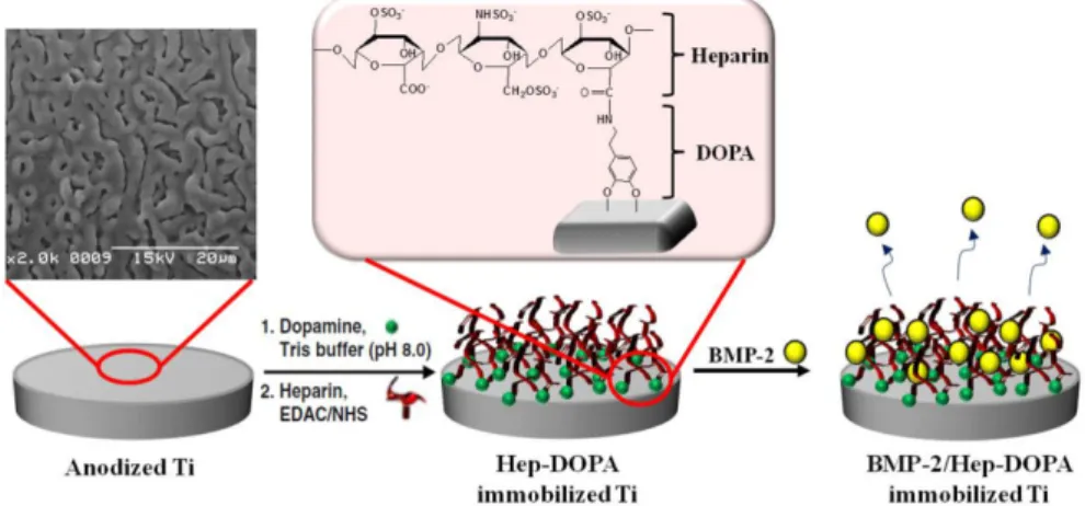
Commercially pure-Titanium 을 사용하여 길이 7.0 mm, 직경 3.5 mm 의 실험용 임플란트(Cowellmedi Co., Pusan, Korea) 18개를 제작하 였다. 모든 임플란트의 표면을 양극산화처리(Cowellmedi Co., Pusan, Korea) 하였고, 임플란트 플랫폼에서 하방으로 2.5 mm 위치에 레이저를 사용하여 식립 기준선을 표시하였다. 양극산 화 임플란트에 rhBMP-2를 코팅하지 않은 집단을 대조군, dip and dry 방법으로 rhBMP-2를 물리적으로 흡착시킨 임플란트 집 단을 BMP군, 화학적 이식 방법으로 3,4-dihydroxyphenylalanine (DOPA)-C-heparin과 rhBMP-2이 고정된 임플란트 집단(Fig. 1)을 Hep-BMP군으로 설정하였다(Table 1).
Fig. 1. Schematic diagram for the immobilization of rhBMP-2 onto anodized Ti-implants coated with heparin and dopamine.
Table 1. Surface treatment of implant fixture used in this study
Group N Surface Treatment
Control 6 Anodized -Ti
BMP 6 rhBMP-2 (0.75 mg/ml)/Anodized-Ti
Hep-BMP 6 rhBMP-2 (0.75 mg/ml)/Hep-DOPA (2 mg/ml)/Anodized-Ti
2) 임플란트 표면의 DOPA-Heparin 처리
DOPA (2 mg/mL)를 포함한 10 mM Tris-HCl 완충용액(pH 8.0)에 양극산화 임플란트를 넣고 암실환경에서 24시간 유지시킨 후 증류수로 세척하고 질소 가스로 건조하였다. 1-ethyl-3-dimethy- laminopropyl carbodiimide (EDAC)와 N-hydrocysuccinimide (NHS)이 포함된 0.1 M morpholinoethanesulfonic acid (MES) 완충용액(pH 5.6) 에 헤파린(2 mg/ml)을 10분간 용해시킨 후, DOPA 개질되어 아민 화된 양극산화 임플란트를 넣고 실온에서 24 시간 유지하였다.
이 과정 동안 EDAC와 NHS에 의해 활성화된 헤파린의 카르복 실 그룹이 임플란트 표면의 1차 아민 그룹과 반응이 일어나 헤 파린이 양극산화 임플란트 표면에 이식된다. 헤파린이 코팅된 임플란트를 증류수로 세척하고 질소 가스로 건조하였다
3) 임플란트 표면 rhBMP-2의 부동화
모든 실험군의 임플란트를 rhBMP-2 (0.75 mg/ml)이 첨가된 0.1 M MES 완충용액(pH 5.6) 에 넣어 실온에서 24 시간 유지하고, 증류수로 세척하였다. 이후 무균 환경의 동결건조기에서 -40℃로 동결 시키고 진공상태에서 20℃를 최고 온도로 하여 건조하였다.
4) 실험 동물
3 마리의 건강한 수컷 beagle 성견(평균 3세, 평균 15 kg)이 이용 되었다. 전남대학교 수의과대학 동물실험 윤리위원회(CNU LACUC-YB-R-2010-10)에의해승인된동물선정관리방식과수술 적프로토콜을따랐다. 실험전2주의적응기간동안23 ±3℃의 온도와 40 ± 10%의 습도가 유지되는 우리에 한 마리씩 수용하 였고, 유동식(Science Diet, Hill's Co., Topeka, KS, USA)을 하루에 2번 제공하였다.
2. 연구 방법
1) 실험 동물의 발치
Atropine sulfate (0.05 mg/kg SC; Dai Han Pharm Co., Seoul, Korea) 와 tiletamine/zolazepam (5 mg/kg IV; Zoletil 50; Virbac, Carros, France) 를 투여한 뒤, 2% isoflurane (Choongwae Co., Seoul, Korea)과 ocxy- gen으로 흡인진정마취하여 실험 동물의 발치를 시행하였다.
Lactated Ringer's solusion을 발치 종료 시까지 5 ml/kg/h 속도로 투여하였고, 수술 전 ampicillin sodium (Penbrook, 20 mg/kg IV;
Chong Kun Dang Co., Seoul, Korea)와 meloxicam (Metacam, 0.2 mg/kg IV; Boehringer Ingelheim Co., Ridgefied, CT, USA)를 정맥주사 하였다. 1:100,000 epinephrine이 포함된 2% lidocaine HCL (Yu-Han Co., Gunpo, Korea) 을 수술부위인 하악 편측 악궁 점막에 1.8 ml 씩 주입하여 국소침윤마취 시행하였다. 하악 제1, 2소구치 그리 고 제1대구치를 발거하였으며, 발치 부위는 4-0 nylon (Mersilk, Ethicon Co., Liningston., UK) 으로 봉합하였다. 발치 후 meloxicam (Metacam, 0.1 mg/kg PO; Boehringer Ingelheim Co., Ridgefied, CT, USA) 을 24 시간마다, amoxicillin (amoxicillin, 20 mg/kg PO; Choongwae Co.,
Seoul, Korea)을 12시간 마다 6일동안 투약하였다.
2) 임플란트 식립
발치 후 2달 뒤에 충분한 치조골의 치유를 확인하고 임플란 트를 식립하였다. 발치를 시행할 때와 같은 방법으로 실험 동 물을 진정 마취 시킨 후 국소 마취를 하였다. 치조정 절개 후 협 설측 전층 판막을 박리하였고, 치조정을 평탄하게 하여 임플 란트 식립을 위한 골 폭을 확보하였다. 임플란트 사이 간격을 3 mm 이상 확보하여 한 마리당 6개씩 총 18개의 실험용 임플란 트를 식립하였다. 출력 토크를 30 Ncm로 설정한 임플란트 수술 용 엔진(NSK Surgic XT, NSK, Tochigi-ken, Japan)을 이용하였으며, 엔진으로 식립이 완료되지 않은 임플란트는 수동토크렌치 (Cowellmedi Co., Pusan, Korea)를 이용하여 치조골 위로 임플란트 가 2.5 mm 돌출 되도록 기준선과 치조정을 일치시켜 식립하였 다. 임플란트에 코팅된 rhBMP-2가 방출되어 이웃한 실험 환경 에 영향을 미칠 수 있기 때문에 split-mouth design으로 한 그룹의 임플란트를 편측 악궁에 식립하였고 다른 그룹의 임플란트를 반대쪽에 식립하였다. 치근단 방사선 검사에 의해 식립된 임 플란트와 치조정과의 배열을 확인한 후 cover screw를 체결하고 흡수성 봉합사 5-0 Goretex (W.L. Gore and Associates, Flagtaff, AZ, USA) 를 사용하여 봉합하였다.
3) 수술 후 관리
실험 동물의 임플란트 식립 직후와 48시간 후에 penicillin G procaine과 penicillin G benzathine (Deasung microbiological labs. Co., Seoul, Korea)을 근육 내 주사하였다(1 mg/5 kg). 치태 조절을 위해 서 술 후 1 주 동안 2% chlorhexidine gluconate를 10 cc 시린지에 넣 어 하루에 2회 구강 내 소독하였다. 봉합사가 제거되기 전까지 임플란트 식립 위치의 점막 건강, 봉합선 접합의 유지, 부종, 조 직 괴사나 감염의 징후를 매일 관찰하였고 임플란트 식립 1주 일 후에 봉합사를 제거하였다. 실험 기간 동안에 유동식 사료 를 제공하였다.
4) 방사선학적 검사
각 군 모두 임플란트 식립 직후, 식립 후 4주 그리고 8주에 진 정 마취하에서 Portable X-ray system (Port-X II, Genoray, Co, SungNam, Kerea)을 이용하여 임플란트의 치근단 방사선 사진을 평행 촬영법으로 촬영하였다. Digital X-ray software (CDX-View, PointNix, Seoul, Korea)를 이용하여 gray scale image를 color coding 한 후 임플란트의 길이와 변연골 수준(임플란트 platform에서 변연골 최상방까지 수직적 거리)을 근원심측에서 각각 측정하 고, 실제 임플란트 길이(7.0 mm)와 비교하여 다음과 같이 변연 골 재생량을 계산하였다.
변연골수준(mm) = 방사선 사진 상 변연골 수준 (mm)
× 7.0 (mm) 방사선 사진 상 임플란트 길이 (mm)
변연골재생량(mm) = 촬영된 변연골 수준 (mm) - 식립 직후 변연골 수준 (mm)
5) 통계 분석
각 군내에서의 시간에 따른 변화와 각 군간 수직적 골 증가 량의 차이를 알아보기 위하여 SPSS ver. 18.0 (SPSS, Chicago, IL, USA)을 이용하여 통계 처리하였다. Kruskal-Wallis test를 통하여 동일한 방사선 촬영 시점에서 군 간 변연골 변화량을 비교 분 석한 후, Mann-Whitney U test로 사후 검증하였으며, 동일 군에서 4주와 8주경 변연골 변화량의 차이를 Mann-Whitney U test를 통하 여 비교하였다. 유의 수준 5%에서 검증하였다.
결과
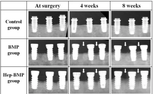
방사선 촬영을 통해 임플란트 주위 변연골의 변화를 확인한 결과, Fig. 2의 화살표 부분에서 살펴볼 수 있는 것처럼 두 실험 군에서는 근원심으로 임플란트 주변을 따라 변연골이 수직적 으로 재생된 반면, 대조군에서는 임플란트 주위 변연골의 변 화를 거의 관찰 할 수 없었다. 각 군별 근원심 변연골의 수직적 재생량에 대한 평균값과 표준편차를 Table 2에 나타내었다. 4주 와 8주경 변연골 재생량을 비교한 결과 두 실험군이 대조군에 비해 유의한 변연골 재생량을 보였다(P<.05). 그러나, Hep-BMP
군과 BMP군의 비교에서 변연골 재생량의 유의한 차이를 보이 지 않았다(P>.05). 모든 집단에서 4주에서 8주로 갈수록 수직적 골 재생량의 평균값은 다소 증가하나, 통계학적 유의성은 없 었다(P>.05) (Fig. 3).
Table 2. Mean (± SD) of vertical bone growth (mm) at 4 weeks and 8 weeks
Group 4 weeks 8 weeks
Control 0.09 (± 0.22) 0.11 (± 0.16)
BMP 1.02 (± 0.72)* 1.11 (± 0.58)*
Hep-BMP 1.29 (± 0.51)* 1.59 (± 0.79)*
(*P<.05, compared with control group)
Fig. 2. Radiographs of the mesio-distal bone height on peri-implant of two experimental groups and control group at 4 weeks and 8 weeks. The two experimental groups showed vertical bone gain at 4 and 8 weeks (arrow), whereas the control group did not show any changes.
Fig. 3. Mean (± SD) Graph of vertical bone growth on peri-implant at 4 weeks and 8 weeks. The BMP group and Hep-BMP group (experimental groups) showed increased vertical bone height. There is no statistically significant difference between the BMP group and Hep-BMP group, whereas the two experimental groups showed a significantly different increase in vertical bone growth compared with the control group (*P<.05).
고찰
BMP와 같은 성장 인자를 임플란트의 표면에 처리하여 추가 적인 골 이식재나 차폐막의 사용없이 수직적인 골 재생의 효 과를 증대시키기 위한 많은 시도가 이루어져 왔다. Titanium porous oxide (TPO) 임플란트 표면에 rhBMP-2를 흡수시켜 쥐의 이 소성 모델에 적용한 Hall 등4의 연구에서 임플란트 표면을 따르 는 골 형성과 골 유도 효과를 확인할 수 있었고, 골 결손 모델을 이용한 Leknes 등26과 Wikesjo¨등27의 동물 실험에서는 rhBMP-2이 코팅된 TPO 임플란트를 치조골 상방으로 5 mm 노출시켜 식립 한 결과를 통해 방사선학적으로 유의한 수직적 골 증대와 조 직학적인 골질의 개선을 보고하였다. 우리의 동물실험에서는 이전 연구28,29에서와 동일한 방법으로 임플란트의 상부를 치조 골 상방으로 2.5 mm 노출시키고, 하부 4.5 mm를 피질골에 식립 하여 종 더 임상적인 상황을 반영하고자 하였다.
임플란트 식립 직후와 4주, 8주 후의 방사선학적 촬영을 통해 임플란트 주위 근원심 변연골의 수직적 재생량 을 비교해 보 았는데, 촬영 시기는 3-4주에 가골을 형성하기 시작하는 골조 직의 재생 과정을 고려하고, 선행 연구에서28rhBMP-2의 초기 방 출로 유도되는 임플란트 주위의 과도한 골 재형성 과정에 의 해 식립시 얻었던 임플란트의 안정도가 2주쯤에 감소되었던 결과를 참고로 하여 선택되어졌다. 방사선학적 분석 결과 변 연골 재생량(평균값 ± 표준편차)은 4주에 대조군은 0.09 ± 0.22 mm, BMP군은 1.02 ± 0.72 mm, 그리고 Hep-BMP군에서는 1.29
± 0.51 mm였으며, 8주에서는 각각 0.11 ± 1.26 mm, 1.11 ± 0.58 mm, 1.59 ± 0.79 mm 였다. 모든 촬영 시점에서 rhBMP-2가 코팅되 지 않은 대조군에 비해 rhBMP-2를 물리적으로 흡착시킨BMP군 과 서방출 유도 위해 헤파린 이용하여 rhBMP-2를 화학적으로 고정시킨 Hep-BMP군에서 유의한 수직적 골 재생량을 나타냈 다(P<.05).
이전 연구에서 임상에서 사용하였을 경우 BMP-2의 사용이 골형성에 유의한 효과를 나타내지 못했다는 보고가 종종 있어 왔다.30우리의 연구에서는 이의 원인 중 하나로 추정되는BMP- 2가 초기에 급격히 방출되어 지속적인 골 형성을 유도하지 못 하는 짧은 반감기의 문제점을 극복하기 위해 헤파린을 이용한 서방출 전달시스템을 적용하여 보았다. 헤파린은 많은 황산기 를 가진 선형의 산성 다당류의 일종으로 성장 인자들에 대한 결합 친화성을 가진 것으로 알려져 왔다.19반면, 임플란트의 재 료로 사용되는 티타늄은 좋은 생체적합성, 우수한 기계적 성 질, 뛰어난 부식 저항성을 가지지만, 생물학적 활성에 대한 특 이성이 없는 무기물질로 성장 인자와 같은 생체분자의 부착은 물리적 흡착법이나 화학적 이식법을 통해서만 가능하다는 것 이 보고되고 있다.31
기존의 rhBMP-2의 화학적 이식 방법을 위해 사용되었던 APTES, EDAC, NHS 등의 화학물질들은 인체에 유해한 것으로 임플란트의 상용화에 걸림돌이 되고 있어2,23본 연구에서는 in vivo 독성실험을 통해 생체 친화적인 것으로 보고된 홍합 접착
메커니즘을 모방한 도파민 표면 개질기법25을 사용하여 헤파 린을 이식하였다. 홍합 접착 단백질에서 특이적으로 높은 함 량을 갖는 도파민은 카테콜과 아민 작용기를 가지는 단분자 물질이다. pH 8.5 완충용액에 다른 산화제의 첨가 없이 공기 중 의 산소기체를 산화제로 하여 티타늄을 넣었다 꺼내면 도파민 의 카테콜이 산화 티타늄 표면에 결합하여 2차 표면 개질이 가 능한 아민기를 제공하게 되어32-34티타늄에 BMP-2를 고정하거 나35헤파린을 이식하는36등의 많은 연구에 이용되고 있다.
헤파린을 이용하여 골형성단백질이 서방출 되는 것을 확인 한 여러 연구들20-24이 있어 왔다. 우리의 이전 연구28에서 양극 산화된 티타늄 표면과 순수한 티타늄 표면에 물리적으로 흡착 시킨 ErhBMP-2는 첫번째 날에 각각 36%와 54.4%의 방출량을, 일 주일 후에는 각각 67%, 78%를 나타내어 양극산화된 티타늄 표 면에서 rhBMP-2이 약간 더 방출이 유지되는 것을 확인 할 수 있 었으나 골재생이 이루어지기 위한 최소 기간인 4주 동안37지속 적으로 방출되지는 않았다. 또 다른 연구22에서는 DOPA-헤파 린을 이용하여 50 ng/ml rhBMP-2를 골이식재(Bio-Oss�)에 고정한 그룹에서의 방출 속도를 측정하여 보았는데 rhBMP-2가 단독으 로 사용된 그룹보다 지속적으로 방출되는 것을 확인할 수 있 었다. Kim 등23,38의 연구에서는 헤파린이 이식된 티타늄 표면에 10 ng/ml와 50 ng/ml의 각기 다른 농도의rhBMP-2를 고정시켜 방 출량을 측정한 결과 첫째날 각각 69%와 26%의 나타내며 4주동 안 서방출 되는 것을 확인하였으며, 28일 동안의 측정 결과를 통해 rhBMP-2의 초기 농도에 따라 다른 농도로 서방출이 이루 어 진다는 것을 보고하였다. Takada 등39과 Zhao 등40의 연구에서 도 rhBMP-2와 헤파린과의 결합이 BMP-2의 반감기를 증가시 켜 서방출을 가능하게 한다는 것을 보고한 바 있다.
본 실험의 방사선학적 평가 통해 측정된 변연골 재생량으로 4주째에서 BMP군은 40.8%, Hep-BMP군은 51.6%의 수직적 골증 대를, 8주째에서는 각각 44.4%, 63.6%의 수직적 골증대를 확인 할 수 있었으나, 두 군간에 유의한 차이는 없는 것으로 나타났 다(P>.05).
본 연구에서 사용한 표준구내방사선 사진을 통한 관찰은 주 기적인 촬영을 통해서 시간에 따른 변연골 변화를 관찰하는데 용이하지만, 관찰부위가 임플란트 근원심부에 한정되어 실제 골흡수가 많은 협측피질골 부위를 평가할 수 없으며, 골량에 대한 평가만 가능하다는 한계를 가진다. 따라서 추후 조직학 적 분석을 통해 근원심 뿐아니라 협설측을 포함한 골질과 골재 생량 및 골-임플란트 계면사이의 골융합 정도를 비교 평가해 보아 헤파린을 이용한 서방출의 효과를 확인해 볼 예정이다.
앞서 서론에서도 언급된 바와 같이 골형성단백질의 적용에 는 표준화되지 못한 농도 또한 문제점으로 지적되고 있다. Koo 등41은 rabbit을 대상으로 한 연구에서 heparin을 이용한 서방출 시 스템은 소량(10 ㎍)의 BMP-2를 적용하였을 때 골유도 효과를 보 이지 않았다고 보고하였고, Rosen 등42의 연구에서는 BMP-2의 농도가 높을 경우 내인성의 BMP inhibitor가 생산됨을 확인하였 고, Kaneko 등43의 연구에서는 BMP-2가 국소적으로 고동도로
존재할 경우 파골 세포를 유도한다고 보고하였다. 본 실험에 서는 선행된 연구결과12,28들을 참고로 하여 0.75 mg/ml의 농도 를 설정하였는데, 이 농도에서는 화학적 고정법과 물리적 흡 착법간의 수직적 골 재생량의 유의성 있는 방사선학적 차이는 보이지 않았으므로, 향후 서방출에 유리한 적정 농도를 표준 화 하기 위한 추가적인 연구가 필요할 것으로 생각된다.
이번 연구 결과는 헤파린에 의한 rhBMP-2의 서방출 효과를 티타늄 시편에서 세포수준으로만 확인할 수 있었던 이전 보고 들과는 달리, rhBMP-2의 서방출을 생체모방적으로 처리한 임 플란트를 골결손 동물 모델에 직접 적용하여 임플란트 주변골 의 수직적 증대 효과를 확인함으로써 앞으로의 임상활용을 위 한 기초자료로서의 의의가 크다고 생각된다.
결론
골형성단백질을 물리적으로 흡착시키거나 서방출 위해 헤 파린을 이용하여 화학적으로 고정시킨 양극산화 임플란트 표 면은 모두 골 결손부에서 임플란트 주변골의 수직적 증대에 효과적이었다. 그러나 방사선학적 관찰의 한계 내에서 골형성 단백질의 이 두가지 적용방법간에는 골증대량에 유의한 차이 가 없었다.
References
1. Junker R, Dimakis A, Thoneick M, Jansen JA. Effects of implant surface coatings and composition on bone integration: a systematic review. Clin Oral Implants Res 2009;20:185-206.
2. Huh JB, Lee JY, Jeon YC, Shin SW, Ahn JS, Ryu JJ. Physical sta- bility of arginine-glycine-aspartic acid peptide coated on anodized implants after installation. J Adv Prosthodont 2013;5:84-91.
3. Rammelt S, Heck C, Bernhardt R, Bierbaum S, Scharnweber D, Goebbels J, Ziegler J, Biewener A, Zwipp H. In vivo effects of coating loaded and unloaded Ti implants with collagen, chondroitin sulfate, and hydroxyapatite in the sheep tibia. J Orthop Res 2007;25:1052-61.
4. Hall J, Sorensen RG, Wozney JM, Wikesjo¨ UM. Bone for- mation at rhBMP-2-coated titanium implants in the rat ectopic model. J Clin Periodontol 2007;34:444-51.
5. Bessa PC, Casal M, Reis RL. Bone morphogenetic proteins in tissue engineering: the road from the laboratory to the clinic, part I (basic concepts). J Tissue Eng Regen Med 2008;2:1-13.
6. Bessa PC, Casal M, Reis RL. Bone morphogenetic proteins in tissue engineering: the road from laboratory to clinic, part II (BMP delivery). J Tissue Eng Regen Med 2008;2:81-96.
7. Mehta M, Schmidt-Bleek K, Duda GN, Mooney DJ. Biomaterial delivery of morphogens to mimic the natural healing cascade in bone. Adv Drug Deliv Rev 2012;64:1257-76.
8. Boyne PJ, Marx RE, Nevins M, Triplett G, Lazaro E, Lilly LC, Alder M, Nummikoski P. A feasibility study evaluating rhBMP- 2/absorbable collagen sponge for maxillary sinus floor aug- mentation. Int J Periodontics Restorative Dent 1997;17:11-25.
9. Hanisch O, Tatakis DN, Boskovic MM, Rohrer MD, Wikesjo¨UM.
Bone formation and reosseointegration in peri-implantitis defects following surgical implantation of rhBMP-2. Int J Oral Maxillofac Implants 1997;12:604-10
10. Howell TH, Fiorellini J, Jones A, Alder M, Nummikoski P, Lazaro M, Lilly L, Cochran D. A feasibility study evaluating rhBMP- 2/absorbable collagen sponge device for local alveolar ridge preser- vation or augmentation. Int J Periodontics Restorative Dent 1997;17:124-39.
11. Sigurdsson TJ, Nygaard L, Tatakis DN, Fu E, Turek TJ, Jin L, Wozney JM, Wikesjo¨UM. Periodontal repair in dogs: evalua- tion of rhBMP-2 carriers. Int J Periodontics Restorative Dent 1996;16:524-37.
12. Huh JB, Park CK, Kim SE, Shim KM, Choi KH, Kim SJ, Shim JS, Shin SW. Alveolar ridge augmentation using an- odized implants coated with Escherichia coli-derived recombi- nant human bone morphogenetic protein 2. Oral Surg Oral Med Oral Pathol Oral Radiol Endod 2011;112:42-9.
13. Stenport VF, Johansson C, Heo SJ, Aspenberg P, Albrektsson T.
Titanium implants and BMP-7 in bone: an experimental model in the rabbit. J Mater Sci Mater Med 2003;14:247-54.
14. Schliephake H, Aref A, Scharnweber D, Bierbaum S, Roessler S, Sewing A. Effect of immobilized bone morphogenic protein 2 coating of titanium implants on peri-implant bone forma- tion. Clin Oral Implants Res 2005;16:563-9.
15. Stadlinger B, Pilling E, Huhle M, Mai R, Bierbaum S, Scharnweber D, Kuhlisch E, Loukota R, Eckelt U. Evaluation of osseointegration of dental implants coated with collagen, chondroitin sulphate and BMP-4: an animal study. Int J Oral Maxillofac Surg 2008;37:54- 9.
16. Wikesjo¨UM, Qahash M, Thomson RC, Cook AD, Rohrer MD, Wozney JM, Hardwick WR. Space-providing expanded poly- tetrafluoroethylene devices define alveolar augmentation at dental implants induced by recombinant human bone morpho- genetic protein 2 in an absorbable collagen sponge carrier. Clin Implant Dent Relat Res 2003;5:112-23.
17. Kim J, Park Y, Tae G, Lee KB, Hwang CM, Hwang SJ, Kim IS, Noh I, Sun K. Characterization of low-molecular-weight hyaluronic acid-based hydrogel and differential stem cell responses in the hydrogel microenvironments. J Biomed Mater Res A 2009;88:967-75.
18. Lee TC, Ho JT, Hung KS, Chen WF, Chung YH, Yang YL. Bone morphogenetic protein gene therapy using a fibrin scaffold for a rabbit spinal-fusion experiment. Neurosurgery 2006;58:373- 80.
19. Gandhi NS, Mancera RL. Prediction of heparin binding sites in bone morphogenetic proteins (BMPs). Biochim Biophys Acta 2012;1824:1374-81.
20. Sasisekharan R, Ernst S, Venkataraman G. On the regulation of fibroblast growth factor activity by heparin-like glycosaminoglycans.
Angiogenesis 1997;1:45-54.
21. Perets A, Baruch Y, Weisbuch F, Shoshany G, Neufeld G, Cohen S. Enhancing the vascularization of three-dimensional porous alginate scaffolds by incorporating controlled release basic fibroblast growth factor microspheres. J Biomed Mater Res A 2003;65:489- 97.
22. Huh JB, Kim SE, Song SK, Yun MJ, Shim JS, Lee JY, Shin SW.
The effect of immobilization of heparin and bone morphogenic protein-2 to bovine bone substitute on osteoblast-like cell's function. J Adv Prosthodont 2011;3:145-51.
23. Kim SE, Song SH, Yun YP, Choi BJ, Kwon IK, Bae MS, Moon HJ, Kwon YD. The effect of immobilization of heparin and bone morphogenic protein-2 (BMP-2) to titanium surfaces on in- flammation and osteoblast function. Biomaterials 2011;32:366- 73.
24. Ishibe T, Goto T, Kodama T, Miyazaki T, Kobayashi S, Takahashi T. Bone formation on apatite-coated titanium with incorporated BMP-2/heparin in vivo. Oral Surg Oral Med Oral Pathol Oral Radiol Endod 2009;108:867-75.
25. Hong S, Kim KY, Wook HJ, Park SY, Lee KD, Lee DY, Lee H.
Attenuation of the in vivo toxicity of biomaterials by poly- dopamine surface modification. Nanomedicine (Lond) 2011;6:793- 801.
26. Leknes KN, Yang J, Qahash M, Polimeni G, Susin C, Wikesjo¨ UM. Alveolar ridge augmentation using implants coated with re- combinant human bone morphogenetic protein-2: radiographic observations. Clin Oral Implants Res 2008;19:1027-33.
27. Wikesjo¨ UM, Qahash M, Polimeni G, Susin C, Shanaman RH, Rohrer MD, Wozney JM, Hall J. Alveolar ridge augmen- tation using implants coated with recombinant human bone morphogenetic protein-2: histologic observations. J Clin Periodontol 2008;35:1001-10.
28. Huh JB, Kim SE, Kim HE, Kang SS, Choi KH, Jeong CM, Lee JY, Shin SW. Effects of anodized implants coated with Escherichia coli-derived rhBMP-2 in beagle dogs. Int J Oral Maxillofac Surg 2012;41:1577-84.
29. Huh JB, Yun MJ, Jeong CM, Shin SW, Jeon YC. Combined ef- fects of rhBMP-2 and rhVEGF coated onto implants on os- seointegration: pilot study. J Korean Acad Prosthodont 2013;51:82- 9.
30. Boyne PJ, Lilly LC, Marx RE, Moy PK, Nevins M, Spagnoli DB, Triplett RG. De novo bone induction by recombinant human bone morphogenetic protein-2 (rhBMP-2) in maxillary sinus floor aug- mentation. J Oral Maxillofac Surg 2005;63:1693-707.
31. Zhoua D, Ito Y. Inorganic material surfaces made bioactive by immobilizing growth factors for hard tissue engineering. RSC Adv 2013;3:11095-106.
32. Lee H, Scherer NF, Messersmith PB. Single-molecule me- chanics of mussel adhesion. Proc Natl Acad Sci USA 2006;103:
12999-3003.
33. Fan X, Lin L, Dalsin JL, Messersmith PB. Biomimetic anchor for surface-initiated polymerization from metal substrates. J Am Chem Soc 2005;127:15843-7.
34. Lee H, Dellatore SM, Miller WM, Messersmith PB. Mussel-in- spired surface chemistry for multifunctional coatings. Science 2007;318:426-30.
35. Lai M, Cai K, Zhao L, Chen X, Hou Y, Yang Z. Surface func- tionalization of TiO2nanotubes with bone morphogenetic pro- tein 2 and its synergistic effect on the differentiation of mesenchymal stem cells. Biomacromolecules 2011;12:1097-105.
36. Lee DW, Yun YP, Park K, Kim SE. Gentamicin and bone mor- phogenic protein-2 (BMP-2)-delivering heparinized-titanium im- plant with enhanced antibacterial activity and osteointegration.
Bone 2012;50:974-82.
37. Kempen DH, Lu L, Hefferan TE, Creemers LB, Maran A, Classic KL, Dhert WJ, Yaszemski MJ. Retention of in vitro and in vivo BMP-2 bioactivities in sustained delivery vehicles for bone tissue engineering. Biomaterials 2008;29:3245-52.
38. Kim SE, Yun YP, Lee JY, Shim JS, Park K, Huh JB. Co-deliv- ery of platelet-derived growth factor (PDGF-BB) and bone morphogenic protein (BMP-2) coated onto heparinized titanium for improving osteoblast function and osteointegration. J Tissue Eng Regen Med 2013 Jan 3.
39. Takada T, Katagiri T, Ifuku M, Morimura N, Kobayashi M, Hasegawa K, Ogamo A, Kamijo R. Sulfated polysaccharides en- hance the biological activities of bone morphogenetic proteins.
J Biol Chem 2003;278:43229-35.
40. Zhao B, Katagiri T, Toyoda H, Takada T, Yanai T, Fukuda T, Chung UI, Koike T, Takaoka K, Kamijo R. Heparin potentiates the in vivo ectopic bone formation induced by bone morphogenetic pro- tein-2. J Biol Chem 2006;281:23246-53.
41. Koo KH, Lee JM, Ahn JM, Kim BS, La WG, Kim CS, Im GI.
Controlled delivery of low-dose bone morphogenetic protein-2 using heparin-conjugated fibrin in the posterolateral lumbar fusion of rabbits. Artif Organs 2013;37:487-94.
42. Rosen V. BMP and BMP inhibitors in bone. Ann N Y Acad Sci 2006;1068:19-25.
43. Kaneko H, Arakawa T, Mano H, Kaneda T, Ogasawara A, Nakagawa M, Toyama Y, Yabe Y, Kumegawa M, Hakeda Y. Direct stimulation of osteoclastic bone resorption by bone morphogenetic protein (BMP)-2 and expression of BMP receptors in mature os- teoclasts. Bone 2000;27:479-86.
ORIGINAL ARTICLE
양극산화 임플란트 표면에 적용된 헤파린과 골형성단백질(rhBMP-2)이 치조골 증대에 미치는 효과: 방사선학적 평가
이소현∙조재영∙윤미정∙전영찬∙허중보∙정창모*
부산대학교 치의학전문대학원 치과보철학교실, 중개치의학연구소
연구 목적: 본 연구는 골형성단백질의 서방출을 위해 헤파린과 골형성단백질 (rhBMP-2)을 화학적으로 고정시킨 양극산화 티타늄 임플란트가 골 결손부에서 임플란트 주 변의 수직적 골증대에 미치는 효과를 방사선학적으로 평가하고자 시행되었다.
연구 재료 및 방법: Pure-Titanium을 사용하여 길이 7.0 mm, 직경 3.5 mm의 실험용 임플란트 18개를 제작하였다. 모든 임플란트를 양극산화처리 하였고, 플랫폼 하방 2.5 mm 에 식립 기준선을 표시하였다. rhBMP-2가 코팅되지 않은 임플란트 집단을 대조군으로, dip and dry 방법으로 rhBMP-2를 물리적 흡착시킨 집단을 BMP군, 3,4-dihydrox- yphenylalanine (DOPA)-heparin을 이식하고 rhBMP-2를 화학적으로 고정시킨 집단을 Hep-BMP군으로 설정하였다. 각 군별 6개씩의 임플란트를 3마리의 비글견 양측 하악에 한 쪽에 3개씩 총 18개를 치조정 상방으로 2.5 mm 노출시켜 식립하였다. 식립 직후와 4주, 8주에 식립부위의 방사선학적 검사가 시행되었고, 각 시기별, 각 군별 임플란트의 근 원심 변연골의 수직적 재생량에 대한 평균값과 표준편차를 얻었다. Kruskal-Wallis test와 Mann-Whitney U test를 이용하여 4주, 8주에서 대조군과 실험군들의 차이를 비교 분석 하고, 유의 수준 5%에서 통계적으로 검정하였다.
결과:방사선학적 관찰 결과 임플란트 근원심 변연골 재생량(평균값 ± 표준편차)은 4주에 대조군은 0.09 ± 0.22 mm, BMP군은 1.02 ± 0.72 mm, Hep-BMP군은 1.29 ± 0.51 mm 였으며, 8주에서는 각각 0.11 ± 1.26 mm, 1.11 ± 0.58 mm, 1.59 ± 0.79 mm였다. 두 실험군 모두 4주와 8주에서 대조군과 비교 시 유의한 수직적 골증대를 나타냈으나(P<.05), Hep-BMP군과 BMP군의 비교에서는 유의한 변연골 재생량 차이를 보이지 않았다(P>.05).
결론: 골형성단백질을 물리적으로 흡착시키거나 서방출 위해 헤파린을 이용하여 화학적으로 고정시킨 양극산화 임플란트 표면은 모두 골 결손부에서 임플란트 주변골 의 수직적 증대에 효과적이었다. 그러나 방사선학적 관찰의 한계 내에서 골형성단백질의 이 두가지 적용방법간에는 수직적 골증대량에 유의한 차이가 없었다. (대한치과 보철학회지 2013;51:307-14)
주요단어: 양극산화 임플란트, 도파민, 헤파린, 골형성단백질
*교신저자: 정창모
626-870 경상남도 양산시 물금읍 범어리 부산대학교 치의학전문대학원 치과보철학교실 055-360-5130: e-mail: [email protected]
원고접수일: 2013년 9월 16일 / 원고최종수정일: 2013년 10월 2일 / 원고채택일: 2013년 10월 8일
2013 대한치과보철학회
이 글은 크리에이티브 커먼즈 코리아 저작자표시-비영리 3.0 대한민국 라이선스에 따라 이용하실 수 있습니다.
c cc
ORIGINAL ARTICLE
*이 논문은 2013년도 정부(교육과학기술부)의 재원으로 한국연구재단의 지원을 받아 수행된 기초연구사업임(NRF-2012R1A1A1005122).