Sang-Hun Shin
Department of Oral and Maxillofacial Surgery, School of Dentistry, Pusan National University, 49 Busandaehak-ro, Mulgeum-eup, Yangsan 50612, Korea
TEL: +82-55-360-5100 FAX: +82-55-360-5104 E-mail: [email protected]
ORCID: https://orcid.org/0000-0002-0467-6206
This is an open-access article distributed under the terms of the Creative Commons Attribution Non-Commercial License (http://creativecommons.org/
licenses/by-nc/4.0/), which permits unrestricted non-commercial use, distribution, and reproduction in any medium, provided the original work is properly cited.
CC
Combined effect of recombinant human bone morphogenetic protein-2 and low level laser irradiation on bisphosphonate-treated osteoblasts
Seok-Young Jeong1, Ji-Un Hong1, Jae Min Song1, In Ryoung Kim2, Bong Soo Park2 , Chul Hoon Kim3, Sang Hun Shin1
Departments of 1Oral and Maxillofacial Surgery and 2Oral Anatomy, School of Dentistry, Pusan National University, Yangsan,
3Department of Oral and Maxillofacial Surgery, Dong-A University Hospital, Busan, Korea
Abstract(J Korean Assoc Oral Maxillofac Surg 2018;44:259-268)
Objectives: The purpose of this study was to evaluate the synergic effect of recombinant human bone morphogenetic protein-2 (rhBMP-2) and low- level laser therapy (LLLT) on bisphosphonate-treated osteoblasts.
Materials and Methods: Human fetal osteoblast cells (hFOB 1.19) were cultured with 100 μM alendronate. Low-level Ga-Al-As laser alone or with 100 ng/mL rhBMP-2 was then applied. Cell viability was measured with MTT assay. The expression levels of receptor activator of nuclear factor kappa-B ligand (RANKL), macrophage colony-stimulating factor (M-CSF), and osteoprotegerin (OPG) were analyzed for osteoblastic activity induc- ing osteoclastic activity. Collagen type and transforming growth factor beta-1 were also evaluated for bone matrix formation.
Results: The results showed that rhBMP-2 and LLLT had a synergic effect on alendronate-treated osteoblasts for enhancing osteoblastic activity and bone matrix formation. Between rhBMP-2 and LLLT, rhBMP-2 exhibited a greater effect, but did not show a significant difference.
Conclusion: rhBMP-2 and LLLT have synergic effects on bisphosphonate-treated osteoblasts through enhancement of osteoblastic activity and bone formation activity.
Key words: Alendronate, Osteoblasts, Bone morphogenetic protein 2, Low-level laser therapy
[paper submitted 2018. 3. 14 / revised 1st 2018. 5. 5, 2nd 2018. 6. 3 / accepted 2018. 6. 8]
Copyright © 2018 The Korean Association of Oral and Maxillofacial Surgeons. All rights reserved.
I. Introduction
Bisphosphonates (BPs) are synthetic drugs that are effec- tive at inhibiting bone resorption. They are used for treatment of various osseous diseases such as osteoporosis, multiple myeloma, and bone metastasis of tumors and other conditions that cause bone fragility1. BP-related osteonecrosis of the jaw (BRONJ) is an undesirable by-product of BPs that appears in the oral and maxillofacial area2,3. The majority of BRONJ cases has been diagnosed in patients administered high doses
of intravenous BPs for treatment/prevention of cancer-related malignances4. A smaller number have been reported in pa- tients prescribed oral BPs for treatment of osteoprorosis5. BPs directly affect osteoclast precursors, resulting in decreased osteoclastogenesis, and indirectly stimulate osteoblasts to produce an inhibitor of osteoclast formation6-8. Osteoblasts and their stromal cells regulate osteoclastogenesis by produc- ing proteins such as macrophage colony-stimulating factor (M-CSF), receptor activator of nuclear factor kappa-B ligand (RANKL), and osteoprotegerin (OPG). RANKL and M- CSF stimulate osteoclast progenitor cells in osteoclasts and promote bone resorption. OPG is a RANKL antagonist that inhibits bone resorption9-11.
Some investigators have studied the effects of various low-level laser therapy (LLLT) protocols on fibroblasts and osteoblast-like cell proliferation in vitro, but few studies have analyzed the effects of these protocols on osteoprogeni- tors present in bone marrow such as mesenchymal cells and osteoclast precursor cells12. It was observed that LLLT had a stimulating effect on decreased cellular activity due to its
biostimulatory characteristics.
Bone morphogenetic protein (BMP) is a subclass of transforming growth factor-beta (TGF-β) superfamilies and induces bone and cartilage formation. Urist first introduced the concept that growth factors can induce bone formation, regardless of the condition of the bone tissue13. To date, there are more than 20 known BMP subgroups, of which BMP-2 is the primary, utilized in many in vitro and in vivo research studies14. Recombinant human BMP-2 (rhBMP-2), a bone healing activating factor, exhibits osteogenic effects and has been used widely in dental clinics. Through a previous study, we know that rhBMP-2 and LLLT have stimulating effects on BP-treated osteoblasts15. In this study, we investigated the synergic effect of rhBMP-2 and LLLT on BP-treated osteo- blasts.
II. Materials and Methods
1. Cell culture
Human fetal osteoblast cells (hFOB 1.19) were purchased from American Type Culture Collection (ATCC, Rockville, MD, USA) and cultured in an incubator at 34oC with a 5%
CO2 atmosphere. The culture medium consisted of a 1:1 mix- ture of Dulbecco’s modified Eagle medium (DMEM) and F12 (Invitrogen, Carlsbad, CA, USA), supplemented with 10% fetal bovine serum (Invitrogen).
2. Alendronate and rhBMP-2 treatment
Cells were cultured on culture dishes and/or in several types of wells for 24 hours, after which the original medium was removed, and the cells were washed with phosphate- buffered saline (PBS). Alendronate (Sigma, St. Louis, MO, USA) stock solution was added to the cells in fresh medium to achieve final drug concentrations of 0, 1, 10, 50, 100, and 200 µM, followed by incubation for 24 to 72 hours. After alendronate application, rhBMP-2 (Cowellmedi, Busan, Korea) was also added to the cells in the medium to achieve concentrations of 0, 10, 25, 50, 75, and 100 ng/mL, followed by incubation for 24 to 72 hours.
3. Low level laser irradiation
The cells (1×10⁵) were seeded in a 24-well plate. Follow- ing alendronate and rhBMP-2 treatment, a gallium-arsenide-
NDLUX, Seoul, Korea) was used for laser irradiation in a dark environment to prevent influences from other light sources. Laser energy was positioned above each well and applied continuously and vertically to the cells. The hand- piece of the laser was fixed at 5 cm above the bottom of the culture plate, and the plate was moved to irradiate one well at a time. Laser irradiation was applied one time to each well for a duration of 15 seconds, and the total energy irradiated to each well was 1.2 J/cm2.
4. MTT assay
The MTT assay was used to monitor cell survivability. A total of 1×10⁴ cells was seeded into each well on a 96-well plate, followed by incubation for 24 hours. Following incu- bation, alendronate was added to the cells, with or without rhBMP-2 and low level laser irradiation, and the cells were incubated for 24 to 72 hours. Afterward, the cells were treat- ed with 500 µg/mL of MTT stock solution and incubated at 34oC in a 5% CO2 atmosphere for 4 hours. The medium was removed, and formazan crystals were dissolved in dimethyl sulfoxide. Cell survivability was monitored with an enzyme- linked immunosorbent assay reader (Tecan, Männedorf, Swit- zerland) at an excitatory emission wavelength of 570 nm.
5. Western blot analysis
Western blot analysis was performed using mouse soluble RANKL (sRANKL), OPG, BMP-2, TGF-β1, osteopontin, poly (ADP-ribose) polymerase (PARP) antibodies, collagen type 1, and rabbit polyclonal anti-human GAPDH antibody (Santa Cruz Biotechnology, Santa Cruz, CA, USA). The cells (1×10⁶) were cultured in 6-well plates. Following incubation with alendronate for 24 hours, aliquots of hFOB cells were either treated or not treated with rhBMP-2 and low-level laser irradiation. The cells were washed twice with ice-cold PBS and centrifuged at 750g for 10 minutes. Total cell proteins were extracted by lysis with RIPA buffer (300 mM NaCl, 50 mM Tris-HCl [pH 7.6], 0.5% TritonX-100, 2 mM PMSF, 2 μg/mL aprotinin, and 2 μg/mL leupeptin) and incubated at 4oC for 1 hour. The lysates were then centrifuged at 14,000g for 15 minutes at 4oC, and sodium dodecyl sulfate (SDS) and sodium deoxycholic acid were added to final concentrations of 0.2%, respectively. The protein concentrations of the cell lysates were determined using a Bradford protein assay kit (Bio-Rad Laboratories, Richmond, CA, USA), with bovine
tein sample of each well was analyzed by 10% SDS-PAGE gel. Following separation, the gels were transferred to a poly- vinylidene fluoride sheet (Amersham GE Healthcare, Little Chalfont, UK) and reacted with each antibody. Immunostain- ing with antibodies was performed by a Super Signal West Femto enhanced-chemiluminescence substrate and detected using an Alpha Imager HP (Alpha Innotech, Santa Clara, CA, USA). Equivalent protein loading for each gel was confirmed by Ponceau S staining. Quantification of protein bands den- sitometry was carried out using ImageJ 1.34s software (Na- tional Institutes of Health, Bethesda, MD, USA).
6. Immunofluorescent staining
hFOB cells were plated on 18 mm coverslips and treated with alendronate with/without rhBMP-2 and low-level laser irradiation for 1 day. The cells were washed twice with pre- warmed PBS and fixed with 4% formaldehyde solution in PBS for 10 minutes at room temperature. The samples were washed two times with PBS and then underwent permeabi- lization with Triton X-100, followed by blocking with 10%
goat serum in PBS. The cells were stained for 30 minutes with fluorescent phalloidin for detection of actin and then incubated with collagen type I (Santa Cruz Biotechnology) in 3% BSA overnight at 4oC. On the following day, the cells were washed with PBS, incubated with FITC-conjugated secondary antibodies (Santa Cruz Biotechnology) in 3%
BSA-PBS for 60 minutes, rinsed in PBS, and finally mounted on slides using ProLong Gold antifade reagent with DAPI (Invitrogen, Eugene, OR, USA). Fluorescent images were observed and analyzed using a laser-scanning confocal mi- croscope (Carl Zeiss, Göettingen, Germany).
7. RNA isolation and RT-PCR
Total RNA was extracted from hFOB cells using spin columns (Rneasy; Qiagen, Hilden, Germany) according to the manufacturer’s instructions. RNA (2 µg) was reverse-
transcribed using a RevertAid First-Stand Synthesis System kit for real-time polymerase chain reaction (RT-PCR; Thermo Fisher Scientific, Pittsburgh, PA, USA) according to the man- ufacturer’s protocol. cDNA was amplified with a PCR Master Mix SYBR Green kit (Applied Biosystems, Warrington, UK), and PCR amplification was performed using a Chromo4 Real-Time PCR Detection System (Bio-Rad Laboratories).
PCR conditions were as follows: incubation at 95oC for 3 minutes, followed by 40 cycles of incubation at 95oC for 15 seconds and 60oC for 30 seconds. After the last cycle, melt- ing curve analysis was performed at 0.5oC intervals between temperatures of 55oC to 95oC. The sense and antisense primer sequences for RANKL, OPG, M-CSF, and the housekeeping gene GAPDH are shown in Table 1.
8. Statistical analysis
Comparative analyses for cell viability and protein and gene expression by western blot and RT-PCR were conducted using ANOVA (post hoc test: Tukey test) and SPSS software (ver. 17.0; SPSS Inc., Chicago, IL, USA). P-values <0.05 were considered statistically significant.
III. Results
1. Cell survivability after treatment with alendronate, rhBMP-2, and LLLT
Cell survivability assays were performed using hFOB cells incubated with alendronate concentrations of 0, 1, 10, 50, 100, and 200 µM for periods of 24, 48, and 72 hours.(Fig.
1) At 24 hours of incubation, cells treated with alendronate concentrations ≥100 µM exhibited significantly decreased survivability. To evaluate the effects of rhBMP-2 on hFOB cells, cell survivability was assayed at 24, 48, and 72 hours following treatment with rhBMP-2 at concentrations of 0, 10, 25, 50, 75, and 100 ng/mL.(Fig. 2) While the results showed significantly increased cell survivability at higher rhBMP-2
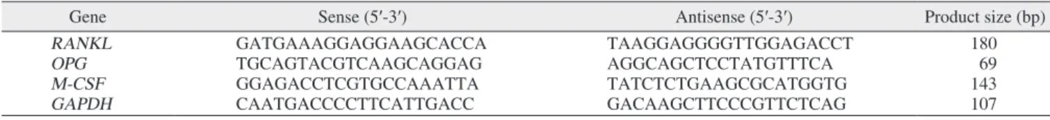
Table 1. Primers used in this study
Gene Sense (5′-3′) Antisense (5′-3′) Product size (bp)
RANKL GATGAAAGGAGGAAGCACCA TAAGGAGGGGTTGGAGACCT 180
OPG TGCAGTACGTCAAGCAGGAG AGGCAGCTCCTATGTTTCA 69
M-CSF GGAGACCTCGTGCCAAATTA TATCTCTGAAGCGCATGGTG 143
GAPDH CAATGACCCCTTCATTGACC GACAAGCTTCCCGTTCTCAG 107
(RANKL: receptor activator of nuclear factor kappa-B ligand, OPG: osteoprotegerin, M-CSF: macrophage colony-stimulating factor)
Seok-Young Jeong et al: Combined effect of recombinant human bone morphogenetic protein-2 and low level laser irradiation on bisphosphonate-treated osteoblasts. J Korean Assoc Oral Maxillofac Surg 2018
concentrations, there was no difference in survivability at the different time points. The effect of rhBMP-2 on alendronate- treated hFOB cells was investigated by performing cell survivability assays using cells treated with alendronate and rhBMP-2 at concentrations of 100 µM and 100 ng/mL, re- spectively, at 24- and 48-hour incubation times.(Fig. 3) At the 24-hour time point, treatment with rhBMP-2 was shown to significantly increase cell survivability. To investigate the
effects of LLLT, cells in the various groups were subjected to LLL irradiation.(Fig. 4) LLLT was shown to increase cell survivability and also demonstrated a synergic effect with rhBMP-2. While LLLT seemed to have a greater effect than rhBMP-2 on cell survivability, the difference was not statisti-
Control 120
100
80
60
0
Cellviability(%)
40
20
Alendronate 24 hr+rhBMP-2 24 hr Alendronate 48 hr+rhBMP-2 24 hr
Alendronate 100 M
Alendronate 100 M +rhBMP-2 100 ng/mL
*
*
Fig. 3. Survivability of human fetal osteoblast cells (hFOB 1.19) following treatment with 100 μM alendronate and recombinant human bone morphogenetic protein-2 (rhBMP-2). *P<0.05, com- pared to the control group; †P<0.05, compared to the alendronate group.
Seok-Young Jeong et al: Combined effect of recombinant human bone morphogenetic protein-2 and low level laser irradiation on bisphosphonate-treated osteoblasts. J Korean Assoc Oral Maxillofac Surg 2018
100
0 10 25 50 75
120
100
80
60
40
Cellviability(%)
Concentrations of rhBMP-2 (ng/mL) 24 hr
48 hr 72 hr
*
Fig. 2. Survivability of human fetal osteoblast cells (hFOB 1.19) following treatment with different concentrations of recombinant human bone morphogenetic protein-2 (rhBMP-2). Cell survival was significantly increased at higher rhBMP-2 concentrations, while no difference was observed at each time point. *P<0.05, compared to the control group.
Seok-Young Jeong et al: Combined effect of recombinant human bone morphogenetic protein-2 and low level laser irradiation on bisphosphonate-treated osteoblasts. J Korean
a b c d e
120
100
80
60
0
Cellviability(%)
40
20
*
*
*,
*,
Fig. 4. Survivability of human fetal osteoblast cells (hFOB 1.19) following treatment with 100 μM alendronate, recombinant hu- man bone morphogenetic protein-2 (rhBMP-2), and low-level laser therapy (LLLT). LLLT was shown to increase cell surviv- ability and also demonstrated a synergic effect with rhBMP-2.
*P<0.05, compared to the control group; †P<0.05, compared to the alendronate group. (a: control, b: alendronate+fresh media, c:
alendronate+rhBMP-2 100 ng/mL, d: alendronate+LLLT [80 mW, 15 seconds], e: alendronate+rhBMP-2/LLLT [80 mW, 15 sec- onds])
Seok-Young Jeong et al: Combined effect of recombinant human bone morphogenetic protein-2 and low level laser irradiation on bisphosphonate-treated osteoblasts. J Korean 200
0 1 10 50 100
120
100
80
60
40
20
Cellviability(%)
Alendronate concentrations ( M) 0
24 hr 48 hr 72 hr
*
Fig. 1. Survivability of human fetal osteoblast cells (hFOB 1.19) following treatment with different concentrations of alendronate.
Cells treated with alendronate concentrations ≥100 µM exhibited significantly decreased survival. *P<0.05, compared to the control group.
Seok-Young Jeong et al: Combined effect of recombinant human bone morphogenetic protein-2 and low level laser irradiation on bisphosphonate-treated osteoblasts. J Korean Assoc Oral Maxillofac Surg 2018
cally significant.
2. Effects on expression of TGF-β1 and BMP-2
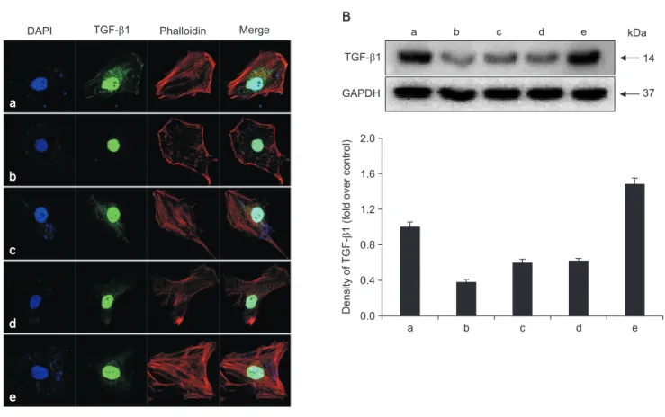
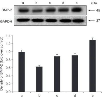
To determine whether alendronate, rhBMP-2, and LLLT altered the expression of differentiation factors, all groups of cells were analyzed for secretion of TGF-β1 and BMP-2.
The results showed that, compared to controls, treatment with 100 μM alendronate (negative control) resulted in decreased expression of both TGF-β1 and BMP-2. However, expression of TGF-β1 and BMP-2 increased following treatment with either rhBMP-2 or LLLT, and the effects of these two agents were similar in magnitude. When rhBMP-2 and LLLT were applied together, the effect was increased, and TGF-β1 ex- pression was increased more than expression of BMP-2. De- position of TGF-β1 was confirmed by immuno-fluorescence microscopy. The staining density around the nucleus de- creased following treatment with alendronate but was restored following treatment with rhBMP-2 and LLLT.(Fig. 5, 6)
3. Effects on expression of collagen type I and osteopontin
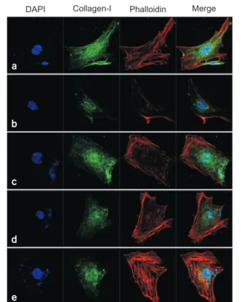
To determine whether alendronate, rhBMP-2, and LLLT have an effect on bone matrix formation, all groups of cells were analyzed for expression of collagen type I and osteo- pontin. The results showed that, compared to the controls, secretion of both collagen type I and osteopontin decreased after treatment with 100 μM alendronate (negative control).
However, the decrease in collagen type I secretion was not statistically significant. The expression of collagen type I increased following application of rhBMP-2 and LLLT, with LLLT having a greater effect on osteopontin expression.
Deposition of collagen type I was confirmed by immunofluo- rescence microscopy. The density of cytoplasm decreased following treatment with alendronate but was restored by treatment with rhBMP-2 and LLLT.(Fig. 7, 8)
a b c d e
2.0
1.6
1.2
0.8
0.4
DensityofTGF-1(foldovercontrol) 0.0 DAPI
A
TGF- 1 Phalloidin Merge
GAPDH TGF- 1
B
a b c d e
a
b
c
d
e
37 kDa
14
Fig. 5. Effects of alendronate, recombinant human bone morphogenetic protein-2 (rhBMP-2), and low-level laser therapy (LLLT) on ex- pression of transforming growth factor-beta1 (TGF-β1). A. Confocal microscopic assay for expression of TGF-β1. B. Western blot assay for expression of TGF-β1. Expression of TGF-β1 increased when following treatment with either rhBMP-2 or LLLT. The staining density around the nucleus decreased following treatment with alendronate but was restored by rhBMP-2 and LLLT. (a: control, b: alendronate 100 µM, c:
alendronate 100 µM+rhBMP-2 100 ng/mL, d: alendronate 100 µM+LLLT [80 mW, 15 seconds], e: alendronate 100 µM+rhBMP-2/LLLT [80 mW, 15 seconds])
Seok-Young Jeong et al: Combined effect of recombinant human bone morphogenetic protein-2 and low level laser irradiation on bisphosphonate-treated osteoblasts. J Korean Assoc Oral Maxillofac Surg 2018
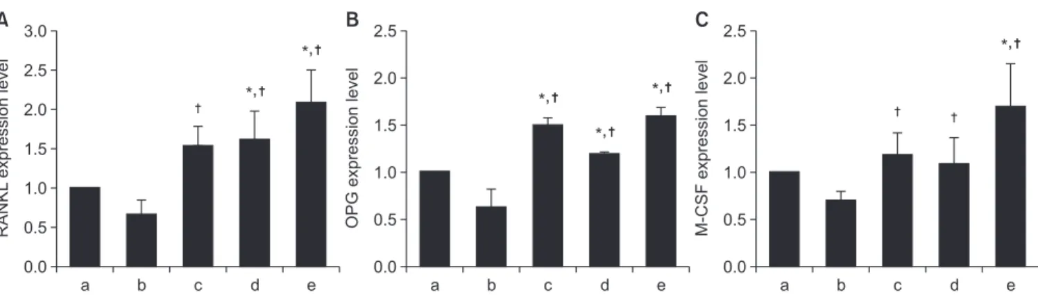
4. Effects on expression of RANKL, OPG, and M-CSF
RT-PCR was performed to investigate the expression of RANKL, OPG, and M-CSF, which are proteins-related to osteoclastogenesis. Compared to the control group, treatment with alendronate resulted in reduced expression of RANKL, OPG, and M-CSF. However, expression of all 3 cytokines increased following application of rhBMP-2 or LLLT. Treat- ment with rhBMP-2 resulted in greater effects on OPG and M-CSF expression, while treatment with LLLT exhibited a greater effect on expression of RANKL. However, the dif- ferences between the effects of rhBMP-2 and LLLT were not statistically significant. The combined application of rhBMP-2 and LLLT resulted in greater increases in cytokine expression than application of either treatment by itself.(Fig.
9)
IV. Discussion
In the present study, we studied the effects of alendronate, the most frequently prescribed oral BP in Korea. The strong inhibition of osteoclast function precipitated by BP therapy can lead to inhibition of normal bone turnover16. The toxic effects of BPs on osteoclasts are well understood and are thought to influence the occurrence of BRONJ. Additional complex mechanisms and events may have a correlation with BRONJ development, and there are various interactions be- tween bone cells that must be considered17. Although the ma- jority of BP studies conducted in vitro has focused on events occurring in osteoclastic lineage cells, recent studies suggest that BPs may also have a stimulatory effect on osteoblasts.
García-Moreno et al.18 reported that, while high concentra- tions of alendronate inhibited osteoblast proliferation, lower concentrations produced no significant effects. This effect may depend on the soluble factors of osteoblasts that inhibit osteoclast formation and activiation19,20.
Two factors supplied by osteoblast lineage cells are of critical importance for osteoclastogenesis: M-CSF and RANKL. M-CSF binds to receptors on osteoclasts to induce differentiation, which ultimately leads to increased plasma calcium level through resorption of bone. M-CSF is required for proliferation and survival of osteoclast precursors, while RANKL is critical for precursor differentiation into mature, multinucleated osteoclasts. RANKL binding to receptor ac- tivator of nuclear factor kappa-B (RANK) results in rapid differentiation of osteoclast precursors to form mature os- teoclasts. In addition to these positive regulators, osteoblast lineage cells produce OPG, which is the major inhibitor of osteoclast differentiation. Specifically, OPG is a member of the tumor necrosis factor receptor family and a soluble decoy receptor that ultimately binds to RANKL by binding and ac- tivating RANK. When OPG binds to RANK, there is rapid cessation of osteoclast formation, activation, and survival, leading to initiation of subsequent bone formation21.
TGF-1 is a member of the TGF-superfamily and one of the primary cytokines involved in growth and remodeling of bone. During bone resorption, active forms of these growth factors are released into the extracellular environment.
Osteopontin has been implicated as an important factor in bone remodeling and is synthesized by a variety of cell types22-25. Specifically, research suggests that osteopontin plays a role in anchoring osteoclasts to the mineral matrix of bones26.
a b c d e
1.4 1.2 1.0
0.6
0.2 DensityofBMP-2(foldovercontrol) 0.0
* GAPDH
BMP-2
a b c d e
0.8
0.4
kDa 45
37
Fig. 6. Effects of alendronate, recombinant human bone morpho- genetic protein-2 (rhBMP-2), and low-level laser therapy (LLLT) on expression of BMP-2. Western blot assay for BMP-2 expression.
When rhBMP-2 and LLLT were applied together, the effect was increased, and transforming growth factor-beta1 expression was increased more than that of BMP-2. (a: control, b: alendronate 100 µM, c: alendronate 100 µM+rhBMP-2 100 ng/mL, d: alen- dronate 100 µM+LLLT [80 mW, 15 seconds], e: alendronate 100 µM+rhBMP-2/LLLT [80 mW, 15 seconds])
Seok-Young Jeong et al: Combined effect of recombinant human bone morphogenetic protein-2 and low level laser irradiation on bisphosphonate-treated osteoblasts. J Korean Assoc Oral Maxillofac Surg 2018
RANKL, OPG, and M-CSF in BP-treated hFOB cells for their effects on osteoclastogenesis. In addition, we surveyed the expression of TGF-β1 and BMP-2 for their roles as dif- ferentiation factors and also investigated the expression of collagen type I and osteopontin as factors involved in forma- tion of bony matrix.
García-Moreno et al.18 evaluated the cytotoxic effects of alendronate on primary human osteoblasts and found no vi- able cells after 48 hours of culture at high alendronate con- centrations. Additionally, synthesis of type I collagen, a vital component of the bony matrix, was completely inhibited. The present study also showed that alendronate had significant effects on osteoblast survival. Cells treated with alendronate for 24 hours at concentrations ≥100 µM exhibited signifi- cantly decreased survival. Additionally, the expression of both TGF-β1 and BMP-2 decreased following treatment with 100 μM alendronate (negative control). The expression of RANKL, OPG, and M-CSF, which are related to osteoclasto- genesis, also decreased. These results suggest that high con-
centrations of alendronate suppress osteoblastic activity and osteoclastogenesis.
BMP-2 is well recognized as most effective for bone gen- eration and has been approved for clinical use by the U.S.
Food and Drug Administration. BMP-2 induces bone and cartilage formation and also plays a key role in osteoblast differentiation. Itoh et al.27 reported that BMP-2 enhanced the survival of purified osteoclasts supported by RANKL, but not by M-CSF. Zheng et al.28 showed that the BMP- 2/7 heterodimer affects every osteoclastogenic event in a complicated dose-dependent manner and could increase the number of osteoclasts at concentrations of 10 to 150 ng/mL.
In our investigation, we assayed the survivability of hFOB-2 cells following 24, 48, and 72 hours of incubation with vari- ous concentrations of rhBMP-2. We found that cell survival was slightly increased at higher rhBMP-2 concentrations, but was not affected by incubation time. Additionally, decreased expression of TGF-β1, BMP-2, RANKL, OPG, and M-CSF recovered to normal level after addition of rhBMP-2.
a b c d e
2.0
1.6
1.2
0.8
0.4
0.0 DensityofcollagentypeI (foldovercontrol) DAPI
A
Collagen-I Phalloidin Merge
GAPDH Collagen-I
B
a b c d e
a
b
c
d
e
kDa 140
37
Fig. 7. Effects of alendronate, recombinant human bone morphogenetic protein-2 (rhBMP-2), and low-level laser therapy (LLLT) on expres- sion of collagen type I. A. Confocal microscopic assay for expression of collagen type I. B. Western blot assay for expression of collagen type I. Expression of collagen type I increased following application of rhBMP-2 and LLLT. The decreased density of cytoplasm with alen- dronate was restored by treatment with rhBMP-2 and LLLT. (a: control, b: alendronate 100 µM, c: alendronate 100 µM+rhBMP-2 100 ng/
mL, d: alendronate 100 µM+LLLT [80 mW, 15 seconds], e: alendronate 100 µM+rhBMP-2/LLLT [80 mW, 15 seconds])
Seok-Young Jeong et al: Combined effect of recombinant human bone morphogenetic protein-2 and low level laser irradiation on bisphosphonate-treated osteoblasts. J Korean Assoc Oral Maxillofac Surg 2018
In the clinical field, diode lasers have been used predomi- nantly for application as LLLT or biostimulation, and many studies have evaluated the efficacy of LLLT on various dis- orders29. In our study, the expression of collagen type I and
BMP increased after treatment with rhBMP-2 and LLLT.
These results were consistent with those of previous studies showing that LLLT promotes osteoblast proliferation and increases BMP expression30-33. Hirata et al.34 demonstrated that LLLT increases type I collagen expression in osteoblas- tic cells. Moreover, we observed significant increases of RANKL and M-CSF expression in cells treated with both LLLT and alendronate compared to alendronate-only treated cells. We suggest that LLLT influences the expression of RANKL, M-CSF, OPG, TGF-β1, and BMP to produce posi- tive effects on bone osteoclastogenesis cell differentiation.
The combined application of rhBMP-2 and LLLT exhibited a synergic effect, especially for TGF-β1.
While the expression of collagen type I and osteopontin slightly increased in each case, there was no evidence of a synergistic effect. This implies that formation of the bony matrix increased. Our study demonstrates the positive ef- fects of rhBMP-2 and LLLT for partially recovering from the inhibitory effects of BPs and provides an experimental basis for the effectiveness of combining rhBMP-2 and LLLT as a treatment modality for BRONJ. In addition, the results showed that combined use of these modalities increased their therapeutic effectiveness compared to use of either modality alone. Therefore, we suggest that combined use of rhBMP-2 and LLLT will be more effective than the separate use of ei- ther modality for treatment of BRONJ.
a b c d e
3.0 2.5 2.0 1.5 1.0 0.5 0.0
RANKLexpressionlevel
a b c d e
2.5
2.0
1.5
1.0
0.5
0.0
OPGexpressionlevel
a b c d e
2.5
2.0
1.5
1.0
0.5
0.0
M-CSFexpressionlevel
A B C
*,
*, *,
*,
*,
*,
Fig. 9. Effects of alendronate, recombinant human bone morphogenetic protein-2 (rhBMP-2), and low-level laser therapy (LLLT) on recep- tor activator of nuclear factor kappa-B (RANKL), osteoprotegerin (OPG), and macrophage colony-stimulating factor (M-CSF) expression as determined by real-time polymerase chain reaction. A. RANKL expression. B. OPG expression. C. M-CSF expression. Treatment with rhBMP-2 resulted in greater effects on OPG and M-CSF expression, while treatment with LLLT had an effect on expression of RANKL.
However, the differences between the effects of rhBMP-2 and LLLT were not statistically significant. *P<0.05, compared to the control group; †P<0.05, compared to the alendronate group. (a: control, b: alendronate 100 µM, c: alendronate 100 µM+rhBMP-2 100 ng/mL, d:
alendronate 100 µM+LLLT [80 mW, 15 seconds], e: alendronate 100 µM+rhBMP-2/LLLT [80 mW, 15 seconds])
Seok-Young Jeong et al: Combined effect of recombinant human bone morphogenetic protein-2 and low level laser irradiation on bisphosphonate-treated osteoblasts. J Korean Assoc Oral
a b c d e
1.4 1.2 1.0
0.6
0.2 0.0 Densityofosteopontin (foldovercontrol)
GAPDH
a b c d e
0.8
0.4 Osteopontin
kDa 66
37
Fig. 8. Effects of alendronate, recombinant human bone morpho- genetic protein-2 (rhBMP-2), and low-level laser therapy (LLLT) on expression of osteopontin. Western blot assay for expression of osteopontin. LLLT exhibited a greater effect on osteopontin ex- pression. (a: control, b: alendronate 100 µM, c: alendronate 100 µM+rhBMP-2 100 ng/mL, d: alendronate 100 µM+LLLT [80 mW, 15 seconds], e: alendronate 100 µM+rhBMP-2/LLLT [80 mW, 15 seconds])
Seok-Young Jeong et al: Combined effect of recombinant human bone morphogenetic protein-2 and low level laser irradiation on bisphosphonate-treated osteoblasts. J Korean Assoc Oral Maxillofac Surg 2018
V. Conclusion
The expression of osteoclastogenesis-related proteins RANKL, OPG, and M-CSF in hFOB cells decreased follow- ing treatment with alendronate and increased following ap- plication of rhBMP-2 and LLLT. Additionally, while alendro- nate suppressed the expression of TGF-β1 and BMP-2, which functions in cell differentiation, this expression recovered fol- lowing combined application of rhBMP-2 and LLLT. Stud- ies on expression of collagen type I and osteopontin, which are related to formation of the bony matrix, showed similar results. In all cases, combined application of rhBMP-2 and LLLT was more effective than application of either modality alone.
ORCID
Seok-Young Jeong, https://orcid.org/0000-0002-9184-713X Ji-Un Hong, https://orcid.org/0000-0002-5736-8573 Jae Min Song, https://orcid.org/0000-0002-4047-2163 In Ryoung Kim, https://orcid.org/0000-0003-0232-0385 Bong Soo Park, https://orcid.org/0000-0002-9799-5627 Chul Hoon Kim, https://orcid.org/0000-0002-2492-174X Sang Hun Shin, https://orcid.org/0000-0002-0467-6206
Authors’ Contributions
S.Y.J. and J.M.S participated in data collection and wrote the manuscript. J.U.H. and C.H.K. participated in the study design and performed the statistical analysis. B.S.P., I.R.K., and S.H.S. participated in the study design and coordination and helped to draft the manuscript. All authors read and ap- proved the final manuscript.
Acknowledgements
This study conducted by 2 year research grant of Pusan National University.
Conflict of Interest
No potential conflict of interest relevant to this article was reported.
References
1. Boonyapakorn T, Schirmer I, Reichart PA, Sturm I, Massenkeil G. Bisphosphonate-induced osteonecrosis of the jaws: prospective study of 80 patients with multiple myeloma and other malignan- cies. Oral Oncol 2008;44:857-69.
2. Marx RE. Pamidronate (Aredia) and zoledronate (Zometa) induced avascular necrosis of the jaws: a growing epidemic. J Oral Maxil- lofac Surg 2003;61:1115-7.
3. Lee JK, Kim KW, Choi JY, Moon SY, Kim SG, Kim CH, et al.
Bisphosphonates-related osteonecrosis of the jaw in Korea: a pre- liminary report. J Korean Assoc Oral Maxillofac Surg 2013;39:9- 4. Woo SB, Hellstein JW, Kalmar JP. Narrative [corrected] review: 13.
bisphosphonates and osteonecrosis of the jaws. Ann Intern Med 2006;144:753-61.
5. Mavrokokki T, Cheng A, Stein B, Goss A. Nature and frequency of bisphosphonate-associated osteonecrosis of the jaws in Australia. J Oral Maxillofac Surg 2007;65:415-23.
6. Rodan GA, Fleisch HA. Bisphosphonates: mechanisms of action. J Clin Invest 1996;97:2692-6.
7. Sahni M, Guenther HL, Fleisch H, Collin P, Martin TJ. Bisphos- phonates act on rat bone resorption through the mediation of osteo- blasts. J Clin Invest 1993;91:2004-11.
8. Murakami H, Takahashi N, Sasaki T, Udagawa N, Tanaka S, Na- kamura I, et al. A possible mechanism of the specific action of bisphosphonates on osteoclasts: tiludronate preferentially affects polarized osteoclasts having ruffled borders. Bone 1995;17:137-44.
9. Aubin JE, Triffitt JT. Mesenchymal stem cells and the osteoblast lineage. In: Bilezikian JP, Raisz LG, Rodan GA, eds. Principles of bone biology. 2nd ed. New York: Academic; 2002.
10. Tay JY, Bay BH, Yeo JF, Harris M, Meghji S, Dheen ST. Identifi- cation of RANKL in osteolytic lesions of the facial skeleton. J Dent Res 2004;83:349-53.
11. Lacey DL, Timms E, Tan HL, Kelley MJ, Dunstan CR, Burgess T, et al. Osteoprotegerin ligand is a cytokine that regulates osteoclast differentiation and activation. Cell 1998;93:165-76.
12. Khadra M, Lyngstadaas SP, Haanaes HR, Mustafa K. Effect of laser therapy on attachment, proliferation and differentiation of human osteoblast-like cells cultured on titanium implant material.
Biomaterials 2005;26:3503-9.
13. Urist MR. Bone: formation by autoinduction. Science 1965;150:
893-9.
14. Chen D, Zhao M, Mundy GR. Bone morphogenetic proteins.
Growth Factors 2004;22:233-41.
15. Kwon TK, Song JM, Kim IR, Park BS, Kim CH, Cheong IK, et al. Effect of recombinant human bone morphogenetic protein-2 on bisphosphonate-treated osteoblasts. J Korean Assoc Oral Maxillo- fac Surg 2014;40:291-6.
16. Marx RE. Clinical concerns of alendronate use. J Oral Maxillofac Surg 2008;66:1322.
17. Rustemeyer J, Bremerich A. Bisphosphonate-associated osteone- crosis of the jaw: what do we currently know? A survey of knowl- edge given in the recent literature. Clin Oral Investig 2009;14:59- 18. García-Moreno C, Serrano S, Nacher M, Farré M, Díez A, Mari-64.
ñoso ML, et al. Effect of alendronate on cultured normal human osteoblasts. Bone 1998;22:233-9.
19. Vitté C, Fleisch H, Guenther HL. Bisphosphonates induce osteo- blasts to secrete an inhibitor of osteoclast-mediated resorption.
Endocrinology 1996;137:2324-33.
20. Nishikawa M, Akatsu T, Katayama Y, Yasutomo Y, Kado S, Kugal N, et al. Bisphosphonates act on osteoblastic cells and inhibit os- teoclast formation in mouse marrow cultures. Bone 1996;18:9-14.
21. Marini H, Minutoli L, Polito F, Bitto A, Altavilla D, Atteritano M, et al. OPG and sRANKL serum concentrations in osteopenic, post-
menopausal women after 2-year genistein administration. J bone Miner Res 2008;23:715-20.
22. Choi ST, Kim JH, Kang EJ, Lee SW, Park MC, Park YB, et al.
Osteopontin might be involved in bone remodelling rather than in inflammation in ankylosing spondylitis. Rheumatology (Oxford) 2008;47:1775-9.
23. Ashizawa N, Graf K, Do YS, Nunohiro T, Giachelli CM, Meehan WP, et al. Osteopontin is produced by rat cardiac fibroblasts and mediates A(II)-induced DNA synthesis and collagen gel contrac- tion. J Clin Invest 1996;98:2218-27.
24. Murry CE, Giachelli CM, Schwartz SM, Vracko R. Macrophages express osteopontin during repair of myocardial necrosis. Am J Pathol 1994;145:1450-62.
25. Ikeda T, Shirasawa T, Esaki Y, Yoshiki S, Hirokawa K. Osteo- pontin mRNA is expressed by smooth muscle-derived foam cells in human atherosclerotic lesions of the aorta. J Clin Invest 1993;92:2814-20.
26. Reinholt FP, Hultenby K, Oldberg A, Heinegård D. Osteopontin: a possible anchor of osteoclasts to bone. Proc Natl Acad Sci U S A 1990;87:4473-5.
27. Itoh K, Udagawa N, Katagiri T, Iemura S, Ueno N, Yasuda H, et al. Bone morphogenetic protein 2 stimulates osteoclast differentia- tion and survival supported by receptor activator of nuclear factor- kappaB ligand. Endocrinology 2001;142:3656-62.
28. Zheng Y, Wang L, Zhang X, Zhang X, Gu Z, Wu G. BMP2/7 het- erodimer can modulate all cellular events of the in vitro RANKL- mediated osteoclastogenesis, respectively, in different dose pat- terns. Tissue Eng Part A 2012;18:621-30.
29. Goldman L, Goldman B, Van Lieu N. Current laser dentistry. La- sers Surg Med 1987;6:559-62.
30. Stein A, Benayahu D, Maltz L, Oron U. Low-level laser irradiation promotes proliferation and differentiation of human osteoblasts in vitro. Photomed Laser Surg 2005;23:161-6.
31. Renno AC, McDonnell PA, Parizotto NA, Laakso EL. The effects of laser irradiation on osteoblast and osteosarcoma cell prolifera- tion and differentiation in vitro. Photomed Laser Surg 2007;25:275- 32. Kiyosaki T, Mitsui N, Suzuki N, Shimizu N. Low-level laser 80.
therapy stimulates mineralization via increased Runx2 expression and ERK phosphorylation in osteoblasts. Photomed Laser Surg 2010;28 Suppl 1:S167-72.
33. Shin SH, Kim KH, Choi NR, Kim IR, Park BS, Kim YD, et al.
Effect of low-level laser therapy on bisphosphonate-treated osteo- blasts. Maxillofac Plast Reconstr Surg 2016;38:48.
34. Hirata S, Kitamura C, Fukushima H, Nakamichi I, Abiko Y, Ter- ashita M, et al. Low-level laser irradiation enhances BMP-induced osteoblast differentiation by stimulating the BMP/Smad signaling pathway. J Cell Biochem 2010;111:1445-52.