I. Introduction
Although recombinant human bone morphogenetic protein- 2 (rhBMP-2), which is a potent osteoinductor, has long been considered a promising avenue for bone regeneration, and rhBMP-2 has been commercially available for the treatment of oral-maxillofacial defects, the issue of the carrier system is an ongoing topic that has not yet been solved. Like other growth factors, rhBMP-2 requires a delivery system that cre- ates optimal conditions for cellular and vascular growth, cel- lular attachment, and release kinetics 1-4 .
Calcium phosphates, such as hydroxyapatite (HA) and β-tricalcium phosphate (β-TCP), have been deemed to be suitable candidates for rhBMP-2 delivery systems because Jae-Jun Ryu
Department of Dentistry, Korea University Anam Hospital, 73 Inchon-ro, Seongbuk-gu, Seoul 02841, Korea
TEL: +82-2-920-6837 FAX: +82-2-921-7348 E-mail: [email protected]
ORCID: http://orcid.org/0000-0001-6903-5955 Hyon-Seok Jang
Department of Dentistry, Korea University Ansan Hospital, 123 Jeokgeum-ro, Danwon-gu, Ansan 15355, Korea
TEL: +82-31-412-5370 FAX: +82-31-401-7125 E-mail: [email protected]
ORCID: http://orcid.org/0000-0002-8493-4657
*These authors contributed equally to this work as first co-authors.
This is an open-access article distributed under the terms of the Creative Commons Attribution Non-Commercial License (http://creativecommons.org/licenses/by-nc/4.0/), which permits unrestricted non-commercial use, distribution, and reproduction in any medium, provided the original work is properly cited.
CC
Demineralized dentin matrix combined with recombinant human bone morphogenetic protein-2 in rabbit calvarial defects
In-Woong Um 1, *, Suk-Hyun Hwang 2, *, Young-Kyun Kim 3 , Moon-Young Kim 4 , Sang-Ho Jun 5 , Jae-Jun Ryu 5 , Hyon-Seok Jang 6
1 R&D Institute, Korea Tooth Bank, Seoul, 2 Department of Medicine, Korea University Graduate School, Seoul,
3 Department of Oral and Maxillofacial Surgery, Section of Dentistry, Seoul National University Bundang Hospital, Seongnam,
4 Department of Oral and Maxillofacial Surgery, College of Dentistry, Dankook University, Cheonan,
5 Department of Dentistry, Korea University Anam Hospital, Seoul,
6 Department of Dentistry, Korea University Ansan Hospital, Ansan, Korea
Abstract (J Korean Assoc Oral Maxillofac Surg 2016;42:90-98)
Objectives: The aim of this study was to compare the osteogenic effects of demineralized dentin matrix (DDM) combined with recombinant human bone morphogenetic protein-2 (rhBMP-2) in rabbit calvarial defects with DDM and anorganic bovine bone (ABB) combined with rhBMP-2.
Materials and Methods: Four round defects with 8-mm diameters were created in each rabbit calvaria. Each defect was treated with one of the fol- lowing: 1) DDM, 2) ABB/rhBMP-2, or 3) DDM/rhBMP-2. The rhBMP-2 was combined with DDM and ABB according to a stepwise dry and dip ly- ophilizing protocol. Histological and microcomputed tomography (µCT) analyses were performed to measure the amount of bone formation and bone volume after 2- and 8-week healing intervals.
Results: Upon histological observation at two weeks, the DDM and ABB/rhBMP-2 groups showed osteoconductive bone formation, while the DDM/
rhBMP-2 group showed osteoconductive and osteoinductive bone formation. New bone formation was higher in DDM/rhBMP-2, DDM and ABB decreasing order. The amounts of bone formation were very similar at two weeks; however, at eight weeks, the DDM/rhBMP-2 group showed a two- fold greater amount of bone formation compared to the DDM and ABB/rhBMP-2 groups. The µCT analysis showed markedly increased bone volume in the DDM/rhBMP-2 group at eight weeks compared with that of the DDM group. Notably, there was a slight decrease in bone volume in the ABB/
rhBMP-2 group at eight weeks. There were no significant differences among the DDM, ABB/rhBMP-2, and DDM/rhBMP-2 groups at two or eight weeks.
Conclusion: Within the limitations of this study, DDM appears to be a suitable carrier for rhBMP-2 in orthotopic sites.
Key words: Demineralized dentin matrix, Recombinant human bone morphogenetic protein-2, Microcomputed tomography, Histomorphometric analysis
[paper submitted 2016. 1. 20 / revised 2016. 3. 8 / accepted 2016. 3. 13]
Copyright Ⓒ 2016 The Korean Association of Oral and Maxillofacial Surgeons. All rights reserved.
This study was supported by a grant from the Korean Health Technology R&D Project, Korea Health Industry Development Institution, Republic of
Korea (HI15C3136).
and cleaned by removing foreign substances such as restora- tion materials, root canals, and prosthetics, as well as soft tis- sues of the periodontium, pulp, and caries. After the cleaned tooth was divided into crown and root, the root portion was collected and prepared for partially DDM as reported previ- ously. Next, 0.3- to 0.8-mm crushed particles were soaked in a distilled water and hydrogen dioxide solution, and the remaining foreign substances were removed by ultrasonic cleaning. The cleaned particles were dehydrated with ethyl alcohol and defatted with an ethyl ether solution. The par- ticles were then decalcified for two hours in 0.6 N HCl 11 .
2. Preparation of ABB combined with rhBMP-2 and DDM combined with rhBMP-2
ABB and DDM were combined with rhBMP-2 by the dip- dry method. Briefly, 0.01 mg of rhBMP-2 (CowellBMP;
Cowellmedi) were added to 1 g of ABB particles, frozen in a deep freezer at –70 o C and then fixed in a lyophilizer (ILShin Lab, Seoul, Korea) following the manufacturer’s instructions.
Alternatively, 2 mg/mL of rhBMP-2 (CowellBMP) were fixed to 0.03 g of DDM by a stepwise dip and dry lyophiliz- ing protocol 11-13 .
3. Surgical procedures
Twelve male rabbits (body weight, 2.50-3.00 kg) were used in this study and were maintained in cages at an ambient room temperature of 21 o C with ad libitum access to water and a standard laboratory pellet diet. Animal selection and man- agement, surgical protocol, and preparation followed routines approved by the Institutional Animal Care and Use Commit- tee (BA1303-124-016-03). The animals were anesthetized with an intramuscular injection (5 mg/kg body weight) of a 4:1 solution of ketamine hydrochloride (Ketalar; Yuhan Co., Seoul, Korea) and xylazine (Rompun; Bayer Korea, Seoul, Korea). The surgical site was shaved and scrubbed with io- dine. For the calvarial defect model, an incision was made in the sagittal plane across the cranium, and a full-thickness flap was reflected to expose the calvarial bone. Standardized circular transosseous defects 8 mm in diameter were created on the cranium using a trephine drill (3i Implant Innovations Inc., Palm Beach Gardens, FL, USA) and copious saline irri- gation. In total, four round defects with 8 mm diameters were created in each rabbit calvaria. Respective groups received the following treatments: 1) DDM control (0.03 g), 2) ABB/
rhBMP-2 (1.0 g), and 3) DDM/rhBMP-2 (0.03 g). All surgi- of their space-providing properties. Biphasic calcium phos-
phate (BCP) in particular, which is a specific ratio of HA and β-TCP, has well-documented osteoconductive properties 5 .
However, for the commercially available rhBMP-2 in the maxillofacial region, the only approved carrier is absorbable collagen sponges (ACSs). Several studies have supported this carrier system 6,7 . Triplett et al. 8 reported the results of multi- center randomized clinical trials of rhBMP-2/ACS applica- tion for sinus floor elevation and showed that its efficacy and safety were comparable to those of an autogenous bone graft.
On the other hand, a report 9 in 1998 suggested that root dentin prepared from extracted teeth could be recycled for use as a carrier of rhBMP-2 because it induces new bone formation in the periodontium, while a later report 10 in 2005 showed that the osteoinductive matrices of human demineral- ized dentin matrix (DDM) particles could be effective as a carrier of rhBMP-2 for bone engineering. Since then, DDM powder has shown great potential as an effective carrier for rhBMP-2 based on in vitro and in vivo studies in 2014 and 2015 11,12 . To date, ectopic bone formation in vivo has dem- onstrated the possibility of DDM as a suitable carrier for rhBMP-2. Because DDM is composed of mainly type I col- lagen and HA, it might be more advantageous than ACSs in alveolar bone repair due to its structural integrity 11 .
The aim of this study is to investigate DDM as a suitable carrier for rhBMP-2 in order to establish efficient and safe approaches for bone regeneration in rabbit calvarial defects.
The first objective was to investigate whether the potential bone forming capacity of DDM/rhBMP-2 was greater than that of DDM, as measured using osteoinductivity and osteo- conductivity. The second objective was to evaluate whether DDM has greater potential as an rhBMP-2 carrier than does anorganic bovine bone (ABB).
II. Materials and Methods
The rhBMP-2 was provided by the Research and Devel- opment Institute of Cowellmedi (Busan, Korea). Two types of scaffolds were selected as carriers of rhBMP-2; AutoBT (human DDM; Korea Tooth Bank, Seoul, Korea) and ABB (Geistlich, Seoul, Korea). DDM and ABB were combined with rhBMP-2 to form DDM/rhBMP-2 (n=12) and ABB/rh- BMP-2 (n=12), respectively.
1. Preparations of DDM
An extracted human tooth was soaked in 70% ethyl alcohol
formalin solution for imaging. The images were produced us- ing a SkyScan 1172 CT system (Bruker microCT, Kontich, Belgium) in a high-resolution scanning mode (pixel matrix, 683×2,000×1,048; pixel size, 10.89 µm). The X-ray source was set at 70 kV and 141 mA for shooting with the aid of a 0.5-mm thick aluminum filter to optimize the contrast. The 360 o rotational angle of specimens was set at 0.5 o and 590 ms of exposure time.(Fig. 2. A) Three-dimensional images were produced using a CT reconstruction program (NRecon;
Bruker microCT), and the bone volume (BV) per tissue vol- ume (TV) was obtained using a reconstructed image analysis program (CTAn, dataviewer; Bruker microCT).
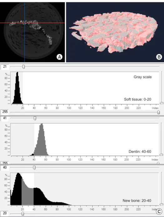
To determine new BV (NBV), the differences in brightness in the images were used. The BV and DDM volume were calculated within the range of the volume of interest (8×1 mm) in the form of a disk inside the skull defect, and this dif- ference was calculated as the NBV ratio.(Fig. 2. B)
To discriminate new bons from graft materials, images of graft materials were produced separately, and Hounsfield units (HU) were calculated to analyze the range of HU val- ues. The HU values considered new bone were set based on a value range of D3 bone of 20-40.(Fig. 2. C) Mean and standard deviation were calculated for all histomorphometric and µCT measurements. Comparative statistics of nonpara- metric variables were compared using the Mann-Whitney U- test, with P<0.05 used as the threshold of significance. All cal sites underwent primary closure using 4-0 Monosyn (gly-
conate absorbable monofilament; B. Braun, Aesculap, PA, USA). The animals were sacrificed at two and eight weeks after implantation for radiologic and histologic evaluation.
(Fig. 1)
4. Histomorphometric analysis
For histological analysis, the specimens were procured en bloc and fixed in 10% buffered formalin for 10 days, decal- cified in 10% formic acid for 14 days, and then embedded in paraffin. From serial sections of 5 µm thickness through the centers of the circular calvarial defects, two sections that contained each central portion were selected and stained with H&E.(Fig. 1. B) For histomorphometric analysis, the speci- mens were observed though an optical microscope at 100
× magnification. The digital images were obtained using a digital camera attached to the microscope, and the bone ratio of the area of newly formed bone (%) to total calvarial defect area was determined using an image analysis program (Kappa ImageBase Metreo; Kappa Optronics GmbH, Gleichen, Ger- many).(Fig. 1. B)
5. Microcomputed tomography ( µCT) analysis
Calvarial en bloc specimens were fixed in 10% buffered
A B
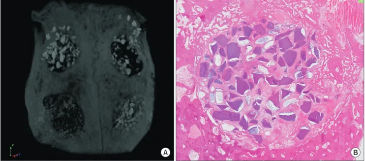
Fig. 1. A. Microcomputed tomography image of an en bloc rabbit calvarial defect. Top left: DDM, top right: DDM, bottom left: ABB/rh- BMP-2, bottom right: DDM/rhBMP-2. B. Histologic specimen of DDM at two weeks (H&E staining, ×100). Note the defect border, where DDM particles were grafted. (DDM: demineralized dentin matrix, ABB: anorganic bovine bone, rhBMP-2: recombinant human bone mor- phogenetic protein-2)
In-Woong Um et al: Demineralized dentin matrix combined with recombinant human bone morphogenetic protein-2 in rabbit calvarial defects. J Korean Assoc Oral Maxillofac Surg 2016
2) ABB/rhBMP-2 at two weeks
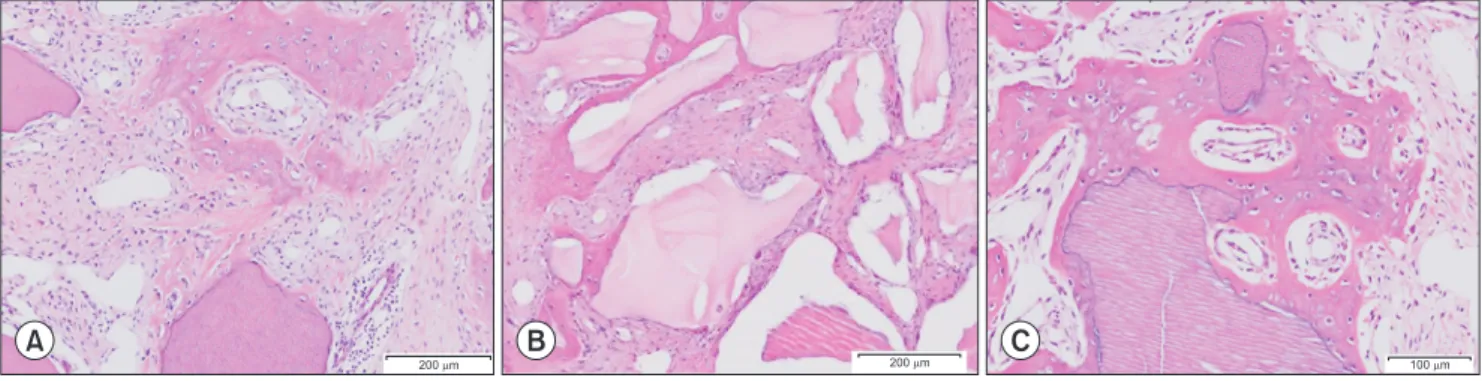
New bone was formed and migrated from the bone defect margin to the surface of the ABB scaffold. The cells around the ABB were stabilized, and spaces were filled with dense, fibrous connective tissues.(Fig. 3. B)
3) DDM/rhBMP-2 at two weeks
The newly formed bone around the DDM had very similar cellular and vascular activities to the bone that was formed and migrated from the defect bone margin.(Fig. 3. A) There were osteoinductive deposits of osteoids as well as pheno- typic transformation of osteoblast-like cells on the surfaces of the osteoids. There was also abundant vascular proliferation, which provided evidence of the remodeling capacity in the data analysis was completed using the GraphPad Prism 5.0
(GraphPad Software, San Diego, CA, USA).
III. Results
1. Histological findings
1) DDM at two weeks
New bone was formed and migrated from the bone defect margin that has abundant osteocytes and blood vessels. All of the fibroblasts around the scaffold and new osteoids were stimulated and transformed phenotypically into osteoblast- like cells. Subsequently, the newly formed bone from the bone defect margin created bridges.(Fig. 3. A)
Fig. 2. A. Microcomputed tomography image of calvarial defect, cross sec- tion at two weeks after demineralized dentin matrix (DDM) implantation. B.
Three-dimensional reconstruction of grafted area as volume of interest (8×1 mm) in the form of a disk from the red line (in Fig. 2. A). C. Quantification of new bone volume at two weeks after DDM implantation using NRecon re- construction software (NRecon v.1.4.4;
SkyScan).
In-Woong Um et al: Demineralized dentin matrix combined with recombinant human bone morphogenetic protein-2 in rabbit calvarial defects.
J Korean Assoc Oral Maxillofac Surg 2016
A B
C
Gray scale
Soft tissue: 0-20
Dentin: 40-60
New bone: 20-40
3) DDM/rhBMP-2
At two weeks, the amount of new bone formation was very similar to those of the DDM and ABB/rhBMP-2 groups, and it was mainly formed and migrated from the defect margin.
The newly formed bone had been deposited directly onto the DDM/rhBMP-2 surface; osteocytes were visible in the lacu- nae, and osteoblasts were lined up along the bony trabeculae.
At eight weeks, new bone was extending remarkably to form an anastomosing network of trabeculae between the DDM/
rhBMP-2 particles.(Table 1, Fig. 4)
3. New bone formation measured by µCT
The increased BV was remarkable from two to eight weeks in DDM/rhBMP-2, increasing by 87.14% compared to those of the DDM (+27.5%) and ABB/rhBMP-2 (–17.9%) groups, which showed similar or decreased amounts of BV from two to eight weeks. The results demonstrated that NBV had increased in the DDM and DDM/rhBMP-2 groups by the end of eight weeks, compared with a decrease in NBV in the ABB/rhBMP-2 group. There were no statistically significant differences among the groups with P<0.05 using the Mann- Whitney U-test.(Table 2, Fig. 5)
IV. Discussion
The aim of this study was to evaluate the bone forming ca- pacity of DDM loaded with rhBMP-2 in an orthoskeletal site.
The first objective was to establish that the potential bone forming capacity of DDM/rhBMP-2 was greater than that of newly deposited osteoids on the surface of DDM/rhBMP-2.
(Fig. 3. C)
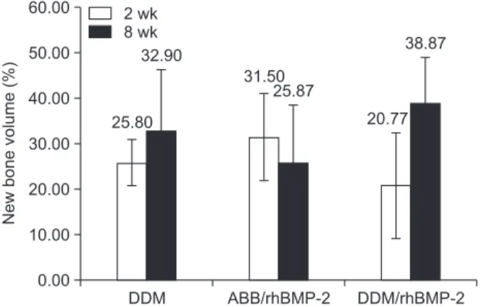
2. Histomorphometric findings of new bone formation
The implantation of the scaffolds combined with rhBMP-2 showed significantly increased bone formation during the period from two weeks to eight weeks. There were no statis- tically significant differences with P<0.05 using the Mann- Whitney U-test.(Table 1, Fig. 4)
1) DDM
After two weeks, minimal amounts of new bone had formed from the bone defect margins toward the central por- tion. Most of the area in a given defect was filled with fibrous connective tissue. At eight weeks, the bone formation and maturation had increased to a greater extent compared with that in the two-week specimens. New bone growth was ob- served both from the margins and around the DDM scaffold.
(Table 1, Fig. 4)
2) ABB/rhBMP-2
At two weeks, new bone was seen at the periphery of the defect margin and around the ABB particles. The immature bone tissue was not only interconnected between particles, but also partially encircled the ABB particles. At eight weeks, resorption of the ABB was not observed, and bone formation was increased compared with the two-week specimens. The bony tissues around the particles were progressively intercon- nected.(Table 1, Fig. 4)
A B C
200 m 200 m 100 m