Effect of recombinant human bone morphogenetic protein-2 on bisphosphonate-treated osteoblasts
Taek-Kyun Kwon1,*, Jae-Min Song1,*, In-Ryoung Kim2, Bong-Soo Park2, Chul-Hoon Kim3, In-Kyo Cheong1, Sang-Hun Shin1
Departments of 1Oral and Maxillofacial Surgery and 2Oral Anatomy and Cell Biology, School of Dentistry, Pusan National University, Yangsan, 3Department of Dentistry, Dong-A University Hospital, Busan, Korea
Abstract(J Korean Assoc Oral Maxillofac Surg 2014;40:291-296)
Objectives: Bisphosphonate-related osteonecrosis of the jaw (BRONJ) is a side effect of bisphophonate therapy that has been reported in recent years.
Osteoclastic inactivity by bisphosphonate is the known cause of BRONJ. Bone morphogenetic protein-2 (BMP-2) plays an important role in the devel- opment of bone. Recombinant human BMP-2 (rhBMP-2) is potentially useful as an activation factor for bone repair. We hypothesized that rhBMP-2 would enhance the osteoclast-osteoblast interaction related to bone remodeling.
Materials and Methods: Human fetal osteoblast cells (hFOB 1.19) were treated with 100 μM alendronate, and 100 ng/mL rhBMP-2 was added.
Cells were incubated for a further 48 hours, and cell viability was measured using an MTT assay. Expression of the three cytokines from osteoblasts, receptor activator of nuclear factor-κB ligand (RANKL), osteoprotegerin (OPG), and macrophage colony-stimulating factor (M-CSF), were analyzed by real-time polymerase chain reaction and enzyme-linked immunosorbent assay.
Results: Cell viability was decreased to 82.75%±1.00% by alendronate and then increased to 110.43%±1.35% after treatment with rhBMP-2 (P<0.05, respectively). OPG, RANKL, and M-CSF expression were all decreased by alendronate treatment. RANKL and M-CSF expression were increased, but OPG was not significantly affected by rhBMP-2.
Conclusion: rhBMP2 does not affect OPG gene expression in hFOB, but it may increase RANKL and M-CSF gene expression.
Key words: Bone morphogenetic protein-2, Alendronate, Osteoblasts, Macrophage colony-stimulating factor, RANK ligand
[paper submitted 2014. 7. 29 / accepted 2014. 9. 2]
oral and maxillofacial surgery, it is one of the most difficult diseases to treat1.
Alendronate pharmacologically inhibits farnesyl diphos- phate synthase in the mevalonate pathway, which is essential for the prenylation of proteins in osteoclasts. This ultimately causes mechanical inhibition of osteoclast adhesion on the bone margin where absorption takes place through osteoclast apoptosis. The effect of BPs on osteoclasts could also arise, at least in part, from modulation of the synthesis of resorption- promoting or resorption-inhibiting factors by osteoblasts2. Osteoblast/stromal cells regulate osteoclastogenesis by pro- ducing proteins such as macrophage colony-stimulating fac- tor (M-CSF), receptor activator of nuclear factor-κB ligand (RANKL), and osteoprotegerin (OPG). Through cell-to-cell contact of osteoblast/stromal cells with osteoclasts, RANKL and M-CSF induce osteoclast progenitor cells to differentiate into osteoclasts3.
I. Introduction
Bisphosphonates (BPs) are used to treat osteoporosis, bone metastasis and other conditions involving fragile bone. Oral BPs are used primarily in the treatment of osteoporosis, and alendronate is one of the most potent antiosteoporotic agents known. As a side effect of using BPs, BP-related osteonecro- sis of the jaw (BRONJ) was reported in 2003; in the field of Sang-Hun Shin
Department of Oral and Maxillofacial Surgery, Pusan National University Dental Hospital, 20 Geumo-ro, Mulgeum-eup, Yangsan 626-770, Korea TEL: +82-55-360-5113 FAX: +82-55-360-5104
E-mail: [email protected]
*These authors contributed equally to this study as first authors.
This is an open-access article distributed under the terms of the Creative Commons Attribution Non-Commercial License (http://creativecommons.org/licenses/by-nc/3.0/), which permits unrestricted non-commercial use, distribution, and reproduction in any medium, provided the original work is properly cited.
CC
Copyright Ⓒ 2014 The Korean Association of Oral and Maxillofacial Surgeons. All rights reserved.
This work was supported by 2-Year Research Grant of Pusan National University.
assay (ELISA) reader (Tecan, Männedorf, Switzerland) at a 570 nm excitatory emission wave length. In cells treated with 100 μM alendronate, cell viability was approximately 80% of the control. Consequently, that concentration was selected for the experimental group.
4. ELISA
Expression levels of the three proteins, RANKL, OPG and M-CSF, were measured with an ELISA kit (Quantikine;
R&D Systems, Minneapolis, MN, USA) according to the manufacturer’s instructions. Briefly, cultured hFOB 1.19 cells were uniformly seeded into 6-well culture dishes at a concen- tration of 2×10⁵ cells/well. When the cells were adherent 24 hours later, the original medium was replaced with medium containing 100 μM alendronate for 48 hours (alendronate group) after which 100 ng/mL BMP-2 (Cowellmedi, Busan, Korea) was added, and the cells were incubated for a further 48 hours (alendronate+rhBMP-2 group). The supernatants were then collected from each group, and an ELISA was used to determine the RANKL, OPG and M-CSF contents of each sample. The sample values were read from the standard curve set at 450 nm with 540-570 nm wavelength correction and with the measurement units expressed as pg/mL. All samples were assayed simultaneously.
5. Real-time polymerase chain reaction (RT-PCR)
The hFOB 1.19 cells were subjected to RNA extraction using spin columns (Rneasy; Qiagen, Hilden, Germany) according to the manufacturer’s instructions. RNA (2 µg) was reverse- transcribed, using the RevertAid First-Strand Synthesis Sys- tem kit for RT-PCR (Thermo Fisher Scientific, Pittsburgh, PA, USA) according to the manufacturer’s protocol. The cDNA was amplified with the PCR master mix SYBR Green kit (Applied Biosystems, Warrington, UK), and PCR ampli- fication was performed using the Chromo4 Real-Time PCR Detection System (Bio-Rad Laboratories Inc., Hercules, CA, USA). The running conditions were as follows: denaturing at 95°C for 3 minutes, 40 cycles of amplification at 95°C for 15 seconds and 60°C for 30 seconds. After the final cycle, a melting curve analysis was performed at 55°C-95°C intervals in 0.5°C steps. The sense and anti-sense primer sequences of OPG, RANKL, M-CSF, and the housekeeping gene glycer- aldehyde 3-phophate dehydrogenase (GAPDH) are listed in Table 1.
Bone morphogenetic protein (BMP), a subgroup of the transforming growth factor-β (TGF-β) superfamily4, induces the formation of bone and cartilage5-10. To date, there are more than 20 known BMP subgroups, of which BMP-2 is the primary subgroup used as a clinical treatment in many in vitro and in vivo studies11. Recombinant human BMP-2 (rh- BMP-2) is an activation factor for bone healing and has been used in dentistry due to its osteogenic effects.
The aim of the present study was to explore the effect of rhBMP-2 on the regulation of OPG, RANKL, and M-CSF in osteoclastogenesis in human osteoblast treated with alendro- nate.
II. Materials and Methods
1. Reagents
The following reagents were commercially obtained: Pro- Long Gold Antifade Reagent with DAPI, from Molecular Probes (Eugene, OR, USA); Dulbecco’s modified Eagle’s medium F12 (DMEM/F12) and fetal bovine serum (FBS), from Gibco (Gaithersburg, MD, USA); Alendronate, Dimeth- yl sulfoxide (DMSO), Hoechst 33342, RNase A, proteinase K, aprotinin, leupeptin, Triton X-100, PMSF and thiazolyl blue tetrazolium bromide (MTT), from Sigma (St. Louis, MO, USA); SuperSignal West Femto enhanced-chemilu- minescence Western blotting detection reagent, from Pierce (Rockford, IL, USA).
2. Cell culture
An hFOB 1.19 human fetal osteoblast cell line was pur- chased from the American Type Culture Collection (Rock- ville, MD, USA). The cells were maintained at 34°C with 5%
CO2 in DMEM/F12 with 4 mM L-glutamine, 1.5 g/L sodium bicarbonate, 4.5 g/L glucose and 1.0 mM sodium pyruvate supplemented with 10% FBS, under an air atmosphere.
3. Cell viability assay
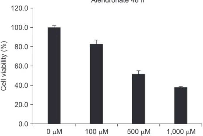
A total of 1×10⁴ cells were seeded in a 96-well plate, incu- bated for 24 hours, and treated with 0, 100, 500 or 1,000 μM concentrations of alendronate. The cells were then treated with 500 μg/mL of MTT stock solution and incubated at 34°C in a 5% CO2 atmosphere for 4 hours. The medium was aspirated, and the formazan crystals were dissolved in DMSO. Cell vi-
2. Effects of alendronate and rhBMP-2 on OPG expression
To evaluate OPG expression, RT-PCR and an ELISA were performed. OPG expression in the alendronate group was significantly decreased (P=0.012) as compared to the control group. In the alendronate+rhBMP-2 group, the OPG level increased relative to alendronate group, but this increase was not significant (P=0.482).(Fig. 2, Tables 3, 4)
6. Statistical analysis
Groups were comparatively analyzed by one way ANOVA.
Tukey’s post hoc test was used for cell viability and RT-PCR (SPSS Statistics 17.0; SPSS Inc., Chicago, IL, USA). P<0.05 was considered statistically significant.
III. Results
1. Viability of hFOB 1.19 cells
An MTT assay was performed at 48 hours with 0, 100, 500, 1,000 μM alendronate concentrations.(Fig. 1) Alendro- nate inhibited cellular viability at 100 μM (P<0.05). When the cells were exposed to concentrations above 100 μM, there was a significant decrease in cell viability (P<0.05).
To investigate the reverse effects of rhBMP-2 on cell vi- ability, cell viability assays were repeated with application of rhBMP-2 to cells treated with 100 μM alendronate after 48 hours.(Table 2) Cell viability significantly increased relative to the control and alendronate alone group (P<0.05).
Table 1. Primers used in this study
Gene Sense (5′-3′) Antisense (5′-3′)
RANKL OPG M-CSF GAPDH
GATGAAAGGAGGAAGCACCA TGCAGTACGTCAAGCAGGAG GGAGACCTCGTGCCAAATTA CAATGACCCCTTCATTGACC
TAAGGAGGGGTTGGAGACCT AGGCAGCTCCTATGTTTCA TATCTCTGAAGCGCATGGTG GACAAGCTTCCCGTTCTCAG
(RANKL: receptor activator of nuclear factor-κB ligand, OPG: osteoprotegerin, M-CSF: macrophage colony-stimulating factor, GAPDH:
glyceraldehyde 3-phophate dehydrogenase)
Taek-Kyun Kwon et al: Effect of recombinant human bone morphogenetic protein-2 on bisphosphonate-treated osteoblasts. J Korean Assoc Oral Maxillofac Surg 2014
Table 2. Effects of alendronate and recombinant human bone morphogenetic protein-2 (rhBMP-2) on cell viability
Groups Cell viability (%)
Control Alendronate rhBMP-2
Alendronate+BMP-2 P-value
100±0.67 82.75±1.001 113.50±1.631,2 110.43±1.351,2
0.000
1Statistical significance (P<0.05) compared with control group.
2Statistical significance (P<0.05) compared with alendronate group.
Values are presented as mean±standard deviation.
Taek-Kyun Kwon et al: Effect of recombinant human bone morphogenetic protein-2 on bisphosphonate-treated osteoblasts. J Korean Assoc Oral Maxillofac Surg 2014
Fig. 1. Cell viability of various alendronate concentrations in hFOB 1.19.
Taek-Kyun Kwon et al: Effect of recombinant human bone morphogenetic protein-2 on bisphosphonate-treated osteoblasts. J Korean Assoc Oral Maxillofac Surg 2014
Cellviability(%)
0 M 120.0
100.0 80.0 60.0 40.0 20.0 0.0
Alendronate 48 h
100 M 500 M 1,000 M
Fig. 2. Effect of alendronate and rhBMP-2 on OPG expression.
ELISA of OPG expression. (rhBMP-2: recombinant human bone morphogenetic protein-2, OPG: osteoprotegerin, ELISA: enzyme- linked immunosorbent assay)
Taek-Kyun Kwon et al: Effect of recombinant human bone morphogenetic protein-2 on bisphosphonate-treated osteoblasts. J Korean Assoc Oral Maxillofac Surg 2014
OPG(pg/mL)
Control 350.0
300.0 250.0 200.0 150.0 100.0 50.0
0.0
Alendronate rhBMP 2 Alendronate +rhBMP 2
3. Effects of alendronate and rhBMP-2 on RANKL expression
RT-PCR and an ELISA were also performed to evaluate RANKL. In the alendronate group, RANKL expression was significantly decreased (P=0.003) as compared to the control.
In the alendronate+rhBMP-2 group, RANKL expression was significantly increased relative to the alendronate alone group (P=0.003).(Fig. 3, Tables 3, 4)
4. Effects of alendronate and rhBMP-2 on M-CSF expression
To determine whether alendronate and rhBMP-2 treatment change the expression of M-CSF, RT-PCR and an ELISA
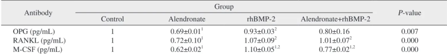
Table 3. Expression of OPG, RANKL and M-CSF in RT-PCR
Antibody Group
P-value
Control Alendronate rhBMP-2 Alendronate+rhBMP-2
OPG (pg/mL) RANKL (pg/mL) M-CSF (pg/mL)
1 1 1
0.69±0.011 0.72±0.101 0.62±0.021
0.93±0.032 1.07±0.092 1.10±0.051,2
0.80±0.16 1.01±0.072 0.77±0.021,2
0.007 0.000 0.000
(OPG: osteoprotegerin, RANKL: receptor activator of nuclear factor-κB ligand, M-CSF: macrophage colony-stimulating factor, RT-PCR: real-time polymerase chain reaction, rhBMP-2: recombinant human bone morphogenetic protein-2)
1Statistically significant (P<0.05) compared with control group.
2Statistically significant (P<0.05) compared with alendronate group.
Values are presented as mean±standard deviation.
Taek-Kyun Kwon et al: Effect of recombinant human bone morphogenetic protein-2 on bisphosphonate-treated osteoblasts. J Korean Assoc Oral Maxillofac Surg 2014
Table 4. Post hoc test result of OPG, RANKL and M-CSF in RT- PCR
Antibody Group P-value1
OPG
RANKL
M-CSF
Control
Alendronate rhBMP-2 Control
Alendronate rhBMP-2 Control
Alendronate rhBMP-2
Alendronate rhBMP-2
Alendronate+rhBMP-2 Alendronate+rhBMP-2 Alendronate+rhBMP-2 Alendronate
rhBMP-2
Alendronate+ rhBMP-2 Alendronate+rhBMP-2 Alendronate+rhBMP-2 Alendronate
rhBMP-2
Alendronate+rhBMP-2 Alendronate+rhBMP-2 Alendronate+rhBMP-2
0.012*
0.800 0.100 0.482 0.346 0.003*
0.580 0.999 0.003*
0.667 0.000*
0.020*
0.000*
0.002*
0.000*
(OPG: osteoprotegerin, RANKL: receptor activator of nuclear factor- κB ligand, M-CSF: macrophage colony-stimulating factor, RT-PCR:
real-time polymerase chain reaction, rhBMP-2: recombinant human bone morphogenetic protein-2)
1Tukey test.
*P<0.05.
Taek-Kyun Kwon et al: Effect of recombinant human bone morphogenetic protein-2 on bisphosphonate-treated osteoblasts. J Korean Assoc Oral Maxillofac Surg 2014
Fig. 3. Effect of alendronate and rhBMP-2 on RANKL expression.
ELISA of RANKL expression. (rhBMP-2: recombinant human bone morphogenetic protein-2, RANKL: receptor activator of nuclear factor-κB ligand, ELISA: enzyme-linked immunosorbent assay) Taek-Kyun Kwon et al: Effect of recombinant human bone morphogenetic protein-2 on bisphosphonate-treated osteoblasts. J Korean Assoc Oral Maxillofac Surg 2014
Fig. 4. Effect of alendronate and rhBMP-2 on M-CSF expression.
ELISA of M-CSF expression. (rhBMP-2: recombinant human bone morphogenetic protein-2, M-CSF: macrophage colony-stimulating factor, ELISA: enzyme-linked immunosorbent assay)
Taek-Kyun Kwon et al: Effect of recombinant human bone morphogenetic protein-2 on bisphosphonate-treated osteoblasts. J Korean Assoc Oral Maxillofac Surg 2014
RANKL(pg/mL)
Control 800
600
400
200
0
Alendronate rhBMP 2 Alendronate rhBMP 2
+
M-CSF(pg/mL)
Control 500
400
300
200
100
0
Alendronate rhBMP 2 Alendronate rhBMP 2
+
inhibits osteoclast differentiation and suppresses RANKL and that addition of M-CSF mediates osteoclast differentiation in a bone-marrow-derived macrophage culture, with no direct BPs-cytoxicity effect on the osteoclasts.
Although the reasons for these differences have not been completely elucidated, the distinct effects of various BPs (pamidronate, zoledronate, and alendronate), use at different concentrations, and use of different cell lines (human vs. rat and primary vs. cancer) clearly play a role15,18.
BMP-2 is well recognized as the most effective bone- induction treatment19-23 and has been approved by the U.S.
Food and Drug Administration. There is evidence that BMP-2 can directly influence osteoclastogenesis through osteoblast/stromal cells24-27. Itoh et al.27 reported that BMP- 2 enhanced the survival of purified osteoclasts supported by RANKL but not by M-CSF. In our present investigation, we found that a lower dose of rhBMP-2, 100 ng/mL, was able to induce osteoclast potency, indicating that at lower con- centrations, BMP-2 can have osteoinductive potency28. The alendronate+rhBMP-2 group showed significant elevation in expression levels of RANKL and M-CSF relative to the alen- dronate alone group. Our results show that BMP-2 induced expression of M-CSF but that RANKL was otherwise sup- pressed by alendronate. This suggest that decreased RANKL expression by BPs can be corrected by treatment of BMP-2.
V. Conclusion
This study was conducted to establish whether alendronate and rhBMP-2 influence the mRNA levels and protein expres- sion levels of M-CSF, RANKL, and OPG in osteoblasts. Our results indicate that alendronate suppressed the expression of M-CSF, RANKL, and OPG and that rhBMP-2 increased BP-impaired RANKL, M-CSF, and OPG expression levels.
Together with the previous results, the present study provides an experimental basis for the clinical effects of rhBMP-2 as a BRONJ-treatment modality.
Conflict of Interest
No potential conflict of interest relevant to this article was reported.
References
1. Marx RE. Pamidronate (Aredia) and zoledronate (Zometa) induced avascular necrosis of the jaws: a growing epidemic. J Oral Maxil-
was decreased significantly compared with the control group (P=0.000). In the alendronate+rhBMP-2 group, the M-CSF level was decreased significantly compared with the control group but increased significantly compared with the alendro- nate group (P=0.002).(Fig. 4, Table 3)
IV. Discussion
We investigated the effects of rhBMP-2 on osteoblasts treated with alendronate, since alendronate is the most widely prescribed oral BP and appears most likely to cause BRONJ12. The effects of BPs on osteoclasts are well un- derstood, and the effects of BPs on osteoclastic toxicity are thought to influence the occurrence of BRONJ. Although the majority of in vitro BPs studies have focused on BPs actions in osteoclastic lineage cells, recent studies have suggested that the presence of osteoblastic lineage cells is required for the anti-resorptive effects of BPs13. An understanding of the effects of alendronate on hFOB cells, particularly in the OPG/RANKL system, however, is lacking. To address this lack of data, the current study investigated the expression of RANKL, OPG and M-CSF in BP-treated hFOB cells.
Here, alendronate was shown to have a strong negative dose-dependent influence on the viability of osteoblasts.
MTT assays performed at 48 hours with 0, 100, 500, and 1,000 μM alendronate concentrations revealed that the 100 μM concentration significantly reduced cell viability, with cell viability being approximately 80% of the control group.
García-Moreno et al.14 reported that high concentrations of alendronate inhibit osteoblast proliferation in primary hFOB cells and indicated that at lower concentrations the drug did not significantly inhibit proliferative effects compared to con- trols (≤10-5 M). The results of the current study are in agree- ment with García-Moreno et al.14 and further suggest that alendronate at higher concentrations affects proliferation and viability of hFOB cells.
We determined that expression of OPG, RANKL and M- CSF was significantly decreased in the alendronate-treated group. In contrast, in a human osteoblast-culture study, Koch et al.15 reported that zoledronate and ibandronate significantly induced RANKL expression.
Several studies have evaluated the impact of BPs on osteo- clastogenesis. Sudhoff et al.16 reported that osteoclasts were suppressed and osteoclast apoptosis was induced in mouse bone-marrow cell (BMC) culture by the addition of M-CSF, RANKL and zolendronic acid. Kwak et al.17, having investi- gated a BMC/osteoblast co-culture, reported that risedronate
18. Idris AI, Rojas J, Greig IR, Van't Hof RJ, Ralston SH. Amino- bisphosphonates cause osteoblast apoptosis and inhibit bone nodule formation in vitro. Calcif Tissue Int 2008;82:191-201.
19. Suh DY, Boden SD, Louis-Ugbo J, Mayr M, Murakami H, Kim HS, et al. Delivery of recombinant human bone morphogenetic protein-2 using a compression-resistant matrix in posterolateral spine fusion in the rabbit and in the non-human primate. Spine (Phila Pa 1976) 2002;27:353-60.
20. Kübler NR, Würzler KK, Reuther JF, Sieber E, Kirchner T, Se- bald W. Effect of different factors on the bone forming properties of recombinant BMPs. Mund Kiefer Gesichtschir 2000;(4 Suppl 2):S465-9.
21. Uludag H, D'Augusta D, Palmer R, Timony G, Wozney J. Char- acterization of rhBMP-2 pharmacokinetics implanted with bio- material carriers in the rat ectopic model. J Biomed Mater Res 1999;46:193-202.
22. Linde A, Hedner E. Recombinant bone morphogenetic pro- tein-2 enhances bone healing, guided by osteopromotive e-PTFE membranes: an experimental study in rats. Calcif Tissue Int 1995;56:549-53.
23. Yamamoto M, Takahashi Y, Tabata Y. Controlled release by bio- degradable hydrogels enhances the ectopic bone formation of bone morphogenetic protein. Biomaterials 2003;24:4375-83.
24. Toth JM, Boden SD, Burkus JK, Badura JM, Peckham SM, McKay WF. Short-term osteoclastic activity induced by locally high concentrations of recombinant human bone morphogenetic protein-2 in a cancellous bone environment. Spine (Phila Pa 1976) 2009;34:539-50.
25. Kanatani M, Sugimoto T, Kaji H, Kobayashi T, Nishiyama K, Fu- kase M, et al. Stimulatory effect of bone morphogenetic protein-2 on osteoclast-like cell formation and bone-resorbing activity. J Bone Miner Res 1995;10:1681-90.
26. Kaneko H, Arakawa T, Mano H, Kaneda T, Ogasawara A, Nak- agawa M, et al. Direct stimulation of osteoclastic bone resorption by bone morphogenetic protein (BMP)-2 and expression of BMP receptors in mature osteoclasts. Bone 2000;27:479-86.
27. Itoh K, Udagawa N, Katagiri T, Iemura S, Ueno N, Yasuda H, et al. Bone morphogenetic protein 2 stimulates osteoclast differentia- tion and survival supported by receptor activator of nuclear factor- kappaB ligand. Endocrinology 2001;142:3656-62.
28. Zhu W, Kim J, Cheng C, Rawlins BA, Boachie-Adjei O, Crystal RG, et al. Noggin regulation of bone morphogenetic protein (BMP) 2/7 heterodimer activity in vitro. Bone 2006;39:61-71.
lofac Surg 2003;61:1115-7.
2. Kim HK, Kim JH, Abbas AA, Yoon TR. Alendronate enhances osteogenic differentiation of bone marrow stromal cells: a prelimi- nary study. Clin Orthop Relat Res 2009;467:3121-8.
3. Aubin J, Liu F, Bilezikian J, Raisz L, Rodan G. Principles of bone biology. 2nd ed. San Diego: Academic Press; 1996.
4. Urist MR. Bone: formation by autoinduction. Science 1965;150:
893-9.
5. Reddi AH. Bone and cartilage differentiation. Curr Opin Genet Dev 1994;4:737-44.
6. Reddi AH. Role of morphogenetic proteins in skeletal tissue engi- neering and regeneration. Nat Biotechnol 1998;16:247-52.
7. Bostrom MP, Asnis P. Transforming growth factor beta in fracture repair. Clin Orthop Relat Res 1998;(355 Suppl):S124-31.
8. Barnes GL, Kostenuik PJ, Gerstenfeld LC, Einhorn TA.
Growth factor regulation of fracture repair. J Bone Miner Res 1999;14:1805-15.
9. Bessa PC, Casal M, Reis RL. Bone morphogenetic proteins in tis- sue engineering: the road from the laboratory to the clinic, part I (basic concepts). J Tissue Eng Regen Med 2008;2:1-13.
10. Bessa PC, Casal M, Reis RL. Bone morphogenetic proteins in tis- sue engineering: the road from laboratory to clinic, part II (BMP delivery). J Tissue Eng Regen Med 2008;2:81-96.
11. Chen D, Zhao M, Mundy GR. Bone morphogenetic proteins.
Growth Factors 2004;22:233-41.
12. Marx RE. Clinical concerns of alendronate use. J Oral Maxillofac Surg 2008;66:1322.
13. Sahni M, Guenther HL, Fleisch H, Collin P, Martin TJ. Bisphos- phonates act on rat bone resorption through the mediation of osteo- blasts. J Clin Invest 1993;91:2004-11.
14. García-Moreno C, Serrano S, Nacher M, Farré M, Díez A, Mari- ñoso ML, et al. Effect of alendronate on cultured normal human osteoblasts. Bone 1998;22:233-9.
15. Koch FP, Merkel C, Ziebart T, Smeets R, Walter C, Al-Nawas B.
Influence of bisphosphonates on the osteoblast RANKL and OPG gene expression in vitro. Clin Oral Investig 2012;16:79-86.
16. Sudhoff H, Jung JY, Ebmeyer J, Faddis BT, Hildmann H, Chole RA. Zoledronic acid inhibits osteoclastogenesis in vitro and in a mouse model of inflammatory osteolysis. Ann Otol Rhinol Laryn- gol 2003;112:780-6.
17. Kwak HB, Kim JY, Kim KJ, Choi MK, Kim JJ, Kim KM, et al.
Risedronate directly inhibits osteoclast differentiation and inflam- matory bone loss. Biol Pharm Bull 2009;32:1193-8.