1/4 https://vetsci.org
ABSTRACT
Tumor incidence in wild mammals is reportedly very low. Wild nutria, a large rodent, is known to carry many infectious diseases, but rarely exhibits neoplastic diseases. We necropsied a male wild nutria and found a large nodular mass in the left inguinal region, adjacent to the penis. Histopathologically, the mass was diagnosed as preputial gland adenoma. Spontaneous preputial gland adenomas are extremely rare in all animals. Moreover, reports of tumors in nutrias have been limited to adenocarcinomas of the lungs and uterus, as well as subcutaneous fibromas. Here, we describe preputial gland adenoma in a wild nutria.
Keywords: Adipophilin; nutria; preputial gland; sebaceous glands
Male rodents have several accessory sex glands that include the prostate, seminal vesicles, ampullary glands, bulbourethral glands, and preputial glands. The preputial glands are paired modified sebaceous glands that are located in subcutaneous tissue lateral to the penis. Notably, the preputial glands are the source of chemical signals, and thus, secrete various substances, including pheromones, which affect reproduction and sexual behavior [1]. The growth and secretory activity of preputial glands are regulated by testosterone and the pituitary hormones; importantly, administration of testosterone causes hypertrophy and hyperplasia of acinar cells in male rats [2]. Primary neoplasms in preputial glands have been reported in laboratory mice and rats [2-5]. However, the prevalence of these neoplasms is very low in other animals.
The nutria (Myocastor coypus) is a large rodent native to South America; it is a semiaquatic herbivore that has been spread to the wild of many countries by humans. The nutria inhabits wild locations in Korea, and is designated as a pest because of its high reproductivity, lack of natural predators, and propensity to cause damage to crops and plants. As a result, an eradication campaign is underway, coordinated by the Korean Ministry of the Environment. Previously, a group of euthanized nutrias underwent necropsy for disease survey, and Capillaria hepatica and Fasciola hepatica infections were revealed [6,7]. Wild nutrias are known as reservoir hosts for various pathogens and parasites in many countries [6].
J Vet Sci. 2020 Jan;21(1):e1
https://doi.org/10.4142/jvs.2020.21.e1 pISSN 1229-845X·eISSN 1976-555X
Case Report
Received: Jul 19, 2019 Revised: Sep 20, 2019 Accepted: Sep 24, 2019
*Corresponding author:
Il-Hwa Hong
Department of Veterinary Pathology, College of Veterinary Medicine, Gyeongsang National University, 501 Jinju-daero, Jinju 52828, Korea.
E-mail: [email protected]
© 2020 The Korean Society of Veterinary Science
This is an Open Access article distributed under the terms of the Creative Commons Attribution Non-Commercial License (https://
creativecommons.org/licenses/by-nc/4.0) which permits unrestricted non-commercial use, distribution, and reproduction in any medium, provided the original work is properly cited.
ORCID iDs Joo-Yeon Kong
https://orcid.org/0000-0002-5319-7412 Il-Hwa Hong
https://orcid.org/0000-0002-2991-2690 Funding
This work was supported by a grant (NRF- 2015R1C1A1A01055527) funded by the National Research Foundation of Korea.
Conflict of Interest
The authors declare no conflicts of interest.
Author Contributions
Conceptualization: Hong IH, Yeon SC ; Data curation: Hong IH, Kong JY, Yeon SC; Formal analysis: Hong IH, Kong JY, Kim HS; Funding
Joo-Yeon Kong 1, Hyo-Seok Kim1, Seong-Chan Yeon2, Jin-Kyu Park3, Kyu-Shik Jeong3, Il-Hwa Hong 1,4,*
1 Department of Veterinary Pathology, College of Veterinary Medicine, Gyeongsang National University, Jinju 52828, Korea
2 Department of Veterinary Clinical Sciences and Research Institute for Veterinary Science, College of Veterinary Medicine, Seoul National University, Seoul 08826, Korea
3 Department of Veterinary Pathology, College of Veterinary Medicine, Kyungpook National University, Daegu 41566, Korea
4Institute of Animal Medicine, Gyeongsang National University, Jinju 52828, Korea
Preputial gland adenoma in a wild nutria (Myocastor coypus): a case report
Pathology
acquisition: Hong IH; Investigation: Hong IH, Kong JY, Kim HS; Methodology: Kong JY, Kim HS; Supervision: Hong IH; Validation: Park JK, Jeong KS; Writing - original draft: Hong IH;
Writing - review & editing: Hong IH, Park JK, Jeong KS.
However, neoplastic lesions have rarely been reported in nutrias. Here, we describe the first histopathological finding of a preputial gland adenoma in a wild nutria.
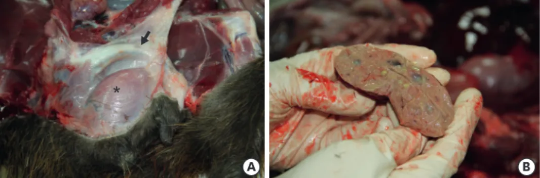
A group of euthanized nutrias from the eradication campaign of the Korean Ministry of the Environment were brought to our laboratory for necropsy. One of these nutrias (sex: male, body weight: 7.5 kg, body length: 98 cm) had a large mass in the inguinal region, adjacent to the penis. The oval-shaped mass was larger than 6 × 3 cm and exhibited yellowish exudate (Fig. 1). For microscopic examination, the mass was fixed in 10% neutral buffered formalin and processed in a routine manner with a graded ethanol series and xylene. The mass was then embedded in paraffin wax, sectioned at 4 μm, and stained with hematoxylin and eosin (H&E). For immunohistochemistry (IHC) analysis, monoclonal anti-adipophilin antibody (sc-377429; Santa Cruz Biotechnology, USA) was used to detect sebaceous cells. Hydrogen peroxide solution (3%) was used to inhibit endogenous peroxidase activity. The antigen- antibody complex was labeled with an avidin-biotin peroxidase complex solution (Vector Laboratories, USA) and a DAB substrate kit (Invitrogen, USA). Slides were then counter- stained with Mayer's hematoxylin. Sectioned slides of canine cutaneous sebaceous gland adenoma were used as positive control for adipophilin staining.
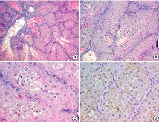
Microscopically, the mass was well-demarcated and composed of reserved basal cells and secretory cells, which contained an oily and waxy matter that comprised a type of sebaceous gland. The gland had a long excretory duct with a wide lumen lined by hyperplastic
squamous epithelium. The excretory ducts demonstrated a variety of shapes and sizes;
occasionally, they were filled with keratinized cell debris (Fig. 2A). The tumor cells had a central round nucleus with one or (rarely) 2 large nucleoli. The cytoplasm contained lipid vacuoles of various sizes (Fig. 2A and B). Hyperplastic basal cells typically surrounded the foci of the sebaceous cells, forming lobulations of various sizes. Mitotic figures were occasionally visible. Infiltration of mononuclear inflammatory cells around secretory ducts was observed in some areas. To confirm the identity of the sebaceous cells, anti-adipophilin was used for IHC. Adipophilin is an adipocyte differentiation-related protein expressed in intracytoplasmic lipid droplets of sebocytes [8]; here, it was expressed in the cytoplasmic lipid vacuoles of sebocytes, but not in basal cells (Fig. 2D). As a result of these findings, preputial gland adenoma was diagnosed in this wild nutria.
Although any neoplasm in accessory genital glands is quite rare in animals, spontaneous preputial gland adenoma is a relatively frequent tumor among neoplasms of the accessory genital glands in the Fischer 344 rat [3-5]. Preputial gland adenoma in male Fischer 344 rats
2/4 https://vetsci.org https://doi.org/10.4142/jvs.2020.21.e1
Preputial gland adenoma of a nutria
*
B A
Fig. 1. Necropsy of a male wild nutria. (A) The ventral aspect of the animal. The ventral skin has been peeled back, revealing a large muscle-covered mass (asterisk) lateral to the penis (arrow). (B) Cut surface of the mass.
Yellowish exudates are observed.
may occur as a result of aging [3]. Injection of 1,2-dimethylhydrazine has been shown to induce adenoma/carcinoma in preputial or clitoral glands (female counterpart of preputial glands) of CBA and BALB/c mice [9]. Tumor incidence in wild mammals is reportedly very low. In nutria, moreover, only a few cases have been reported, involving adenocarcinoma in the lungs and uterus, as well as subcutaneous fibroma [10]. Here, we have described preputial gland adenoma in a wild nutria.
REFERENCES
1. Marchlewska-Koj A, Pochroń E, Sliwowska A. Salivary glands and preputial glands of males as source of estrus-stimulating pheromone in female mice. J Chem Ecol 1990;16:2817-2822.
PUBMED | CROSSREF
2. Rudmann D, Cardiff R, Chouinard L, Goodman D, Küttler K, Marxfeld H, Molinolo A, Treumann S, Yoshizawa K; INHAND Mammary, Zymbal's, Preputial, and Clitoral Gland Organ Working Group.
Proliferative and nonproliferative lesions of the rat and mouse mammary, Zymbal's, preputial, and clitoral glands. Toxicol Pathol 2012;40:7S-39S.
PUBMED | CROSSREF
3. Coleman GL, Barthold W, Osbaldiston GW, Foster SJ, Jonas AM. Pathological changes during aging in barrier-reared Fischer 344 male rats. J Gerontol 1977;32:258-278.
PUBMED | CROSSREF
4. Reznik G, Ward JM. Morphology of hyperplastic and neoplastic lesions in the clitoral and preputial gland of the F344 rat. Vet Pathol 1981;18:228-238.
PUBMED | CROSSREF
3/4 https://vetsci.org https://doi.org/10.4142/jvs.2020.21.e1
Preputial gland adenoma of a nutria
B A
D C
Fig. 2. Microscopic examination of the mass in a male wild nutria. (A) The mass is composed of reserved basal cells and secretory cells containing oily and waxy matter, which constitute a type of sebaceous gland. The gland has a long excretory duct with a wide lumen lined by hyperplastic squamous epithelium. (B, C) The tumor cells have a central round nucleus with one or (rarely) 2 large nucleoli. The cytoplasm contains small to large vacuoles.
(D) Adipophilin is expressed in intracytoplasmic lipid vacuoles in sebaceous cells. Hematoxylin and eosin (A-C);
immunohistochemistry of adipophilin (D) (all scale bar = 100 µm).
5. Mitsumori K, Elwell MR. Proliferative lesions in the male reproductive system of F344 rats and B6C3F1 mice: incidence and classification. Environ Health Perspect 1988;77:11-21.
PUBMED | CROSSREF
6. Hong IH, Kang SY, Kim JH, Seok SH, Lee SK, Hong SJ, Lee SY, Park SJ, Kong JY, Yeon SC.
Histopathological findings in wild Nutrias (Myocastor coypus) with Capillaria hepatica infection. J Vet Med Sci 2017;78:1887-1891.
PUBMED | CROSSREF
7. Kim HS, Kong JY, Kim JH, Yeon SC, Hong IH. A case of fascioliasis in a wild nutria, Myocastor coypus, in Republic of Korea. Korean J Parasitol 2018;56:375-378.
PUBMED | CROSSREF
8. Gang DG, Sim CH, Lee TJ, Kong JY, Hong IH. Sebaceous cell differentiation in a canine oral papilloma. J Vet Diagn Invest 2018;30:569-571.
PUBMED | CROSSREF
9. Turusov VS. Morphology and histogenesis of anal region and clitoral gland tumors induced in mice by 1,2-dimethylhydrazine. J Natl Cancer Inst 1980;64:1161-1167.
PUBMED
10. Ratcliffe HL. Incidence and nature of tumors in captive wild mammals and birds. Cancer Res 1933;17:116-135.
CROSSREF
4/4 https://vetsci.org https://doi.org/10.4142/jvs.2020.21.e1
Preputial gland adenoma of a nutria