재활치료과학 제 9 권 제 4 호
Therapeutic Science for Rehabilitation Vol. 9. No. 4. 2020.
https://doi.org/10.22683/tsnr.2020.9.4.007
The Effects of Simultaneous Application of Peripheral Nerve Sensory Stimulation and Task-Oriented Training to Improve Upper Extremity Motor Function After Stroke: Single Blinded Randomized Controlled Trial
Kim, Sun-Ho*, Ph.D., O.T., Won, Kyung-A**, M.S., O.T., Jung, Eun-Hwa***, Ph.D., O.T.
Abstract
Objective : This study aimed to investigate the effect of simultaneous application of peripheral nerve sensory stimulation and task-oriented training on the improvement of upper extremity motor function after stroke.
Methods : This study included 29 patients with hemiplegia. The 14 subjects were in the peripheral nerve sensory stimulation and task-oriented training group for 4 weeks (30 min/d, 5 d/wk), while the 15 control group subjects underwent only task-oriented training for the same duration. The outcome measures were the percentage of voluntary baseline muscle contractions of the wrist and shoulder and Box and Block Test, grip and pinch strength, and Action Research Arm Test.
Results : After 4 weeks, muscle activity of extensor carpi radialis, flexor carpi radialis and grip strength and Action Research Arm Test were significantly higher in the experimental group.
Conclusion : Simultaneous application of the peripheral nerve sensory stimulation and task-oriented training was found to be superior to task-oriented training for improving upper extremity motor function of adults with stroke.
Key Words : Peripheral nerve sensory stimulation, Stroke, Task-oriented training, Upper extremity
교신저자 : 김선호([email protected]) || 접수일: 2020.01.28 || 심사일: 2020.01.29
|| 게재승인일: 2020.02.25
*Dept. of Occupational Therapy, Young Kwang Rehabilitation Hospital, Department Head
**Incheon Metropolitan Dementia Center, Occupational Therapist
***Dept. of Occupational Therapy, FAR East University, Professor
I. Introduction
Stroke is a neurological disorder that affects the arteries leading to and within the brain. It occurs when a blood supply to part of the brain is ruptured or reduced. Sudden consciousness loss and paralysis occur when brain cells die during a stroke (Chang, Tung, Wu, & Su, 2006). Most stroke patients experience upper extremity dyskinesia due to abnormalities in muscle tone or motor paresis. This is a main problem that affects performance of everyday activities (Raghavan, 2007).
There have been various studies to induce efficient movement, including using more of the upper extremity on the contralesional hemisphere for recovery of upper limb movement disorder in stroke patients. Previous studies have reported that recovery of the central nervous system in stroke patients is facilitated by neuroplasticity, and that it changes the functional connectivity of brain to promote functional recovery (Gerloff et al., 2006). Based on this understanding of neuroplasticity, various electrical stimulation treatments such as electromyogram-triggered neuromuscular stimulation (EMG-stim), functional electronic stimulation (FES), and peripheral nerve sensory stimulation (PNS) have been applied to promote the recovery of the upper extremity motor function in stroke patients (Bolton, Cauraugh, & Hausenblas, 2004; Celnik, Hummel, Harris-Love, Wolk, & Cohen, 2007; Mangold, Schuster, Keller, Zimmermann- Schlatter, & Ettlin, 2009).
PNS is a treatment that induces the reorganization of the sensory motor cortex by providing low-frequency stimulation at a constant interval (at which muscle contraction does not occur) in the peripheral nerve territories (Deuchert et al., 2002). Previous studies have reported that this repetitive low-frequency electrical stimulation is effective in activating cortical motor activity as well as in improving hand function and sensory function
in stroke patients. The advantage of PNS is that there is no fatigue, pain, or discomfort in the muscles (Celnik et al., 2007; Conforto et al., 2010; Ikuno et al., 2012; Sullivan & Hedman, 2004). It is also low cost and easy to apply in clinical practice.
It is useful as a supplementary treatment to promote upper extremity motor function recovery because it does not require the patient’s voluntary participation or concentration during stimulation, unlike other electro-stimulation treatments (Klaiput & Kitisomprayoonkul, 2009). Recent studies of upper extremity function in stroke have been conducted with interventions such as task-based training using PNS, which requires less exercise and cognition than EMG-stim and FES, and its effectiveness has been reported (Ikuno et al., 2012).
Task-oriented training (TOT) has been widely used in clinical practice. It is based on neuroplasticity through brain activation, which combines motor control and motor learning for functional recovery and motor improvement in patients with neurological impairment (Thieme et al., 2012).
When TOT and PNS are combined, there is a synergy that enhances activation of the corticospinal tract, as the peripheral afferent input from the sensory nerve-branching skin is stimulated (Lin et al., 2012; Saito et al., 2013). Ikuno et al. (2012) investigated this approach in 22 stroke patients by combining peripheral sensory stimulation of the median and ulnar nerves with task-oriented training for one week.
As a result, there was a significant improvement of hand function in the group in which the peripheral sensory nerve stimulation was performed compared to that in the group with only task performance training. Fleming et al. (2015) reported a positive effect on upper extremity function in an experimental group that underwent TOT and PNS to the median, ulnar, and radial nerves compared to the effects in a control group that only performed the task-oriented training.
However, studies considering the clinical usefulness of
Patient assessed for eligibility (n=41)
Excluded (n=12)
Not meeting inclusion criteria (n=8) Declined to participate (n=2)
Other reasons (n=2)
Randomized (n=29)
Allocated to EG (n=14)
(Occupation based-bilateral upper extremity training)
Allocated to CG (n=14)
(Task oriented-bilateral upper extremity training)
Pre test: Muscle activity, Strength(pinch, grip), BBT, ARAT
Intervention: A total of 20 sessions, 5 days a week for 4 weeks
Drop out (n=4) Declined to participate (n=1)
Discharge (n=3) Other reasons (n=0)
Drop out (n=5) Declined to participate (n=0)
Discharge (n=5) Other reasons (n=0)
Post test: Muscle activity, Strength(pinch, grip), BBT, ARAT
Measure within 24 hours after intervention (n=10) Measure within 24 hours after intervention (n=10)
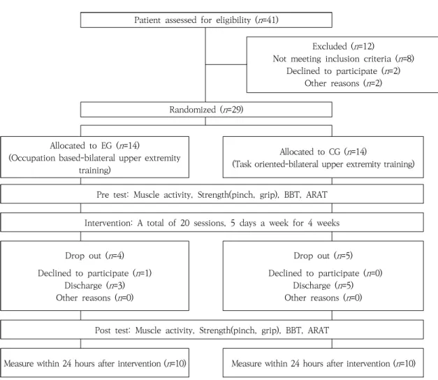
Figure 1. Flow Diagram of Subjects in the Study
ARAT=Action Research Arm Test; BBT=Box and Block Test; CG=Control group; EG=Experimental group; PNS=Peripheral nerve sensory stimulation; TOT=Task-oriented training
combined PNS and task-oriented training are limited. Most studies have validated the effectiveness of the PNS-only intervention, and studies that combine PNS and TOT have limitations in clinical application with intervention times that are too long because the interventions were applied sequentially (Conforto, Cohen, dos Santos, Scaff, & Marie, 2007; Fleming et al., 2015; Ikuno et al., 2012; Koesler, Dafotakis, Ameli, Fink, & Nowak, 2009; Wu, Seo, & Cohen, 2006). In addition, there were limitations to generalization due to the lack of RCT studies or short intervention periods.
In conclusion, studies on peripheral sensory nerve stimulation in korean stroke patients are insufficient, and
studies that simultaneously conducted two interventions to reduce the intervention time are also insufficient.
This study aimed to investigate the effect of simultaneous application of PNS and TOT on the improvement of upper extremity motor function after stroke.
II. Methods
1. Participants
29 patients who met the criteria for the study were
selected from 41 patients who were admitted to Y hospital
who were considered to be able to participate in the study.
The inclusion criteria and exclusion criteria for this study were as follows. They were screened by a assessor.
The inclusion criteria were as follows: Patients diagnosed with stroke via MRI/CT medical record; Patients aged 20 years and older; Patients with an onset at least 12 months prior to the study; Patients without cognitive impairments (MMSE-K
≧24) (Folstein, Folstein, & McHugh, 1975);
Patients who have a Brunnstrom stage of the shoulder and hand of 3 or more; Patients who understand the study’s purpose and can provide consent for participation.
Exclusion criteria were as follows: Patients who have had previous strokes or have other neurological or surgical conditions; Patients who recently participated in other rehabilitation research or drug experiments; Patients who had seizures repeatedly during treatment; Patients with visual perception problems (severe unilateral or visual field defects); Patients with contraindications to electrical stimulation.
This was a single-blind study, and subjects were allocated randomly to the experimental or control group using a random number generated by computer software.
The study was conducted after approval from the Ethics Committee of Yonsei University School (Approval number.: 1041849-201608-BM-042-02).
All subjects provided informed consent before study inclusion according to the Declaration of Helsinki 2004.
Selected patients were randomly assigned to 14 patients in the experimental group (EG) and 15 in the control group (CG). During the intervention, 4 patients in the EG and 5 in the CG were dropped out because of the reasons such as rejection of treatment, discharge. The results of the final 20 patients were analyzed (Figure 1).
2. Protocol
This study was conducted from September 2017 to August 2018 and recruitment took place from September 1 to 30. A simple randomization method using a computer randomization program was used to assign 14 subjects to experimental group, and 15 to control group. All sessions were conducted by one occupational therapist with six years of clinical experience.
EG performed PNS combined with TOT, and the training was conducted a total of 20 sessions, 5 days a week for 4 weeks. In the CG, only TOT was performed, and the training was conducted similarly to the EG. During the study period, all participants in the EG and CG continued to receive conventional physical therapy, occupational therapy, and medication. Physical therapy included central nervous system exercise therapy such as NDT. Occupational therapy performed fine motor control training, strength exercise, and did not include exercise similar to this study intervention.
The assessments were conducted one-on-one within the hospital’s occupational therapy room.
Pre-assessment was conducted within 24 hours before the intervention and post-assessment was performed within 24 hours after the intervention. Two occupational therapists with more than three years of experience conducted all the assessments in a fixed order. muscle activity, hand dexterity and hand strength, functional level of the affected upper extremity were assessed at pretreatment, post treatment.
3. Intervention
In this study, we used FES-1000-FES EMG (Cyber
Medic,
Seoul, Korea) to provide PNS to the participants. It has
two channels in total, and can be controlled with frequency
Task Methods
Press the bell
1. The task involved pressing two bells with the palm of the hand. Two bells were spaced 30 cm apart and 50 cm in front of the center of the participant’s body.
2. The task involved pressing five bells with the palm of the hand. The bells were placed 50 cm in front of the center of the participant’s body .
3. The task involved reaching the bell target in the upper, lower, left, and right positions while in the sitting position and pressing the bell with the palm.
Moving a rubber ball The task involved taking a 6.5 cm diameter rubber ball and putting it in a basket with a diameter of 10 cm (the basket location was moved in various directions).
Moving the plastic bottle The task involved reaching and moving five plastic bottles of different thickness, height, and weight (the range of movement did not extend past the desk).
Moving a peg The task involved taking a cylindrical peg with a diameter of 2.5 cm and a height of 7 cm and putting it in a basket placed on the box, while in a sitting position.
Table 1. Content of Task-Oriented Training of 1
–200 Hz, output current of 0
–99 mA, and pulse width of 20
㎲-50
㎲. It is also possible to set the treatment mode, output waveform (monophasic/biphasic), and treatment time from 0
–60 min. It has an independent channel method that can control the intensity of each channel’s stimulation according to the stimulation site. Peripheral sensory nerve stimulation was provided to the affected upper extremity of each stroke patient using a neuromuscular electrical stimulation therapy device. The location of the electrode was attached to the median nerve and ulnar nerve on the affected side (Kim & Park, 2016). The electrode for the median nerve was attached to the wrist branch of the flexor carpi radialis and the palmaris longus wrist tendon. The electrode for the ulnar nerve was attached to the flexor carpi ulnaris, branching from the ulnar nerve and attaching to the wrist (Burridge
& Ladouceur, 2001). The electrode used was a square disposable electrode of 5 x 5 cm. The stimulus was provided for 30 min at a frequency of 10 Hz, a pulse width of 10
㎲
, and an on-off duty cycle to ensure that there was no pain or visual contraction of the muscle (Ikuno et al., 2012).
Task-oriented training (TOT) was performed simultaneously for 30 min to provide peripheral sensory nerve stimulation. TOT used in this study involved a total of six items, including “Press the bell 1-3”, “Moving a rubber ball”,
“Moving the plastic bottle”, and “Moving a peg”. The
intervention method was based on the study by Kim, Park, Jung, & Yoo (2016) (Table 1). The difficulty of the task was controlled by the number, direction, size, and distance of the target, according to the ability of the participant.
TOT was performed in a sitting position in a chair with a backrest, and both legs were flexed at 90° in the hip, knee, and ankle joints.
4. Outcome measures
1) Mini-Metal State Examination (MMSE-K)
MMSE-K was used to assess the cognitive abilities required to perform tasks when enrolling the participants.
MMSE was developed by Folstein et al. (1975). In 1989, it was translated into Korean and standardized. MMSE-K is widely used in clinical practice to easily assess the cognitive level of patients with brain damage and older adults. It consists of 12 items from a total of 6 categories, and less than 24 points out of 30 points is considered an indication of cognitive impairment. In this study, patients with score of 24 or more were enrolled.
2) Percentage of reference voluntary muscle contractions (% RVC)
The surface electromyography equipment was an
ME6000 (Mega Electronics Ltd., Kuopio, Finland), with
a total of eight channels. In this study, five channels were used. The EMG signal sampling rate was 1000 Hz, and a band pass filter of 10
–500 Hz and a notch filter of 60 Hz were used. The EMG signals collected on the ME6000 were converted into digital signals and then filtered and signal processed using the MegaWin software 3.0 on connected personal notebooks. Surface electromyography (sEMG) is a test that measures the electrical activity of muscles by using electrodes that are attached to the skin.
In this study, it was used to measure muscle activity of the flexor carpi radialis (FCR), extensor carpi radialis (ECR), and anterior fibers of the deltoid (AD).
An electrode was attached to the ECR, which caused the wrist and fingers to extend. The placement was on the muscle belly with supination and pronation of the elbow at 90°, with the participant in a sitting position, where there was maximal contraction of the wrist. An electrode was attached to the FCR, which caused bending of the wrist and fingers. Similar to the ECR, this electrode was attached to the muscle belly with supination and pronation of the elbow at 90°, with the participant in a sitting position, where there was maximal flexion of the wrist. An electrode was attached to the AD where it attaches to the posterior surface of the triceps nodule, at a point one finger wide (Riek, Carson, & Wright, 2000).
In this study, the percentage of reference voluntary muscle contractions (% RVC) was used as the muscle activity. The measurements were taken with the participants sitting in a comfortable chair sitting in a separate, quiet space without noise interference. The posture for taking measurements was such that the waist was bent back and maintained at 90 degrees, and the subjects were cushioned on the knee so that they would feel comfortable. In this state, the muscle activity was measured in the resting position for a period of 15 s, and finger flexion and extension, and the reaching position were measured for 10 s. This procedure was repeated three times and the muscle activity was
calculated as the mean value (Cook, Burgess-Limerick,
& Papalia, 2004). This value was entered into a text file and normalized to % RVC using MATLAB software. The
% RVC values
were compared before and after the intervention, based on the muscle activity in the resting position, when the specific actions (finger extension and bending, and arm lifting) were performed. Muscle activity was considered to increase as the % RVC values increased.
3) Box and Block Test (BBT)
Box and Block Test (BBT) was used to evaluate the dexterity of the affected upper extremity of the participant in this study. BBT measures the number of blocks that are successfully moved from one side of the subject to the other over a period of 60 s (Chanubol et al., 2012).
The test-retest reliability was high at overall 0.98, right to left at 0.94, and right to left at 0.94. The inter-reliability was high at 1.00 on the right side and 0.99 on the left side. Concurrent validity has also been reported to be high (Mathiowetz, Volland, Kashman, & Weber, 1985).
4) Strength
Hand grip strength was measured using a Jamar dynamometer (Sammons Preston, Inc., Bolingbrook, IL, USA) to determine the change in grip strength in the affected extremity. The lateral pinch was measured using pinch gauze (SAEHAN, Korea) (Mathiowetz et al., 1985).
The Jamar dynamometer range can be adjusted from 3.45 to 8.57 cm and can measure up to 90 kg. The pinch gauze can measure up to 20 kg. The measurement posture involved the patients sitting in a chair with a 90° elbow flexion, with the forearm and wrist in a neutral position.
5) Action Research Arm Test (ARAT)
Action Research Arm Test (ARAT) was used to evaluate
the upper extremity functional level in this study. Tools such
as cylinders, cups, and rods were used to score tasks according
to the degree to which they were performed on grasp, grip,
EG (n=10) CG (n=10) p
Age (mean (SD)) 57.3 (9.01) 61.7 (7.41) .363
Time since injury (months; mean (SD)) 16.9 (6.1) 19.5 (7.89) .377
MMSE-K (mean (SD)) 29.4 (0.70) 29.4 (1.07) .605
Brunnstrom stage (mean (SD))
Shoulder 4.6 (0.52) 4.8 (0.63) .483
Hand .483
Gender, n (%)
Male 4 (40) 4 (40)
1.000
Female 6 (60) 6 (60)
Paresis, n (%)
Right 5(50) 5 (50)
1.000
Left 5(50) 5 (50)
Dominant hand, n (%)
Right 9 (90) 9 (90)
1.000
Left 1 (10) 1 (10)
Stroke type, n (%)
Infarction 7 (70) 4 (40)
Hemorrhage 3 (30) 6 (60) .369
Baseline
Muscle activity (%RVC) (mean (SD))
ECR 1088.09 (251.39) 1183.1 (272.27) .364
FCR 1391.24 (282.34) 1889.41 (626.61) .028
*AD 2921.82 (442.77) 2881.82 (476.2) .850
Pinch strength (lbs) (mean (SD)) 9.9 (2.92) 13.7 (4.42) .037
*Grip strength (lbs) (mean (SD)) 24.73 (4.3) 26.68 (3.59) .364
BBT (mean (SD)) 24.8 (3.08) 24.9 (3) .938
ARAT (mean (SD)) 27 (6.16) 34.3 (7.8) .028
*Table 2. Characteristics and Baseline Assessment Results of Study Participants pinch, and gross movements. ARAT consists of 19 items and
4 categories, an uses a 4-point scale consisting of 0
–3 points.
The reliability by category was grasp=0.98, grip=0.99, pinch=0.99, and gross movement=0.98 (Lang, Wagner, Dromerick, & Edwards, 2006).
5. Data analysis
The collected data were analyzed using SPSS 21.0 (IBM Corp., Armonk, NY, USA). The homogeneity of the experimental group and the control group before the intervention was verified using the chi-squared (
χ2) and Mann-Whitney U tests. The Wilcoxon signed rank test was used to test for significance before and after intervention. An analysis of covariance was used to compare changes between the groups, and the covariance
was set as a pre-assessment of each group. Statistical significance was set at p<.05.
III. Results
1.
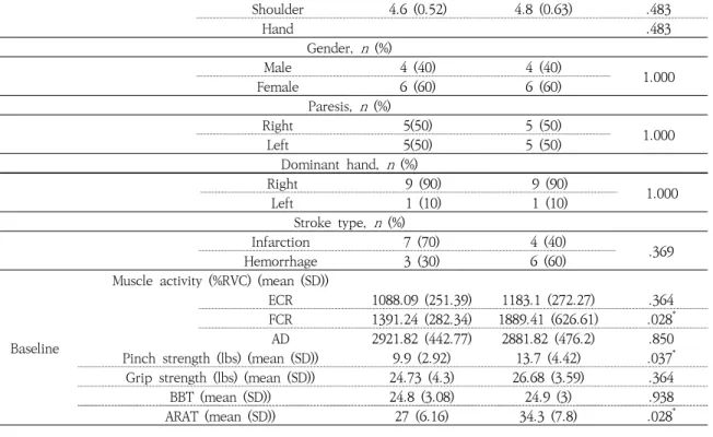
Characteristics and baseline assessment results of study participantsTable 2 shows the results of the homogeneity test for
the general characteristics and baseline assessment results
of the EG and CG in this study. There was no significant
difference in general characteristics between the two
groups. However, in the pre-assessment, there was a
significant difference between the EG and the CG in the
FCR (p=.028), pinch strength (p=.037), and ARAT score
(p=.028) (Table 2).
Baseline Post intervention
Z p
Mean (SD) Mean (SD)
ECR (%)
EG 1088.09 (251.39) 1783.69 (190.74) -2.803 .005
*CG 1183.10(272.27) 1694.31 (370.87) -2.803 .005
*FCR (%)
EG 1391.24 (282.34) 2346.78 (321.47) -2.803 .005
*CG 1889.41 (626.61) 2400.041 (556.59) -2.803 .005
*AD (%)
EG 2921.82 (442.77) 3015.50 (441.52) -1.784 .074
CG 2881.82 (476.20) 3025.50 (440.73) -1.988 .047
*Pinch strength (lbs)
EG 9.9 (2.92) 10.6 (1.96) -1.734 .083
CG 13.7 (4.42) 13.9 (3.81) -0.707 .480
Grip strength (lbs)
EG 24.73 (4.30) 29.88 (4.17) -2.803 .005
*CG 26.68 (3.59) 29.32 (3.59) -2.652 .008
*BBT (sec)
EG 24.8 (3.08) 25.7 (2.90) -2.041 .041
*CG 24.9 (3.00) 25.4 (3.17) -2.236 .025
*ARAT (score)
EG 27.0 (6.16) 34.1 (8.20) -2.818 .005
*CG 34.3 (7.80) 36.9 (6.62) -2.825 .005
**
p
<.05AD=Anterior deltoid; ARAT=Action Research Arm Test; BBT=Box and Block Test; CG=Control Group; ECR=Extensor carpi radialis;
EG=Experimental group; FCR=Flexor carpi radialis; SD=Standard Deviation
Table 3. Changes in Muscle Activity, Hand Strength, BBT, ARAT Within Group 2. Changes in muscle activity, hand
strength, BBT, ARAT.
In the EG, muscle activity was increased in the ECR and the FCR. In the CG, muscle activity was significantly increased in the ECR, FCR, and AD (Table 3).
The differences between the two groups were analyzed using an ANCOVA because of the differences in muscle activity before the intervention. After the intervention, there was a significant difference (p=.041) between the two groups, with 695.60% RVC and 511.20% RVC increase in the muscle activity of the ECR. The muscle activity of the FCR also showed a significant difference (p=.008) between the two groups, with 955.54% RVC in EG and
510.64% RVC in CG after the intervention (Table 4).
The grip and pinch strength tests of the hand showed significant changes both before and after the intervention only in the grip strength (Table 3), and the difference between the two groups was also significant only in the grip strength (p=.015) (Table 4). In both groups, significant changes were observed in the ARAT results before and after the intervention (Table 3), and EG showed a significant improvement (p=.018) (Table 4).
The BBT also showed a significant change in the
pre-and post-intervention groups (Table 3), but the
difference between the two groups was not significant
(Table 4).
Ther apeut ic S ci ence fo r Rehab ilit at io n V o l. 9 . N o . 4 . 2 0 2 0 . 15
Me a n ( SD ) M ea n d if fe re nc e C ov a ri a te C I Fd .f . p EG (n =10) C G (n =10) Mu sc le act ivi ty (% R V C )
EC R 695. 60 (146. 85) 511 .20 (194. 13) 175 .56 (95. 64) 1135. 60 7. 77 345. 3 5 4 .871 1 .041
*FC R 955. 54 (266. 55) 510 .64 (210. 29) 352 .89 (154. 23) 1640. 33 107. 18 59 8. 61 9. 182 1 .008
*A D 93. 68 ( 139. 97) 143 .68 (225. 97) -42. 06 (85. 92) 2901. 82 -213. 23 129 .1 0 .298 1 .592 Gr ip st re n gt h (l b s) 5. 15 (2 .02) 2 .64 (1 .66) 2 .64 (1 .66) 11. 80 0. 47 3. 90 7. 261 1 .015
*Pi nc h st re n gt h (l b s) 0. 70 (1 .25) 0 .20 (0 .92) 0 .20 (0 .92) 25. 71 -1 .1 4 0 .4 9 .69 1 .417 B B T (s ec ) 7. 10 (4 .12) 2 .60 (1 .51) 2 .60 (1 .51) 24. 80 -0 .4 3 1 .2 3 1 .0 2 1 .3 28 A R A T ( sc or e) 0. 90 (1 .10) 0 .50 (0 .53) 0 .50 (0 .53) 30. 65 0. 82 7. 66 6. 828 1 .018
* *p
<.05 AD=Anterior deltoid; ARAT=Action Research Arm Test; BBT=Box and Block Test; CG=Control group; CI=Confidence Interval; d.f=degree of freedom; ECR=Extensor carpi radialis; EG=Experimental group; FCR=Flexor carpi radialisTabl e 4. Diff e renc e Bet w ee n Group s
Ⅳ. Discussion
This pilot study demonstrated the feasibility of simultaneous use of PNS and TOT for recovery of upper limb dysfunction in stroke patients. The results of this study showed that muscle activity, hand grip strength, and upper limb function were significantly higher in the PNS plus TOT group than the TOT group. These results are consistent with the results of previous studies that combined prior PNS and TOT. In addition, these results show that when spontaneous and active training (such as TOT) PNS are combined, the peripheral afferent input from the sensory nerve-branching skin stimulates the activation of the corticospinal tract (Fleming et al., 2015;
Ikuno et
The results of this study showed that the muscles activity of the ECR and FCR in the experimental group and the control group was significantly increased before and after the intervention, and the muscle activity of experimental group was significantly greater than that of control group. Changes in the motor unit due to stroke will modify the pattern of muscular activation, making the muscles unable to selectively move (Wagner, Dromerick, Sahrmann, & Lang, 2007). In this study, there was a difference between the groups of muscle activity because the PNS in experimental group stimulated the direct nerve root which could affect the grasping and releasing function of the hand. This increase in muscle activity implies an increase in the number of muscle fibers forming the exercise unit and induces the growth of muscle fibers through neurological plasticity and changes in myofibrils. This is consistent with previous findings that peripheral nerve stimulation promotes paralyzed wrist muscles and may support the results of this study in that changes in muscle activity in groups that provided additional PNS were greater than TOT (Castel-Lacanal et
al., 2009). On the other hand, TOT, which was a common intervention in this study, included shoulder movements without electrical stimulation in that area, so there was no difference between groups in the AD, despite the improvement effect in each group.
This change in muscle activity may lead to improvement of motor recovery. In this study, hand grip improvements were significant only in grip strength, not in pinch strength. TOT used in this study affected the plasticity of the cerebral cortex, but did not provide a direct challenge to improve hand muscle strength during tasks, and the location where the PNS was applied was also not significant. It seems that pinch and fine motor are not directly related to the tasks involved in the training.
Although TOT used in this study affects cerebral cortex plasticity, it does not provide a direct challenge to improve hand muscle strength during task training, and the location where PNS is applied is also not directly related to pinch and fine motor; therefore, it is considered that the results were not significant in this aspect because there was no direct involvement.
These results support the hypothesis that excitability of the corticospinal tract connected to the hand muscles via PNS is not directly related to the fine motion of the hand (Liu & Au-Yeung, 2017).
In this study, simultaneous application of PNS and TOT
was effective in improving upper extremity function in
stroke patients. As a result of evaluating upper limb
function with ARAT, both groups showed significant
changes based on the pre and post interventions, and
the score increased significantly in EG. PNS improves the
ability to control movement by reducing muscle tone in
stroke patients, allowing for spontaneous movement. In
addition, when task training is provided at the same time,
it significantly increases the activity of the corticospinal
tract (Lin et al., 2012; Saito et al., 2013). Based on previous
al., 2012; Lin et al., 2012; Saito et al., 2013).
studies, it is suggested that TOT with PNS activated the corticospinal tract and affected the restoration of upper extremity motor function after stroke.
The clinical significance of this study is that it can be applied in clinical situations to improve upper limb function of stroke patients. PNS is relatively safe and less fatigue inducing than FES, and can be easily applied with upper extremity training such as TOT. In addition, this approach makes it easier to induce patients to participate in stroke treatment.
There were several limitations in this study. First, the sample size was small. Second, the participants were chronic stroke patients, and shoulder and hand function loss was mild, with a Brunnstrom stage average of 4 or more. In the future, it is necessary to investigate the effect of providing PNS and TOT in patients with severe upper extremity function impairment. Third, it is difficult to confirm whether the treatment effects were maintained because they were evaluated within 24 hours of the intervention. Future studies are needed to confirm that treatment effects are maintained through at least 1-2 weeks of follow-up.
Ⅴ. Conclusion
This study investigated the effect of TOT on stroke patients while providing PNS to induce changes in muscle activity, hand grip, and upper extremity function. These variables are meaningful to the clinician because they are important for rehabilitation goals of stroke patients, including a return to daily life activities. In the future, it is necessary to study the amplification intensity and the delivery time, which can appropriately and efficiently provide the peripheral nerve stimulation to stroke patients. In addition to the TOT, studies on the effects of various upper extremity functional treatments, such
as bilateral upper extremity training and robotic therapy, are needed.
References
Bolton, D. A., Cauraugh, J. H., & Hausenblas, H. A. (2004).
Electromyogram-triggered neuromuscular stimulation and stroke motor recovery of arm/hand function: A meta-analysis. Journal of the Neurological Sciences, 223(2), 121-127. doi:10.1016/j.jns.2004.05.005 Burridge, J. H., & Ladouceur, M. (2001). Clinical and
therapeutic applications of neuromuscular stimulation:
A review of current use and speculation into future developments. Neuromodulation, 4(4), 147 – 154. doi:10.
1046/j.1525-1403.2001.00147.x
Castel-Lacanal, E., Marque, P., Tardy, J., de Boissezon, X., Guiraud, V., Chollet, F., ... Simonetta-Moreau, M.
(2009). Induction of cortical plastic changes in wrist muscles by paired associative stimulation in the recovery phase of stroke patients. Neurorehabilitation and Neural Repair, 23(4), 366-372. doi:10.1177/1545968308322841 Celnik, P., Hummel, F., Harris-Love, M., Wolk, R., & Cohen,
L. G. (2007). Somatosensory stimulation enhances the effects of training functional hand tasks in patients with chronic stroke. Archives of Physical Medicine and Rehabilitation, 88(11), 1369-1376. doi:10.1016/j.apmr.2007.08.001 Chang, J. J., Tung, W. L., Wu, W. L., & Su, F. C. (2006).
Effect of bilateral reaching on affected arm motor control in stroke – with and without loading on unaffected arm.
Disabilitation and Rehabilitation, 28(24), 1507 – 1516.
doi:10.1080/09638280600646060
Chanubol, R., Wongphaet, P., Ot, N. C., Chira-Adisai, W., Kuptniratsaikul, P., & Jitpraphai, C. (2012). Correlation between the action research arm test and the box and block test of upper extremity function in stroke patients.
Journal of the Medical Association of Thailand, 95(4), 590-597.
Conforto, A. B., Cohen, L. G., dos Santos, R. L., Scaff, M.,
& Marie, S. K. N. (2007). Effects of somatosensory stimulation on motor function in chronic cortico-subcortical strokes.
Journal of Neurology, 254(3), 333-339. doi:10.1007/s 00415-006-0364-z
Conforto, A. B., Ferreiro, K. N., Tomasi, C., dos Santos,
R. L., Moreira, V. L., Nagahashi Marie, S. K. N., ... Cohen,
L. G. (2010). Effects of somatosensory stimulation on motor function after subacute stroke. Neurorehabilitation and Neural Repair, 24(3), 263-272. doi:10.1177/1545968 309349946
Cook, C., Burgess-Limerick, R., & Papalia, S. (2004). The effect of upper extremity support on upper extremity posture and muscle activity during keyboard use. Applied Ergonomics, 35(3), 285-292. doi:10.1016/j.apergo.2003.
12.005
Deuchert, M., Ruben, J., Schwiemann, J., Meyer, R., Thees, S., Krause, T., ... Villringer, A. (2002). Event-related fMRI of the somatosensory system using electrical finger stimulation. Brain Imaging, 13(3), 365-369. doi:10.1097/00001756-200203040-0 0023 Fleming, M. K., Sorinila, I. O., Roberts-Lewis, S. F., Wolfe, C. D., Wellwood, I., & Newham, D. J. (2015). The Effect of combined somatosensory stimulation and task-specific training on upper limb function in chronic stroke: A double-blind randomized controlled trial.
Neurorehabilitation and Neural Repair, 29(2), 143-152.
doi:10.1177/1545 968314533613
Folstein, M. F., Folstein, S. E., & McHugh, P. R. (1975).
Mini-mental state: A practice method for grading the cognitive state of patients for the clinician. Journal of Psychiatric Research, 12(3), 189-198.
Gerloff, C., Bushara, K., Sailer A, Wassermann, E. M., Chen, R., Matsuoka, T., ... Hallett, M. (2006). Multimodal imaging of brain reorganization in motor areas of the contralesional hemisphere of well recovered patients after capsular stroke. Brain, 129(3), 791-808. doi:10.1093 /brain/awh713
Ikuno, K., Kawaguchi, S., Kitabeppu, S., Kitaura, M., Tokuhisa, K., Morimoto, S., ... & Shomoto, K. (2012).
Effects of peripheral sensory nerve stimulation plus task-oriented training on upper extremity function in patients with subacute stroke: A pilot randomized crossover trial. Clinical Rehabilitation, 26(11), 999-1009.
doi:10.1177/0269215512441476
Kim, S. H., & Park, J. H. (2016). Systematic review on effect of peripheral sensory nerve stimulation on upper extremity function for stroke patients. Therapeutic Science for Neurorehabilitation, 5(2), 11-22.
Kim, S. H., Park, J. H., Jung, M. Y., & Yoo, E. Y. (2016) Effects of task-oriented training as an added treatment to electromyogram-triggered neuromuscular stimulation on upper extremity function in chronic stroke patients.
Occupational Therapy International, 23(2), 165 – 174.
doi:10.1002/oti.1421
Klaiput, A., & Kitisomprayoonkul, W. (2009). Increased pinch strength in acute and subacute stroke patients after simultaneous median and ulnar sensory stimulation. Neurorehabilitation and Neural Repair, 23(4), 351-356. doi:10.1177/1545968308324227
Koesler, I. B., Dafotakis, M., Ameli, M., Fink, G. R., &
Nowak, D. A. (2009). Electrical somatosensory stimulation improves movement kinematics of the affected hand following stroke. Journal of Neurology Neurosurgery and Psychiatry. 80(6), 614-619. doi:10.1136/jnnp.2008.161117 Lang, C. E., Wagner J. M., Dromerick, A. W., & Edwards,
D. F. (2006). Measurement of upper-extremity function early after stroke: Properties of the action research arm test. Archives of Physical Medicine and Rehabilitation, 87(12), 1605-1610. doi:10.1016/j.apmr.2006.09.003 Lin, K. C., Chen, Y. T., Huang, P. C., Wu, C. Y., Huang,
W. L., Yang, H. W., ... Lu, H. J. (2012). Effect of mirror therapy combined with somatosensory stimulation on motor recovery and daily function in stroke patients: A pilot study.
Journal of the Formosan Medical Association, 113(7), 422-428. doi:10.1016/j.jfma.2012.08.008
Liu, H., & Au-Yeung, S. S. (2017). Corticomotor excitability effects of peripheral nerve electrical stimulation to the paretic arm in stroke. American Journal of Physical Medicine & Rehabilitation, 96(10), 687-693. doi:
10.1097/PHM.0000000000000748
Mangold, S., Schuster, C., Keller, T., Zimmermann-Schlatter, A., & Ettlin, T. (2009). Motor training of upper extremity with functional electrical stimulation in early stroke rehabilitation. Neurorehabilitation and Neural Repair, 23(2), 184-190. doi:10.1177/1545968308324548 Mathiowetz, V., Volland, G., Kashman, N., & Weber, K.
(1985). Adult norms for the box and block test of manual dexterity. American Journal of Occupational Therapy, 39(6), 386 ‐ 391. doi:10.5014/ajot.39.6.386
Raghavan, P. (2007). The nature of hand motor impairment after stroke and its treatment. Current Treatment Options in Cardiovascular Medicine, 9(3), 221-228. doi:10.1007/s 11936-007-0016-3
Riek, S., Carson, R. G., & Wright, A. (2000). A new technique
for the selective recording of extensor carpi radialis longus
and brevis EMG. Journal of Electromyography and
Kinesiology, 10(4), 249-253. doi:10.1016/S1050-6411(00)0
0017-1
Saito, K., Yamaguchi, T., Yoshida, N., Tanabe, S., Kondo, K., & Sugawara, K. (2013). Combined effect of motor imagery and peripheral nerve electrical stimulation on the motor cortex. Experimental Brain Research, 227(3), 333-342. doi:10.1007/s00221-013-3513-5
Sullivan, J. E., & Hedman, L. D. (2004). A home program of sensory and neuromuscular electrical stimulation with upper-limb task practice in a patient 5 years after a stroke. Physical Therapy, 84(11), 1045-1054. doi:1 0.1093/ptj/84.11.1045
Thieme, H., Bayn, M., Wurg, M., Zange, C., Pohl, M.,
& Behrens, J. (2012). Mirror therapy for patients with severe arm paresis after stroke-A randomized controlled trial. Clinical Rehabilitation, 27(4), 314-324. doi:10.11 77/0269215512455651
Wagner, J. M., Dromerick, A. W., Sahrmann, S. A., &
Lang, C. E. (2007). Upper extremity muscle activation during recovery of reaching in subjects with post-stroke hemiparesis. Clinical Neurophysiology, 118(1), 164-176.
doi:10.1016/j.clinph.2006.09.022
Wu, C. W., Seo, H. J., & Cohen, L. G. (2006). Influence of electric somatosensory stimulation on paretic-hand function in chronic stroke. Archives of Physical Medicine and Rehabilitation, 87(3), 351-357. doi:10.1016/j.apmr.2005.
11.019
국문초록
뇌졸중 환자의 상지기능 개선을 위한 말초감각신경자극과 과제 지향적 훈련의 동시 적용 효과: 단일 맹검 무작위대조군실험
김선호*, 원경아**, 정은화***
*영광 재활 병원 작업치료실 실장
**인천광역시 광역치매센터 작업치료사
***극동대학교 작업치료학과 교수
목적
:
본 연구는 뇌졸중 환자들의 상지기능 개선을 위해 말초신경감각자극과 과제지향적 훈련의 동시적용 하여 효과를 알아보는 것이다.
연구방법
:
본 연구는29
명의 편마비 환자를 대상으로 수행하였다.
말초신경감각자극과 과제지향적 훈련을 동시에 적용한 실험군은14
명,
과제지향적 훈련만 실시한 대조군은15
명으로 주5
회,
회기당30
분씩,
총4
주간 진행하였다.
결과측정은 손목과 어깨근육의 자발적 근수축 비율과 상자와 나무토막 검사,
잡기와 쥐기의 근력, Action Research Arm Test
를 사용하여 중재 전・
후로 측정하였다.
결과
: 4
주간의 중재 후 짧은노쪽손목폄근,
노쪽손목굽힘근의 근 활성도와 잡기 근력, Action Research Arm Test
에서 실험군은 대조군 보다 유의한 개선을 나타냈다.
결론
:
말초신경감각자극과 과제지향적 훈련의 동시적용은 과제지향적훈련만 하는 것보다 뇌졸중 환자의 상지기능 개선에 보다 효과적이었다.
주제어