371
Pulmonary Pneumatocele in a Pneumonia Patient Infected with Extended-Spectrum
-Lactamase Producing Proteus mirabilis
Sung Hyeok Ryou, M.D., Jong Wook Bae, M.D., Hyun Jin Baek, M.D., Doo Hyuk Lee, M.D., Sang Won Lee, M.D., Gyu Ho Choi, M.D., Kyu Hyung Han, M.D., Se Weon Kim, M.D., Hyunbeom Kim, M.D. and Goohyeon Hong, M.D.
Department of Internal Medicine, Dankook University Hospital, Dankook University College of Medicine, Cheonan, Korea
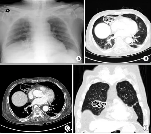
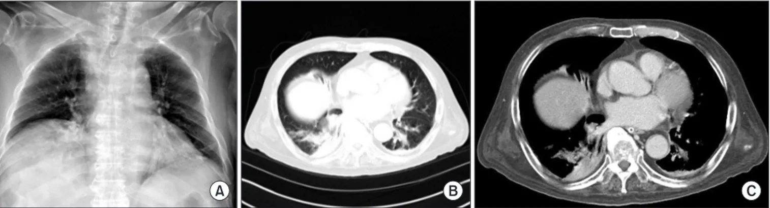
Pulmonary pneumatoceles are air-filled thin-walled spaces within the lung and are rare in adult cases of pneumonia. We report the case of a 74-year-old male who was admitted with a cough and sputum production. He had been treated with oral dexamethasone since a brain tumorectomy 6 months prior. Contrast-enhanced computed tomography (CT) of the chest revealed a large pneumatocele in the right middle lobe and peripheral pneumonic consolidation. Bronchoalveolar lavage was performed; cultures identified extended-spectrum -lactamase (ESBL) producing Proteus mirabilis . A 4-week course of intravenous ertapenem was administered, and the pneumatocele with pneumonia resolved on follow-up chest CT. To the best of our knowledge, this is the first reported case of pulmonary pneumatocele caused by ESBL-producing P.
mirabilis associated with pneumonia.
Keywords: Pneumonia; Proteus mirabilis ; Beta-Lactamase
aureus
3,4. Pneumatoceles are also caused by other organisms such as gram-negative bacilli (especially pseudomonas), but reports of pneumatoceles caused by Proteus mirabilis are few
1,5.
P. mirabilis is often associated with contamination and colo- nization, but it only occasionally associated with severe infec- tions
6. Antimicrobial resistance has been reported increas- ingly for P. mirabilis, and increased resistance of this species to
-lactams, aminoglycosides, and quinolones has become of great concern
6,7.
We here describe a case of pulmonary pneumatocele caused by P. mirabilis in a patient with pneumonia; the organ- ism produced an extended-spectrum -lactamase (ESBL).
Case Report
A 74-year-old male was admitted with a 2-week history of cough and sputum production. He exhibited low-grade fever and general weakness. His medical history included a brain tumor; tumorectomy of the right frontal cortex and basal gan- glia had been performed, followed by concurrent chemora- Copyright © 2015
The Korean Academy of Tuberculosis and Respiratory Diseases.
All rights reserved.
Introduction
Pulmonary pneumatoceles are thin-walled gas-filled cavi- tary lesions of the lung parenchyma
1, and are common in infants and young children with pneumonia, but unusual in adults
2. Most often, pneumatoceles develop as complications of acute pneumonia, commonly caused by Staphylococcus
CASE REPORT
http://dx.doi.org/10.4046/trd.2015.78.4.371ISSN: 1738-3536(Print)/2005-6184(Online) • Tuberc Respir Dis 2015;78:371-374
Address for correspondence: Goohyeon Hong, M.D.
Division of Allergy and Pulmonary Medicine, Department of Internal Medicine, Dankook University Hospital, Dankook University College of Medicine, 119 Dandae-ro, Dongnam-gu, Cheonan 31116, Korea Phone: 82-41-550-3870, Fax: 82-41-556-3256
E-mail: [email protected] Received: Apr. 23, 2015 Revised: Jun. 15, 2015 Accepted: Jun. 19, 2015
cc