with interlobular septal thickening in both lower lobes, right middle lobe and left lingular division. He had been initially diagnosed with lipoid pneumonia and was kept under observation. However, his chest lesion showed continuous progression and a video-associated thoracoscopy was performed His pulmonary lesion was confirmed histologically to be a BALT(bronchial associated lymphoid tissue) lymphoma. We report a case of a BALT lymphoma, which was initially misdiagnosed as lipoid pneumonia. (Tuberc Respir Dis 2007; 63: 194-199)
Key Words: BALT lymphoma, Marginal zone lymphoma, Non-Hodgikin's lymphoma, Lipoid pneumonia.
Address for correspondence: Seung Soo Sheen, M.D.
Department of Pulmonary and Critical Care Medicine, Ajou University School of Medicine, Wonchon-dong, Suwon, 443-721, Korea
Phone: 82-31-219-5122, Fax: 82-31-219-5124 E-mail: [email protected]
Received: Jun. 14. 2007 Accepted: Aug. 6. 2007
서 론
림프절 외 변연부 B-세포 림프종(mucosal-associated lymphoid tissue lymphoma)은 비호지킨스 림프종의 한 소그룹으로 가장 흔한 호발 부위는 위장관이고 그 외 폐, 침샘, 눈물샘, 결막, 갑상선, 흉선, 유방 및 방광 등에 발생한다1. 폐에 발생한 림프절 외 변연부 B-세 포 림프종(BALT lymphoma)은 폐에서 원발하는 비 호지킨스 림프종의 70% 이상을 차지하지만 전체 림 프종의 1% 미만을 차지할 만큼 흔한 질환은 아니다2,3. BALT lymphoma의 증상은 대부분 무증상이고 흉부 방사선 촬영에서 우연히 발견되는 경우가 많다. 예후 는 비교적 양호하나 아직 치료법은 정립되지 않은 상 태이다. 저자들은 흉부 방사선 촬영상 지속되는 폐침
윤 소견으로 내원하여 림프절 외 변연부 B-세포 림프 종으로 진단 받은 증례를 보고하는 바이다.
증 례
환 자: 50세 남자
주 소: 호흡곤란, 발열 및 마른기침
현병력: 상기 환자는 3년 전 호흡곤란, 발열 및 기 침을 주소로 내원하여 흉부 방사선 촬영을 시행하였 고 우중엽 및 설상엽에 경질(consolidation) 소견이 보 여 시행한 흉부 전산화 단층 촬영 상 리포이드(lipoid) 폐렴이 의심되어 추적 관찰하던 중 내원 2일전부터 호흡곤란, 발열 및 마른기침이 악화되어 내원하였다.
과거력 및 가족력: 혈변으로 입원치료 하였고, 가족 력은 특이소견 없었다.
진찰소견: 내원 당시 혈압 100/70 mmHg, 맥박 110 회/분, 호흡수 24 회/분, 체온 38.4℃였고, 급성 병색을 보였으며, 흉부 청진 상 양 하폐야에서 수포음이 청진 되었고, 다른 특이소견은 없었다.
검사실 소견: 말초 혈액 검사에서 백혈구 8,090 /mm3(호중구 0.3%, 림프구 11.3%, 단핵구 2.8%, 호산
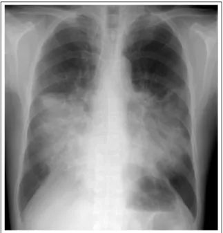
Figure 1. Chest radiography shows the aggravated consolidation compared with previous study in right middle lobe, left lingular division and both lower lobes.
Figure 2. CT scan shows aggravated air-space con- solidation , ground glass opacities with interstitial thickening of interlobular septa in right middle lobe, left lingular division and both lower lobes.
구 0.1%), 혈색소 12.6 g/dL, 혈구용적 39.4%, 혈소판 234,000 /mm3, 적혈구 침강 속도(ESR) 61 mm/hr, C- 반응단백(CRP) 5.71 mg/dL이었고 말초 혈액 도말검 사는 정상이었다. 소변 검사 상 특이소견 없었으며, 생화학 검사에서 BUN 10.3 mg/dL, 크레아티닌 1.0 mg/dL 총 단백 7.3 g/dL, 알부민 3.7 g/dL. 총 빌리루 빈 0.8 mg/dL, AST 23 IU/L, ALT 9 IU/L, LDH 228 IU/L이었다. 동맥혈 가스검사상 pH 7.439, PaCO2
35.1 mmHg, PaO2 60.5 mmHg, HCO3 22.9 mmol/L, SaO2 92 %이었다. 객담 세포학적 검사 및 항산균 도 말검사는 모두 음성이었고, 혈액배양검사 및 객담 배 양검사도 모두 음성이었다.
방사선 소견: 단순 흉부 방사선 촬영 상 우중엽 및 왼쪽 설상엽에 경질화(consolidation) 소견이 관찰되 었다. 흉부 전산화 단층 촬영 상 우중엽, 설상엽, 양 폐 하엽에 간유리 음영과 소엽사이 및 소엽내의 간질 비 후 및 점상 경화(patchy consolidation)가 관찰되었다 (Figure 1, 2).
기관지 내시경: 점막 충혈 외에 특이 소견 없었고, 기관지 세척 검사 상 성상은 탁하였고, 단백질 599 mg/dL,당 4 mg/dL, 세포수 6,320 /μL(호중구 7%, 림
프구17%, 단핵구 72%, 호산구 0%)였고, 그람 염색, 항산균 염색 및 배양, 세포학적 검사상 특이소견 없었 다.
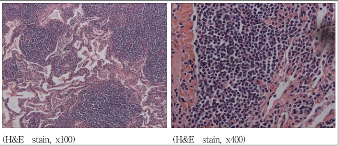
병리 검사: VATS(video-associated thoracoscopy) 로 조직검사 시행하였고, 림프절 외 변연부 B세포 림 프종(Extranodal marginal zone B-cell lymphoma)로 진단되었다(Figure 3, 4, 5).
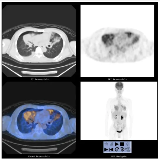
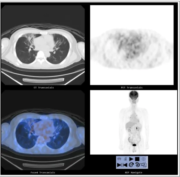
경 과: 림프절 외 변연부 B세포 림프종(BALT lymphoma) 진단 하에 병기검사를 진행하였다. 골수 흡인 검사 상 골수 침범이 있었고, 양성자 방출 단층 촬영(PET CT) 상 양 폐하, 양 폐문(hilar), 우 기관방 (paratracheal), 분기즐하(subcarinal) 림프절 침범으 로 IV 병기에 합당하였다. 이에 따라 RCHOP(ritu- ximab, cyclophosphamide, adriamycin, vincristine, and prednisolon)으로 항암 화학요법 6주기를 시행하 였고 임상양상 및 흉부방사선 및 양성자 방출 단층 촬 영 소견은 호전 되었다(Figure 6, 7).
고 찰
BALT(bronchus-associated lymphoid tissue)는 세기관지의 점막하층에 위치한 림프의 집합체로 기도 점막의 면역에 중요한 역할을 한다. 태생기 2, 3분기
Figure 4. Immunohistochemical stain for CD 20 shows positive reation reactivity (x400).
Figure 5. Immunohistochemical stain for CD 5 shows negative reation (x400).
에 출현하여 출생 이후 급격한 발달을 하다 나이가 들 면서 점차 소실되나 지속적인 항원의 자극이나 IL-4 의 분비가 BALT의 과증식을 유발하고 림프종을 일 으키는 것으로 추정하고 있다4.
그에 대한 근거로 Helicobacter pylori 에 의한 만 성 위염, C형 간염 바이러스, Borrelia burgdorferi 감 염, 하시모토(Hashimoto's) 갑상선염, 쇼그렌 (Sjogren's) 증후군 등이 다양한 MALT(mucosa associated lym- phoid tissue) 림프종을 일으킨다는 예가 보고 되었고, BALT 림프종 또한 만성 염증 질환, 자가면역 질환 및 흡연과 관계되어 유병률이 높은 것이 보고 되었으 나 명확한 항원은 밝혀진 것이 없다5,6.
MALT 림프종은 1983년 Isaacson과 Wright 등에
의해 처음 기술되었고, 1994년 REAL(real European- American lymphoma) classification에서 비호지킨스 림프종의 한 소그룹으로 분류되었고, 1997년 WHO 분류에서 MALT 림프종이란 용어대신 'marginal zone B-cell lymphoma of MALT type'으로 명명하 였다7-9.
최근 발생 빈도가 증가하는 이유는 과거에 가성 림 프종(pseudolymphoma) 또는 만성염증으로 진단되었 던 질환이 면역조직화학염색이 점차 발달하면서 MALT 림프종으로 재분류되었기 때문이다2. 면역조 직형은 비특이적이나 CD19, 20, 21, 35를 표현하고 비 호지킨스 림프종의 다른 부류인 소림프구성 림프종 및 외투세포성 림프종에서 표현되는 CD5와 여포성
Figure 6. PET CT shows multiple FDG uptake in right middle lobe, left lingular division, both lower lobe, both hilar, right paratracheal and subcarinal lymph node.
림프종에서 표현되는 CD10은 표현하지 않는다10. 진단 당시 1/3가량이 무증상이고 우연히 흉부 방사 선 촬영에서 발견되며 기침, 호흡 곤란, 흉통, 객혈 등 의 비특이적인 호흡기 증상을 호소하고 림프종에서 흔히 관찰되는 B-증상(발열, 체중 감소, night sweating)은 20-40%정도로 흔하지 않다11,12. 본 증례 에서는 기침, 호흡곤란, B-증상을 호소하였다.
방사선적 소견은 다양한 양상을 나타내며 주로 중 심부 또는 변연부의 폐실질 경화 또는 공동이나 석회 화가 없는 폐포내 결절 및 다발성 소결절이 관찰된다.
흔하지는 않지만 무기폐, 흉막 삼출 등도 나타나며 폐 문 및 종격동 림프절 전이는 드물다12. 본 증례에서 관 찰된 양 폐야에 간유리 음영, 간질 비후 및 경화 등의 방사선 소견은 리포이드 폐렴이나 폐포 단백증(pul- monary alveolar proteinosis) 등에서 특징적으로 보
이는 소견으로 처음에 BALT 림프종으로 진단되지 못하였다. 따라서 확진을 위해서는 조직학적 진단이 중요하며 기관지 내시경 및 세침 흡입검사를 시행할 수 있으나, 진단율이 비교적 낮아 VATS(Video- assisted thoracostomy) 또는 개흉폐생검술 등이 요 구된다11.
BALT lymphoma는 병변이 국한되어 보이더라도, 몸 전체의 MALT로 전이가 가능하여 진단 당시 흉부 및 복부 전산화 단층 촬영, 골수 검사, 양성자 방출 단 층 촬영 등의 적극적인 병기 검사가 필요하다. 불량한 예후를 시사 하는 소견으로는 골수 침범, 높은 종양 부하량(high tumor burden), 낮은 수행능력상태 (ECOG) 및 높은 LDH와 β2-micoglobulin이며 드물 게 고등급 B 세포 림프종으로 진행하는 경우가 있다13. 국한된 병변을 가지고 있는 경우 수술적 절제가 우
Figure 7. PET CT shows the decreased FDG uptake compared with previous study in right middle lobe and leff lingular division. The subcarinal lymph node is disappeared in FDG uptake after 6th chemotherapy.
선이고, 국소적 방사선 치료나 항암 치료가 대체요법 으로 시행될 수 있고, 완전 절제가 되지 않은 경우 추 가적인 치료로 시행 될 수 있다. 병변이 크거나 전이 된 경우 CVP(cyclophosphamide, vincristine and prednisolon), CHOP(cyclophosphamide, adriamycin, vincristine and prednisolon) 등의 복합 화학요법이 시행되었고, rituximab의 사용이 치료에 좋은 결과를 가져올 수 있다13.
본 증례는 리포이드 폐렴으로 오인되어 진단과 치 료가 늦었고, 높은 종양 부하량, 골수 침범이 있었던 비교적 예후가 좋지 않은 그룹으로 rituximab을 추가 한 전신 복합 화학 요법(RCHOP)을 시행하였으며 현재 6주기 시행 후 부분 관해 상태에서 경과 관찰 중이다.
요 약
기관에 발생한 림프절 외 변연부 B-세포 림프종 (BALT lymphoma)은 폐에서 원발하는 비호지킨스 림프종의 70% 이상을 차지하지만 전체 림프종의 1%
미만을 차지할 만큼 드문 질환으로 흉부 단순 촬영에 서 우연히 발견되는 경우가 많고, 예후는 비교적 양호 하나 아직 확립된 치료법은 없는 상태이다. 저자들은 흉부 방사선 촬영 상 지속되는 폐침윤 소견을 주소로 내원 후 리포이드 폐렴으로 오인되어 진단과 치료가 늦어 골수 침범까지 진행된 폐에 발생한 림프절 외 변 연부 B-세포 림프종을 1예를 경험하여 보고하는 바이다.
Tsuji M, et al. Bronchial infiltration with diffuse large B-cell lymphoma. Leuk Res 2006;30:1319-22.
4. Varoczy L, Gergely L, Illes A. Diagnostics and treatment of pulmonary BALT lymphoma: a repert on four cases. Ann Hematol 2003;82:363-6.
5. Kang MJ, Lee JM, Lee SJ, Son JW, Kim DG, Lee MG, et al. A case of Broncus-Associated Lymphoid Tissue lymphoma in the lung of the patient with primary Sjogren's Syndrome. Tuberc Respir Dis 2002;52:
179-85.
6. Kurtin PJ, Myers JL, Adlakha H, Strickler JG, Lohse C, Pankratz VS, et al. Pathologic and clinical features of primary pulmonary extranodal marginal zone B-cell lymphoma of MALT type. Am J Surg Pathol 2001;25:
997-1008.
Classification of lymphoid neoplasms: a proposal from the International Lymphoma Study Group. Blood 1994;84:1361-92.
10. Matsushima AY, Hamele-bena D, Osborne BM. Fine- needle aspiration biopsy findings in marginal zone B cell lymphoma. Diagn Cytopathol 1999;20:190-8.
11. Ferraro P, Trastek VF, Adlakha H, Deschamps C, Allen MS, Pairolero PC. Primary non-Hodgkin's lymphoma of lung. Ann Thorac Surg 2000;69:993-7.
12. Takamori M, Noma S, Kobashi Y, Inoue T, Gohma I, Mino M, et al. CT findings of BALTOMA. Radiat Med 1999;17:349-54.
13. Ahmed S, Siddiqui AK, Rai KR. Low-grade B-cell bronchial associated lymphoid tissue(BALT) lym- phoma. Cancer Invest 2002;20:1059-68.