Vol. 10, No. 1, March, 2003
ꠏꠏꠏꠏꠏꠏꠏꠏꠏꠏꠏꠏꠏꠏꠏꠏꠏꠏꠏꠏꠏꠏꠏꠏꠏꠏꠏꠏꠏꠏꠏꠏꠏꠏꠏꠏꠏꠏꠏꠏꠏꠏꠏꠏꠏꠏꠏꠏ
<접수일:2002년 7월 29일, 심사통과일:2002년 10월 15일>
※통신저자:고 은 미
서울특별시 강남구 일원동 50번지 삼성서울병원 내과
Tel:02) 3410-3439, Fax:02) 3410-3849, E-mail:[email protected] 이 연구는 한국학술진흥재단 연구비(KRF-1999-003-F00172)의 보조로 이루어졌음.
류마티스 섬유모세포양 활막세포에서 저산소증이 시토카인 생산에 미치는 영향
성균관대학교 의과대학 삼성서울병원 내과학교실, 삼성생명과학연구소*, 중앙대학교 의과대학 내과학교실**
안중경․전찬홍․고재현․김진희*․최화정*
안광성*․차훈석․유석희**․고은미
= Abstract =
Effect of Hypoxia on Cytokine Production in Rheumatoid Fibroblast-Like Synoviocytes
Joong Kyung Ahn, M.D., Chan Hong Jeon, M.D., Jae Hyun Koh, M.D., Jin-Hee Kim*, Hwa-Jung Choi*, Kwang-Sung Ahn, Ph.D.*, Hoon-Suk Cha, M.D., Suk Hee Yu, M.D.**, Eun-Mi Koh, M.D.
Department of Medicine, Samsung Medical Center, Sungkyunkwan University School of Medicine, Samsung Biomedical Research Institute*
Department of Internal Medicine, Graduate School of Medicine Chung-Ang University College of Medicine**, Seoul, Korea
Objective: Rheumatoid arthritis (RA) is a chronic inflammatory disease characterized by increased production of cytokines, proliferation of fibroblast-like synoviocytes (FLS) and joint destruction. It is well known that the involved joints in RA are hypoxic. Hypoxia may play a role in the pathogenesis of RA. We thought that hypoxia might alter the production of cytokines by FLS and these changes could affect the biologic behaviors of FLS. Based on that, we investigated whether hypoxia affects the production of cytokines in FLS and the effect of these changes on matrix metalloproteinases (MMPs) expression.
Methods: Fibroblast-like synoviocytes from human rheumatoid synovial tissue obtained duringjoint replacement surgery were cultured in vitro. Hypoxic culture was performed by
서 론
류마티스 관절염은 염증성 시토카인과 chemokine 을 생산하는 활막세포의 종양성 증식을 특징으로 하 는 질환이다1). 류마티스 관절염은 T 세포 매개성 질 환으로 알려져 있지만 대식세포나 섬유모세포양 활 막세포(fibroblast-like synoviocytes: FLS)와 같은 비 T 세포들이 류마티스 관절염의 발병에 중요한 역할을 한 다. 즉, 활성화된 섬유모세포양 활막세포가 자발적으 로 다양한 시토카인을 생산하여 관절에서 기질 분해 효소인 matrix metalloproteinases (MMPs), cathepsin, ag- grecanase 등을 분비하게 할 뿐 아니라 신혈관 형성 을 유도하여 주변 조직으로의 성장과 침윤을 유발할 수 있다2). 최근 류마티스 관절염에서 조절되지 않는 신혈관 형성이 염증 반응 및 면역학적 질병 경과에 중심적 역할을 하는 것으로 알려져 있으며3,4), 이러 한 신혈관 형성에 관여하는 시토카인으로 IL-1, IL-6, IL-8, TGF-β, 혈관 내피세포 성장인자(vascular endo- thelial growth factor: VEGF) 등이 연구되고 있다5-10). 류마티스 관절염에 의해 침범된 관절은 물리적, 대 사적 요인에 의해 저산소 상태를 유지하고 있다11,12). 이에 대한 근거로는 류마티스 관절염 시 관절강내의 산소 분압이 낮으며 pH가 저하되고 이산화탄소 농 도가 증가하였다13). 또한 정상 활액에 비해 젖산 농 도가 높고14,15), hypoxia-inducible factor (HIF)의 발현 이 증가되어 있는 것이 보고되고 있다16). 각종 세포에 서 저산소증에 의하여 VEGF가 증가됨은 이미 잘
알려져 있어8,9,17) 류마티스 관절염에서도 관절강내 저산소증이 단독으로 또는 시토카인과 함께 VEGF 를 증가시켜 신혈관 형성에 관여하는 것으로 생각할 수 있다. 즉, 류마티스 관절염의 질병 초기 혹은 진 행 과정에서 형성되는 국소적인 저산소증은 활막세 포의 변화를 유도하며, 이로 인하여 활막세포의 생 물학적인 특성이 변화하여 질병 진행 과정에 관여할 수 있다. 그러나 저산소증이 류마티스 관절염의 병 태 생리에 어떠한 역할을 하며, 섬유모세포양 활막 세포에 어떤 영향을 미치는가에 대한 연구는 많이 이루어지지 않았다. 본 연구에서는 류마티스 섬유모 세포양 활막세포에서 저산소증에 의하여 시토카인의 생산이 변화되는가를 알고자 하였고 변화된 시토카 인이 활막세포에 미치는 영향을 살펴보고자 하였다.
대상 및 방법
1. 류마티스 섬유모세포양 활막세포의 배양
활막 조직은 미국류마티스학회(American College of Rheumatology)의 진단기준18)에 맞는 류마티스 관 절염 환자 5명에서 관절 치환술을 통해 얻었다. 먼 저 환자로부터 떼어낸 활막 조직을 phosphate buffered saline (PBS, pH 7.5)으로 두 번 씻고 아주 잘게 잘라 낸 후 1 mg/ml collagenase (GIBCO BRL, Rockville, MD, USA)를 섞은 후 37oC에서 2시간 동안 교반하 며 배양했다. 배양 후 얻어진 상층액의 세포는 70μm cell strainer (Becton Dickinson, Franklin Lakes, NJ)로 incubating cells in BBLⓇ Gaspak pouchTM anaerobic system. After incubation under hypoxic condition for 24 hr, the concentrations of various cytokines in culture supernatants were determined by ELISA. To determine the effect of highly expressed cytokines on MMP expression, we performed ELISA of MMP-1, MMP-2 and MMP-3 in cultured FLS, after stimulation with respective cytokines.
Results: In hypoxic state, IL-6, IL-8 and vascular endothelial growth factor (VEGF) con- centrations were significantly increased compared to those in normoxic condition. However, there were little differences in IL-1, IL-2, IL-4, TNF-α and TGF-β. Stimulation of FLS with IL-6 and IL-8 showed the increased concentrations of MMP-1, MMP-2 and MMP-3.
Conclusion: Hypoxic environment of rheumatoid synovium might affect FLS to produce proinflammatory and proangiogenic cytokine such as IL-6 and IL-8. These cytokines again could stimulate MMPs production in FLS leading to joint destruction.
ꠏꠏꠏꠏꠏꠏꠏꠏꠏꠏꠏꠏꠏꠏꠏꠏꠏꠏꠏꠏꠏꠏꠏꠏꠏꠏꠏꠏꠏꠏꠏꠏꠏꠏꠏꠏꠏꠏꠏꠏꠏꠏꠏꠏꠏꠏꠏꠏꠏꠏꠏꠏꠏꠏꠏꠏꠏꠏꠏꠏꠏꠏꠏꠏꠏꠏꠏꠏꠏꠏꠏꠏꠏꠏꠏꠏꠏꠏꠏꠏꠏꠏꠏꠏꠏꠏ Key Words: Rheumatoid arthritis, Fibroblast-like synoviocytes, Hypoxia, IL-6, IL-8, Metallopro-
teinase
여과한 후 4 ml의 Ficoll/Paque (Pharmacia Biotech, Uppsala, Sweden)를 넣어 상층액에 층이 생기면 원심 분리를 20oC, 400 g에서 30분간 실시하였다. 원심분 리 후 경계층을 4배의 RPMI 1640 (BioWhittaker, Walk- ersville, MD)으로 재현탁시킨 후 다시 원심분리를 25 g에서 10분씩 세 번 시행하였다. 이렇게 하여 분 리된 세포는 마지막으로 20% heat-inactivated horse serum (GIBCO BRL)이 포함된 α-minimum essential medium (α-MEM; Irvine Scientific, Santa Ana, CA)으 로 현탁시킨 다음 well당 106개의 세포가 들어갈 수 있도록 24 well에 분주하여 키웠다. 이후 10% fetal bovine serum (FBS), penicillin 50 unit/ml, streptomycin 50μg/ml, fungizon 100μg/ml이 포함된 Dulbecco's Modified Eagle Medium (DMEM; Life Technologies, Gaithersburg, NY)으로 바꾸어 37oC, 5% CO2 배양기 에서 배양한 후 배지는 3일마다 교체하였다. 3번 또 는 4번의 계대배양한 섬유모세포양 활막세포를 실험 에 사용하였다.
2. 저산소 배양 조건의 확립
저산소 조건을 만들기 위해 BBLⓇ Gaspak pouchTM anaerobic system (Becton Dickinson)을 사용하였다. 먼 저 pouch 내부를 저산소증으로 만들기 위해 활성화 한 용해액을 pouch의 터널을 통해 넣은 다음 그 pouch 안에 세포가 80% 자란 배양 플라스크를 넣고 봉합 막대나 열-봉합(heat-sealing)기를 이용하여 밀폐 시켰다. 밀폐시킨 pouch를 37oC 배양기에 넣은 후 2 시간이 경과하면 Gaspak pouch anaerobic indicator strip의 색깔이 푸른색에서 하얀 색으로 변하는데, 이 는 총 산소농도가 2% 이하이며 이산화탄소 농도는 4% 이상의 저산소증을 나타내는 것이다.
3. 효소 면역 측정법(Enzyme-Linked Immuno- sorbent Assay: ELISA)
류마티스 관절염 환자로부터 얻은 섬유모세포양 활막세포의 상층액(supernatants)에서 IL-1, IL-2, IL-4, IL-6, IL-8, TNF-α, TGF-β, VEGF의 양을 sandwich ELISA kit (R&D Systems, Minneapolis, MN)를 이용 하여 kit에서 제시한 방법에 따라 측정하였다. 섬유 모세포양 활막세포는 세포배양기 바닥 전체를 한 겹 의 세포로 덮기 직전에 10% FBS를 포함한 신선한
배지로 교환한 후 24시간 동안 각각 저산소증과 정 상 산소 상태에서 배양하였다. 그리고 상층액을 모 아서 검사하기 전까지 -70oC에서 보관하였다.
4. IL-6, IL-8 처리에 따른 metalloproteinases의 생산 분석
IL-6와 IL-8에 의한 섬유모세포양 활막세포의 matrix metalloproteinases 생산에 어떤 변화가 일어나 는지를 분석하기 위하여 10% FBS가 포함된 DMEM 에서 자라고 있는 섬유모세포양 활막세포를 혈청이 없는 DMEM으로 교환한 후, IL-6 (R&D Systems), IL-8 (R&D Systems)을 각각 0.8 ng/ml, 0.5 ng/ml씩 첨가하고 24시간 동안 배양하였다. 배양 후 상층액 을 분리하여 sandwich ELISA kit (R&D Systems)를 이용하여 total MMP-1, MMP-2, MMP-3의 농도를 측 정하였다.
5. 통계 분석
모든 통계처리는 SPSS (ver. 10.0)를 이용하여 처 리하였다. 정상 산소 상태와 저산소증에서 배양된 류마티스 섬유모세포양 활막세포의 상층액에서 측정 한 시토카인의 농도의 차이에 대한 유의성은 연구대 상의 수가 적고 자료의 정규성도 인정할 수 없어서 비모수 검정 기법인 Mann-Whitney U 검정을 시행하 였다. p값이 0.05 이하인 경우 통계적으로 유의하다 고 판정하였다.
결 과
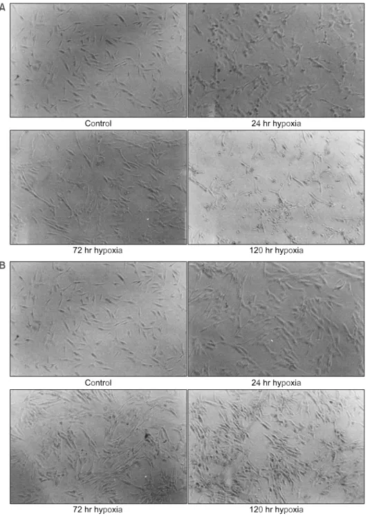
1. 저산소증에서 섬유모세포양 활막세포의 형태
정상 산소 상태와 저산소증에서 배양된 섬유모세 포양 활막세포를 현미경으로 비교 관찰 하였다. 혈 청이 없는 배지(serum-free culture media)를 이용하여 저산소증에서 24시간 동안 배양한 경우 세포의 변화 를 관찰할 수 없었으나, 저산소증을 120시간 동안 지속한 경우에는 활막세포의 세포고사를 관찰할 수 있었다(그림 1). 하지만 혈청이 있는 배지를 사용한 경우에는 저산소증에서 120시간을 배양하여도 세포 가 지속적으로 성장하는 것을 관찰할 수 있었다(그 림 2). 이러한 연구 결과를 바탕으로 본 실험에서는 저산소증에 의한 다양한 시토카인 변화를 측정하기
Fig. 1. Morphologic changes of fibroblast-like synoviocytes in hypoxic condition. Fibroblast-like synoviocytes were cultured under BBLⓇ Gaspak pouchTM anaerobic system with serum-free media (A) and serum containing media (B), respectively. After incubation for varying length of time, these cells were examined under light microscopy (40×). Fibroblast-like synoviocytes under hypoxic condition with serum-free media survived for 72 hr but died after 120 hr.
위해 혈청이 없는 배지를 이용하여 저산소증에서 24 시간 배양된 활막세포를 이용하였다.
2. 저산소증에서 섬유모세포양 활막세포에서의 VEGF 생산
3명의 류마티스 관절염 환자에서 얻은 섬유모세포 양 활막세포를 정상 산소 상태와 저산소증에서 24시
간 동안 배양한 후 상층액에서 VEGF의 농도를 측 정하였다(그림 2). 그 결과, 과거 보고와 같이8-10,17,19)
정상 산소 조건에서 배양한 경우에 비해 저산소증에 서 VEGF의 농도가 통계적으로 유의하게 증가하여 (p=0.008), 본 실험에서 저산소증이 잘 유발되었음을 알 수 있었다.
Fig. 2. Change of VEGF concentration in rheumatoid fibroblast-like synoviocytes cultured under hypoxic condition. The VEGF concentration in hypoxic condition was increased compared to those in normoxic condition (*statistically signi- ficant difference (p<0.05) by Mann-Whitney U test).
Fig. 4. Effect of cytokines (IL-6 and IL-8) on production of MMP-1 (A), MMP-2 (B) and MMP-3 (C) in rheumatoid fibroblast-like synoviocytes. MMP-1, MMP-2 and MMP-3 productions were induced by either IL-6 or IL-8 (*statistically significant difference (p<0.05) by Mann-Whitney U test).
Fig. 3. Changes of concentrations of various cytokines (IL-1, IL-2, IL-4, IL-6, IL-8, TNF-α and TGF-β) in supernatants of rheumatoid fibroblast-like synoviocytes cultured under hypoxic condition.
The concentrations of IL-6 and IL-8 were significantly increased compared to those in normoxic condition (*statistically significant dif- ference (p<0.05) by Mann-Whitney U test).
3. 저산소증에 의한 섬유모세포양 활막세포에서 의 시토카인 생산의 변화
5명의 류마티스 관절염 환자에서 얻은 섬유모세포 양 활막세포를 정상 산소 상태와 저산소증에서 24시 간 동안 배양한 후 효소 면역 측정법을 이용하여 상 층액에서 IL-1, IL-2, IL-4, IL-6, IL-8, TNF-α, TGF-β의 농도를 측정하였다(그림 3). 그 결과 정상 산소 상태 와 비교하였을 때 저산소증에서 IL-2, IL-4, TGF-β의 농도는 통계적으로 유의한 변화를 보이지 않았다 (p=0.092, 1.000, 0.070). 그리고 IL-1, TNF-α의 농도 는 통계적으로 유의한 차이는 없었지만 저산소증에 서 농도가 증가하는 경향을 보였다(p=0.056, 0.056).
IL-6와 IL-8의 농도는 저산소증에서 정상 산소 상태와 비교하여 통계적으로 유의하게 증가되었다(p=0.008, 0.008).
4. IL-6와 IL-8의 처리에 따른 metalloproteinases 의 생산 변화
저자들은 과거 연구에서 저산소증에 의하여 섬유 모세포양 활막세포의 matrix metalloproteinase 활성도 가 증가됨을 관찰하였다(비출간 자료). 따라서 이번 실험에서 저산소증에 의하여 증가된 IL-6, IL-8이 섬 유모세포양 활막세포의 matrix metalloproteinase 생산 에 어떤 영향을 주는지 알아보고자 하였다.
IL-6와 IL-8으로 처리한 후 배지에서 total MMP-1 과 MMP-2, MMP-3의 농도를 측정한 결과 각각의 시토카인에 의하여 MMP-1, MMP-2, MMP-3의 농도 가 통계적으로 유의하게 증가되는 것을 관찰할 수 있었다(p=0.029, 0.029, 0.029)(그림 4).
고 찰
류마티스 관절염에 의하여 침범된 관절의 관절강 내부가 저산소증이라는 것은 일부 관절 생리학자들 에게는 잘 알려져 있었으나13-15) 이러한 관절내 저산 소증이 류마티스 관절염의 병태 생리에 어떠한 역할 을 하는가에 대하여는 별로 알려진 바가 없다. 최근 류마티스 관절염에서 신혈관 형성의 중요성이 알려 지면서 저산소증에 대한 관심도 늘어나고 있다. 저 산소증의 발생 기전은 급성 염증기에는 조직의 산소
사용이 증가하여 충분한 산소의 공급이 안 되어 일 어난다. 그리고 만성기에는 관절강 내 압력이 증가 되어 모세혈관의 관류압을 넘어서게 되어 허혈 상태 가 발생하게 된다12,20). 또한 운동으로 인한 굴곡 및 신전에 의해 반복적으로 저산소-재관류화가 발생하 여 활막염이 악화될 수도 있다21).
류마티스 활막에서 TNF-α22), IL-123), TGF-β 등10) 이 VEGF의 발현을 증가시키는 인자들로 논의되고 있으며 특히 저산소증은 VEGF의 발현을 증가시키 는 것으로 알려져 있다8-10,17,19)
. 본 연구 결과 중 저 산소증에서 배양한 섬유모세포양 활막세포의 상층액 에서 VEGF 농도가 정상 산소 상태에 비해 세배 이 상 증가되어 이는 다른 연구들과 마찬가지로 저산소 증이 VEGF의 발현을 자극한다는 것을 확인할 수 있었다8-10,17,19)
.
류마티스 관절염의 활막에는 TNF-α, IL-1, IL-6, GM-CSF와 같은 염증성 시토카인, IL-8과 같은 chemo- kine, 그리고 IL-10, TGF-β와 같은 항염증성 시토카 인이 풍부하게 분포되어 있다24). 앞서 말한 바와 같 이 류마티스 관절염에서 관절강 내가 저산소증이므 로 저산소증에 의하여 섬유모세포양 활막세포의 시 토카인의 생산과 분비가 변화될 수 있다고 가정할 수 있다. 따라서 본 연구에서는 정상 산소 상태와 저산 소증에서 각각 류마티스 섬유모세포양 활막세포를 배양한 후 상층액에서 류마티스 관절염과 관련이 있 는 다양한 시토카인의 발현을 살펴보았다.
저산소증에서 IL-2, IL-4, TGF-β의 농도는 정상 산소 상태와 비교하였을 때 증가하지 않았다. VEGF 의 발현을 증가시키는 인자로 알려진 IL-1, TNF-α 는 저산소증에서 증가하는 경향을 보였지만 통계적 으로 유의한 변화는 없었다. 하지만 IL-1이나 TNF-α가 소량 증가하여도 충분히 VEGF의 발현을 증가시킬 가능성은 남아있다. 저산소증에서 IL-6와 IL-8는 통 계학적으로 의미있게 증가하였다. 이런 결과가 일반 적 현상인지 아니면 류마티스 관절염에서만 나타나 는 것인지에 대해서는 골관절염의 활막세포에서 실 험을 하지 않아 논의하기는 어렵다. 본 실험에서 IL-6 가 높게 측정된 것은 이전의 연구와 비슷한 결과를 보인 것으로5,25,26) 이는 IL-6가 국소적으로 류마티스 관절의 활액에서 높은 농도를 유지하면서 활막염을 악화시킬 것으로 생각된다. 한 연구에서 IL-6는 류마
티스 관절염의 활막에서 높은 농도로 측정되었고 만 성 관절염의 조직학적 소견과 활액의 IL-6 농도와 양 의 상관관계를 가지는 것으로 보고하였다25). 이전의 동물 실험에서도 저산소증이 쥐의 신경교세포에서 IL-6의 발현을 증가시키는 것으로 보고된 바 있다26). 또한 활발하게 신혈관 형성이 일어나고 있는 조직에 서 IL-6의 발현이 증가되어 있으며 IL-6를 다양한 세 포에 투여한 경우에 VEGF의 발현이 유도되는 것을 보고하였다5). 따라서 저산소증의 섬유모세포양 활막 세포는 IL-6의 발현을 증가시켜서 자신과 주변의 섬 유모세포양 활막세포에서 VEGF 생산을 증가시키고 신혈관 형성을 유도하여 판누스 형성에 중요한 역할 을 할 가능성이 있다.
이번 연구에서 증가한 IL-8은 chemokine으로 종양 조직에서 신혈관 형성과 세포 분열, 전이를 유도하 는 것으로 알려져 있다7). IL-8을 토끼의 관절강 내에 주입했을 때, 1시간 이내에 중성 백혈구의 급속한 침윤과 함께 급성 염증을 관찰하였다는 보고가 있었 다27). 또한 IL-8은 신혈관을 형성하는 능력을 가지고 있으며, IL-8에 대한 항체를 투여한 경우 신혈관 형 성이 억제되는 것이 보고되었다7).
본 연구에서는 저산소증에 의해 활막세포에서 증 가된 IL-6와 IL-8이 섬유모세포양 활막세포에 미치는 영향을 알아보고자 하였다. 섬유모세포양 활막세포 에 IL-6, IL-8을 투여한 결과, 섬유모세포양 활막세포 에서 MMP-1, MMP-2, MMP-3의 농도가 증가하였다.
여기서 IL-6가 MMPs의 농도를 증가시킨 것은 지금 까지 알려진 결과와는 다른 것이다. 즉, 류마티스 섬 유모세포양 활막세포에서 IL-6는 MMP의 생산과는 관련이 없으며 tissue inhibitor of metalloproteinases (TIMP)의 생산을 촉진하는 것으로 알려져 왔다28-30). 또 IL-6가 IL-1의 MMP-1과 MMP-3에 대한 효과를 증강시켜 류마티스 관절염에서 조직 손상을 일으킨 다고 보고되었다28). 그러나 저자들의 연구에서는 IL-6 자체가 MMP의 생산을 증가시킴을 알 수 있었 다. 이처럼 이전과 다른 결과를 보인 이유는 첫째, 실험 방법의 차이 때문이다. 지금까지의 보고는 모 두 90년대 초의 연구로서 정확히 어떤 종류의 MMP 를 측정하였는지에 대한 명시가 없이 collagenase 활 성도를 측정한 것으로 되어 있다29,30). 또 다른 연구 에서는 pro-MMP를 측정한 것인데 반해28) 본 연구는
효소 면역 측정법을 이용하여 total MMP를 측정하 였기 때문에 이러한 차이가 발생했을 가능성이 있 다. 둘째, 이전의 연구 결과는 류마티스 관절염만을 대상으로 하지 않아 제한점이 있다29,30). 이처럼 본 연구에서는 류마티스 관절염에서 IL-6의 역할이 지 금까지 알려진 바와 다르게 나타났지만 최근에 악성 림프종에서 IL-6가 MMP-2 생산을 증가시켜 악성 종 양의 공격성(aggressiveness)에 중요한 역할을 할 것 이라는 연구가 있어31) 본 연구와 유사한 결과를 보여 주고 있다. IL-8에 의한 MMPs가 증가된 것으로 보 아 IL-8은 류마티스 관절염에서 chemokine으로서의 역할 외에 다른 역할을 할 것으로 생각된다. 악성 질환에서는 이에 대한 증거들이 제시되고 있는데, 흑색종과 방광의 이행세포 암종에서 IL-8은 gelatinase (MMP-2, MMP-9)의 발현을 조절하여 종양세포의 침 윤과 신혈관 형성 및 전이를 촉진하는 것으로 알려 져 있다32,33). 따라서, 저산소 상태에서 증가된 IL-6와 IL-8은 이미 알려진 염증성 시토카인과 chemokine으 로서의 작용 이외에도 MMP의 발현을 증가시켜 류 마티스 관절염의 조직 파괴에 기여를 할 것으로 생 각된다. 결론적으로 저산소증에 의해 섬유모세포양 활막세포에서 VEGF, IL-6, IL-8의 생산이 증가되며 이 증가된 IL-6와 IL-8은 관절 파괴와 관련된 MMP-1, MMP-2, MMP-3를 증가시키기 때문에 관절의 저산 소증은 류마티스 관절염의 병태 생리에 중요한 역할 을 할 가능성이 있다.
결 론
저산소증은 섬유모세포양 활막세포에서 IL-1, IL-2, IL-4, TNF-α, TGF-β의 농도는 증가시키지 않은 반 면에 IL-6, IL-8, VEGF의 농도는 의미 있게 증가시 켰다. IL-6, IL-8의 증가는 섬유모세포양 활막세포에 서 MMP-1, MMP-2, MMP-3의 증가를 유도하였다.
위와 같은 결과는 류마티스 관절염에서 발생하는 저산소증이 IL-6, IL-8의 생산을 유도하여 MMP 농 도를 높여 판누스 형성과 관절 파괴에 관여할 가능 성을 제시하므로 향후 이에 대한 연구가 더 필요할 것으로 생각한다.
REFERENCES
1) Nanki T, Nagasaka K, Hayashida K, Saita Y, Miya- saka N. Chemokines regulate IL-6 and IL-8 pro- duction by fibroblasts-like synoviocytes from patients with rheumatoid arthritis. J Immunol 2001;167:
5381-5.
2) Yamanishi Y, Firestein GS. Pathogenesis of Rheu- matoid arthritis: The role of synoviocytes. Rheum Dis Clin North Am 2001;27:355-71.
3) Coleville-Nash, Seed MP. The current state of angio- statin therapy with special reference to rheumatoid arthritis. Curr Opin Invest Drugs 1993;2:763-813.
4) Firestein GS. Starving the synovium: angiogenesis and inflammation in Rheumatoid arthritis. J Clin Invest 1999;103:3-4.
5) Cohen T, Nahari D, Cerem LW, Neufeld G, Levi BZ.
Interelukin-6 induces the expression of vascular endo- thelial growth factor. J Biol Chem 1996;271:736-41.
6) Koch AE. Angiogenesis: implication for Rheumatoid arthritis. Arthritis Rheum 1998;41:951-62.
7) Xie K. IL-8 and human cancer biology. Cytokine Growth Factor Rev 2001;12:375-91.
8) Fava RA, Olsen NJ, Spencer-Green G, Yeo KT, Yeo TK, Berse B, et al. Vascular permeability factor/
endothelial growth factor (VPF/VEGF): Accumulation and expression in human synovial fluids and rheu- matoid synovial tissue. J Exp Med 1994;180:341-6.
9) Oliver SJ, Banquerigo ML, Brahn E. Suppression of collagen-induced arthritis using an angiogenesis in- hibitor, AGM-1470, and a microtubule stabilizer, taxol. Cell Immunol 1994;157:291-9.
10) Berse B, Hunt JA, Diegel RJ, Morganelli P, Yeo K, Brown F, et al. Hypoxia augments cytokines (trans- forming growth factor-β (TGF-β) and IL-1) induced vascular endothelial growth factor secretion by human synovial fibroblasts. Clin Exp Immunol 1999;115:
176-82.
11) Edmonds SE, Blake DR, Morris CJ, Winyard PG. An imaginative approach to synovitis: the role of hypoxic reperfusion damage in arthritis. J Rheumatol 1993;
37l:S26-31.
12) Edmonds SE, Ellis G, Gaffney K, Archer J, Blake DR.
Hypoxia and the rheumatoid joint: immunological and therapeutic implications. Scand J Rheumatol 1995;101:
S163-8.
13) Treuhaft PS, McCarty DJ. Synovial fluid pH, lactate, oxygen, and carbon dioxide partial pressure in various
joint diseases. Arthritis Rheum 1971;14:475-84.
14) Wallis WJ, Simkin PA, Nelp WB. Low synovial clearance of iodide provides evidence of hypoperfusion in chronic rheumatoid synovitis. Arthritis Rheum 1985;28:1096-104.
15) James MJ, Cleland LG, Rofe AM. Determinants of synovial fluid lactate concentration. J Rheumatol 1992;19:1107-10.
16) Hollander AP, Corke KP, Freemont AJ, Lewis CL.
Expression of hypoxia-inducible factor 1 by macro- phages in the rheumatoid synovium: Implications for targeting of therapeutic genes to the inflamed joint.
Arthritis Rheum 2001;44:1540-4.
17) Jackson JR, Minton JA, Ho ML, Wei N, Winkler JD.
Expression of vascular endothelial growth factor in synovial fibroblasts is induced by hypoxia and inter- leukin-1β. J Rheumatol 1997;24:1253-9.
18) Arnett FC, Edworthy SM, Bloch DA, MaShane DJ, Fries JF, Cooper NS, et al. The American Rheu- matism Association 1987 revised criteria for the classification of the rheumatoid arthritis. Arthritis Rheum 1988;31:315-24.
19) Paleolog EM, Young S, Stark AC, McCloskey RV, Feldmann M, Maini RN. Modulation of angiogenic vascular endothelial growth factor by tumor necrosis factor and interleukin-1 in rheumatoid arthritis.
Arthritis Rheum 1998;41:1258-65.
20) Stevens CR, Williams RB, Farrell AJ, Blake DR.
Hypoxia and inflammatory synovitis: Observation and speculation. Ann Rheum Dis 1991;50:124-32.
21) Blake DR, Merry P, Unsworth J, Kidd BL, Outhwaite JM, Ballard R, et al. Hypoxic-reperfusion injury in the inflamed human joint. Lancet 1989;1:289-93.
22) Feldmann M, Bondeson J, Brennan FM, Foxwell BM, Maini RN. The rationale for the current boom in anti-TNFα treatment. Is there an effective means to define therapeutic targets for the drugs that provide all the benefits of anti-TNFα and minimise hazards? Ann Rheum Dis 1999;8:S127-31.
23) Paleolog EM, Hunt M, Elliot MJ, Feldmann M, Maini RN, Woody JN. Deactivation of vascular endothelium by monoclonal anti-tumor necrosis factor α antibody in rheumatoid arthritis. Arthritis Rheum 1996;39:1082-91.
24) Feldmann M, Brennan FM, Maini RN. Role of cytokines in rheumatoid arthritis. Annu Rev Immunol 1996;14:397-440.
25) Sack U, Kinne RW, Marx T, Heppt P, Bender S, Emmrich F. Interleukin-6 in synovial fluid is closely associated with chronic synovitis in rheumatoid
arthritis. Rheumatol Int 1993;13:45-51.
26) Maeda Y, Matsumoto M, Hori O, Kuwabara K, Ogawa S, Yan SD, et al. Hypoxia/reoxygenation-mediated induction of astrocyte interleukin-6: a paracrine mech- anism potentially enhancing neuron survival. J Exp Med 1994;180:2297-308.
27) Endo H, Akahoshi T, Takagishi K, Kashiwazaki S, Matsushima K. Elevation of interleukin-8 levels in joint fluids of patients with rheumatoid arthritis and the induction by IL-8 of leukocyte infiltration and synovitis in rabbit joint. Lymphokine Cytokine Res 1991;10:245-52.
28) Ito A, Itoh Y, Sasaguri Y, Morimatsu M, Mori Y.
Effects of interleukin-6 on the metabolism of con- nective tissue components in rheumatoid synovial fibroblasts. Arthritis Rheum 1992;35:1197-201.
29) Lotz M, Guerne PA. Interleukin-6 induces the synthesis of tissue inhibitor of metalloproteinases-1/ ery- theroid potentiating activity (TIMP-1/ EPA). J Biol Chem 1991;266:2017-20.
30) Sato T, Ito A, Mori Y. Interleukin 6 enhances the production of tissue inhibitor of metalloproteinases (TIMP) but not that of matrix metalloproteinases by human fibroblasts. Biochem Biophys Res Commun 1990;170:824-9.
31) Kossakowska AE, Edwards DR, Prusinkiewicz C, Zhang MC, Guo D, Urbanski SJ, et al. Interleukin-6 regulation of matrix metalloproteinase (MMP-2 and MMP-9) and tissue inhibitor of metalloproteinase (TIMP-1) expression in malignant non-Hodgkin's lymphomas. Blood 1999;94:2080-9.
32) Luca M, Huang S, Gershenwald JE, Singh RK, Reich R, Bar-Eli M. Expression of interleukin-8 by human melanoma cells up-regulates MMP-2 activity and increases tumor growth and metastasis. Am J Pathol 1997;151:1105-13.
33) Inoue K, Slaton JW, Kim SJ, Perrotte P, Eve BY, Bar-Eli M, et al. Interleukin-8 expression regulates tumorigenicity and metastasis in human bladder cancer.
Cancer Res 2000;60:2290-9.