DOI:10.4078/jkra.2009.16.2.123
<접수일:2009년 5월 25일, 수정일:2009년 6월 1일, 심사통과일:2009년 6월 2일>
※통신저자:박 성 환
서울시 서초구 반포동
가톨릭대학교 의과대학 류마티스내과학교실
Tel:02) 2258-6011, Fax:02) 537-4673, E-mail:[email protected]
본 연구는 과학기술부/한국과학재단 우수연구센터 육성사업의 지원으로 수행되었음(R11-2002-098-05003-0).
류마티스관절염 활막세포에서 TLR3 자극에 따른 MIF 생성의 증가
가톨릭대학교 의과학연구원 류마티스 연구센터1, 가톨릭대학교 의과대학 류마티스내과학교실2
허양미
1ㆍ박성환
2ㆍ박미경
1ㆍ오혜좌
1ㆍ강귀영
2ㆍ조미라
1= Abstract =
Upregulation of Macrophage Migration Inhibitory Factor (MIF) Production by Engagement of Toll-like Receptor 3 (TLR3) on Fibroblast-like
Synoviocyte (FLS) from Patients with Rheumatoid Arthritis
Yang Mi Her1, Sung-Hwan Park2, Mi Kyung Park1, Hye-Jwa Oh1, Kwi Young Kang2, Mi-La Cho1
The Rheumatism Resarch Center, Catholic Research Institute of Medical Science1, Division of Rheumatology, Department of Internal Medicine, College of
Medicine2, The Catholic University of Korea, Seoul, Korea
Objective: Rheumatoid arthritis (RA) is a chronic autoimmune disease. Macrophage migration inhibitory factor (MIF) has been shown to be an important pro-inflammatory cytokine in RA. The aim of this study was to determine if the engagement of toll-like receptor 3 (TLR3) induces the production of MIF in the fibroblast-like synoviocytes (FLS) of patients with RA.
Methods: The expression of inflammatory cytokines (e.g. MIF, IL-6, IL-1β and TNFα) and toll-like receptors (e.g. TLR2, TLR3 and TLR4) in the synovial tissue were quantified by immunohistochemistry. FLS were isolated from the synovial tissues of patients with RA and stimulated with TLR-3 ligand polyI:C, in the presence of a neutralizing antibody against IL-6.
The concentrations of MIF and IL-6 in the culture supernatants from the FLS were measured using sandwich ELISA.
Results: The engagement of TLR3 with PolyI:C increased the production of MIF in FLS. The stimulatory effect of these TLR ligands showed a dose-dependent trend. The combination of
TLR3 and TLR4 synergistically increased the level of MIF and IL-6 production. The addition of neutralizing antibodies against IL-6 abrogated the stimulatory effect of the ligands of TLR3 and TLR4 on the production of MIF.
Conclusion: These results show that TLR3 engagement stimulates the production of MIF and IL-6. Therefore, the TLRs help perpetuate of RA pathogenesis through production of MIF from the FLS in patients with RA, and might provide a new therapeutic approach for the treatment of rheumatoid arthritis.
Key Words: Rheumatoid arthritis, Fibroblast-like synoviocytes, Macrophage migration inhibitory factor, Toll-like receptor engagement
서 론
류마티스관절염은 다발성 관절의 염증 및 파괴를 특징으로 하는 만성 염증성 자가면역 질환으로 병인 은 정확하게 밝혀지지 않았다. 하지만 환경적, 유전 적인 병인에 의해 복합적으로 발생하는 면역 반응 결과 생산되는 사이토카인 같은 매개 물질이 염증세 포의 비정상적인 증가를 초래하여 활막염으로 인한 활막세포의 과도한 증식과 신생 혈관의 생성, 지속 적인 염증 반응을 일으키는 것을 특징으로 한다.
Toll-like receptor (TLR)는 류마티스관절염의 병인 에 관련된 중요한 인자 중 한가지로 생체내로 침입한 미생물의 pathogen-associated molecular pattern (PAMP) 와 결합하여 항원제시세포를 활성화하여 염증 사이 토카인 및 케모카인을 생성하는 유전자를 활성화시 키고 T 림프구를 활성화 시켜 선천면역(innate im- munity) 및 적응면역(adaptive immunity)에 주요한 역 할을 한다.
Polyinosinic-polycytidylic acid (poly(I:C))는 바이러 스성의 이중가닥 RNA (viral double-stranded RNA:
dsRNA)로 TLR3에 의해 인지되며 다양한 세포상에 서 바이러스 감염에 의한 효과를 연구하는데 이용되 어 지고 있다 (1,2). 류마티스관절염 환자의 활막세 포내 TLR3는 과발현되어 있으며 TLR3를 통한 자극 은 활성화된 T 림프구를 끌어당길수 있는 CXC che- mokine IFN-gamma-inducible protein-10 (IP-10/CXCL10), IFNβ, CXCL10, CCL5과 IL-6의 발현증가가 보고되 었다 (3-5). 그러나 TLR2나 TLR4와 같은 다른 TLRs 에 비하여 TLR3는 류마티스관절염의 병인에 관련된 연구가 거의 이루어져 있지 않다
류마티스관절염의 과다한 염증반응은 단핵구/대식 세포와 염증 T 림프구와 같은 면역세포들에 의해 분비되는 케모카인과 사이토카인들과 같은 면역 매 개 물질에 의해 이루어지며 IL-1, IL-6, IL-8, IL-15과 IL-17과 같은 사이토카인들에 의한 질병 활성화가 매우 중요한 병인으로 알려져 있다 (6-8).
대식세포유주 억제인자(Macrophage migration inhi- bitory factor, MIF)는 대식세포의 이동을 억제하는 물 질로 처음에는 주로 활성화된 T 세포에 의해 주로 분비되는 사이토카인으로 밝혀졌지만 (9) 이 후에 많은 연구들을 통하여 다양한 기능들이 밝혀지면서 류마티스관절염을 포함한 면역 매개성 염증 질환의 병인에 있어 중요한 역할을 담당하는 것으로 알려졌 다 (10-17). MIF는 TNF-α 또는 IFN-γ와 같은 염증 성의 자극에 반응하는 대식세포에 의해 유도될 수 있으며, TNF-α, IFN-γ, IL-1β, IL-6, IL-8, nitric ox- ide와 cyclo-oxygenase 2 (COX2)와 같은 염증성 분자 의 생산을 유도한다 (18-22).
류마티스관절염에서의 MIF의 중요성은 많은 연구 들을 통하여 여러가지 증거들이 제시되고있다. MIF 는 활막내 대식세포와 활막세포에서 발현하고 류마 티스관절염 환자의 혈청과 활액내에서 과발현되고 있다. 게다가 류마티스관절염 환자의 활막세포에서 유래된 MIF는 단핵구에 의해 TNF-α를 분비시키고 다시 활막세포를 활성시킨다 (23). 또한 류마티스관 절염 환자의 활막세포에서 MIF는 MMP-1과 MMP3 의 발현을 전사인자 수준에서 증가시키며 IL-8를 유 도하며 뿐만 아니라 최근에는 본 연구진과 다른 연 구진들을 통해 신생혈관형성에도 중요하게 작용하는 것으로 확인되었다 (19,20,24-26). 또한 MIF는 내독소 에 대한 감지를 활성화 시키는 TLR4의 발현을 증가
시킨다 (27).
따라서 MIF와 TLR은 두가지 모두 선천면역와 적 응면역계내에서 중요한 역할을 하며 면역 불균형이 나타나는 류마티스관절염의 병인에 관련된 중요한 요소로써 두 인자간의 관련성 연구는 자가면역질환 의 병인 이해에 중요하다. 그러나 류마티스관절염의 활막세포를 대상으로 한 연구는 부족한 실정이다.
본 연구에서는 류마티스관절염 환자의 활막세포내 TLR2, TLR3 및 TLR4의 발현 패턴을 조사하고, 특 히 TLR3의 리간드인 PolyI:C에 의한 류마티스관절염 의 활막세포에서의 MIF생성에 대한 영향을 조사하 고자 한다.
대상 및 방법 1. 시약
Lipopolysaccharide (LPS) (SIGMA, USA), polyinosinic- polycytidylic acid (PolyI:C) (SIGMA, USA)를 사용 하 였다. 중성화 항체로 anti IL-6 (R&D, USA)를 사용 하였다.
2. 환자
가톨릭대학교 강남성모병원 류마티스센터에서 1987 년에 개정된 미국 류마티스학회(American College of Rheumatology, ACR)의 분류 기준에 만족하는 류마티 스관절염 환자 10명을 대상으로 하였다. 대조 군으 로는 골관절염 환자와 최근 건강검진에서 정상으로 판명된 지원자중 성별과 나이가 맞춰진 대상으로 하 였다. 본 연구는 강남성모병원 임상연구 관리규정과 헬싱키 선언을 준수하여 시행하였다. 활막조직은 관 절치환술을 받은 류마티스관절염 환자로부터 분리 하였다.
3. 면역 조직 화학 염색법
4% 파라포름알데하이드에 고정된 각 활막(류마티 스관절염 환자, 골관절염 환자) 조직을 통상의 방법 대로 파라핀에 포매한 후 절편 기를 이용하여 7 um 절편을 만들어 슬라이드에 붙인 후 헤마톡실린과 에 오신(hematoxylin-eosin) 염색을 하여 광학현미경으로 관찰하였다. 사이토카인의 발현의 측정은 면역조직 화학 염색방법으로 ABC (Vector laborites, Burlingame,
CA, USA) kit를 사용하여 염색하였으며, 슬라이드에 부착된 절편을 자일렌과 에탄올로 탈파라핀과 함수 를 시킨 후 3% H2O2로 내인성 과산화효소를 차단시 키고, 인산화 완충액(Phosphate buffered saline, PBS) 으로 수세한다. 비특이적인 반응을 차단할 목적으로 anti-mouse serum을 30분 반응시킨 후 primary 항체를 MIF (R&D, USA) 1:50, TLR-2 (Santa Cruz, CA) 1:
100, TLR-3 (Santa Cruz, CA) 1:100 TLR-4 (Santa Cruz, CA) 1:100을 다음날 (16∼18 h)까지 반응시 켰다. Primary 항체 반응 후 결합이 안된 항체를 PBS 로 수세하고 바이오틴이 결합된 이차 항체와 과산화 효소가 결합된 streptavidin 반응을 시킨 후 DAB으로 발색 시킨다. Mayer’s 헤마톡실린으로 대조군 염색한 후 수세하고 봉입하여 광학현미경으로 관찰하였다.
4. 활막세포의 분리 및 자극
활막세포는 관절치환술을 받은 류마티스관절염 환 자로부터 얻은 활막 조직을 효소를 이용한 분해를 통해 분리 하였다. 조직은 2∼3 mm 조각으로 잘게 자른 후 Dulbecco’s modified Eagle’s medium (DMEM, Gibco BRL, Carlsbad, CA) 배양액에 4 mg/mL의 농 도의 type 1 collagenase (Worthingion Bio-chemical, Freehold, NJ)와 함께 37oC water bath에서 4시간 동 안 반응 시켰다. 반응이 끝난 조직은 500 g에서 원 심분리 하였고 20% fetal bovine serum (FBS, Gibco), L-gutamin 2 mM, penicillin 100 units/mL과 streptomy- cin 100 ug/mL이 들어있는 DMEM 배양액으로 희석 후 25 cm2 플라스크에 담아 37oC, 5% CO2의 배양기 에서 10일을 배양해준다. 10일 후 붙지 않은 조직은 제거하고 붙은 세포만 20% DMEM 배양액에서 배양 하였다. 배양액은 주 3일마다 교환해 주었고 계대 배양은 면적의 90% 이상으로 세포가 차지했을 때 1:3으로 희석하여 늘려 주었다. 모든 실험의 활막 세포는 5번에서 9번사이의 계대배양한 세포를 사용 하였다. 90% 자란 세포를 TryPLE Express (Gibco, USA)를 이용하여 세포를 띄어준다. 세포를 count하 여 24well pate에 1×10⁴/mL로 분주 하여 준다. 세포 가 80% 정도의 자라면 ITSA가 포함된 serum free DMEM 배양액과 섞어 24well plate에 분주하여 준다.
2시간 전에 anti-IL-6 2 ug/mL를 처리하여 준다. 2시 간이 지난 뒤 LPS (10 ng/mL)와 PloyI:C (1ug/mL, 10
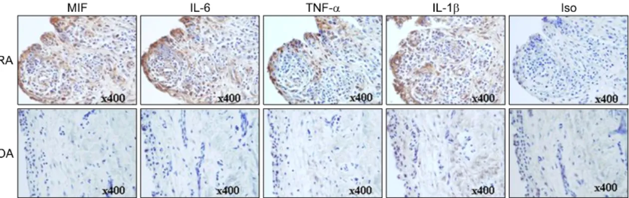
Fig. 1. Increased expression of MIF, IL-6, TNFα and IL-1β in RA synovium. Immunostaining was performed using the specific antibodies against MIF, IL-6, TNFα and IL-1β in synovial biopsy samples from patients with rheumatoid arthritis (RA) and osteoarhtirits (OA). MIF, IL-6, TNFα and IL-1β stained a brown color. All tissues were counterstained with hematoxylin (original magnification ×400).
ug/mL, 50 ug/mL)으로 처리하여 72시간 동안 37oC, 5% 배양기에서 배양하였다.
5. 사이토카인 측정
Sandwich ELISA용 96 well plate (NUNC, Denmark) 에 capture monoclonal antibody MIF 2 ug/mL, IL-6 4 ug/mL, IL-1β 4 ug/mL, TNFα 4 ug/mL (R&D, SA) 로 50 ul/mL씩 넣고, 4oC에 밤새 반응시킨 다음 차단 용액(1% bovine serum albumin (BSA)/0.05% Tween 20)이 함유된 phosphate buffered saline (PBS)을 200 ul/well씩 넣고 실온에서 2시간 반응시킨다. 샘플과 recombinant human MIF, IL-6, IL-1b, TNFa (R&D, USA)을 5,000∼78.125 pg/mL 이용하여 실온에서 2시 간 반응시켰다. Well을 세척용액(0.05% PBST)으로 4 번 세척하고 biotinylated goat-anti-human MIF 200 ng/mL, IL-6 300 ng/mL, IL-1β 100 ng/mL, TNFα 200 ng/mL (R&D, USA)로 50 ul/well씩 넣어 실온에 서 2시간 반응시킨 후 4번 세척하였다. 마지막으로 는 ExtraAvidin-Alkaline Phosphatase conjugate (SIGMA, USA)를 1:2,000으로 희석하여 50 ul/well씩 넣고 실 온에서 2시간 반응시키고 세척 후 PNPP (Fluka, Phos- phate Disodium salt Hexahydrate)/DEA 용액을 1 mg/
mL 농도로 녹여 50 ul/well씩 넣어 20∼30분 후 0.2N NaOH로 반응을 멈추고 405 nm 파장에서 흡광을 측 정하였다.
6. 통계적 유의성의 검증
실험 결과는 평균±표준오차로 표현하였으며 통계 적 유의성은 student’s t-test를 실시하였고 p값이 0.05 이하 일 때 통계적으로 유의하다고 분석하였다.
결 과
1. 류마티스관절염 환자의 활막조직내 염증성 사이 토카인과 TLR2, TLR3와 TLR4의 과발현
류마티스관절염 환자와 대조군으로써 골관절염 환 자의 활막조직에서 MIF를 비롯한 IL-6, TNFα과 IL-1β 및 TLR2, TLR3와 TLR4의 발현 양상을 조사 하였다. 각각의 사이토카인에 특이적인 항체를 사용 하여 염색하였을 때 골관절염 환자보다 류마티스관 절염 환자에서 면역 세포들의 침착의 정도가 높았으 며 그러한 세포에서의 MIF, IL-6, TNFα과 IL-1β의 발현 양상도 류마티스관절염 환자에서 모두 높게 발 현하였다(그림 1).
또한, TLR2, TLR3와 TLR4의 발현 역시 기존보고 와 마찬가지로 류마티스관절염 환자의 활막조직내 높게 발현하였다(그림 2).
2. 류마티스 관절염 환자의 활막 세포에서 PolyI:C에 의한 MIF와 IL-6의 생산량 증가
다음으로 TLR3에 의한 신호를 통해 활막 세포에 서 MIF와 IL-6의 생성을 단백질 수준에서 조사하였
Fig. 2. Increased expression of TLR 2, 3 and 4 in RA synovium. Immunostaining was performed using the specific antibody against TLR 2, 3 and 4 in synovial biopsy samples from patients with RA and OA. TLR 2, 3 and 4 stained a brown color. All tissues were counterstained with hematoxylin (original magnification ×400).
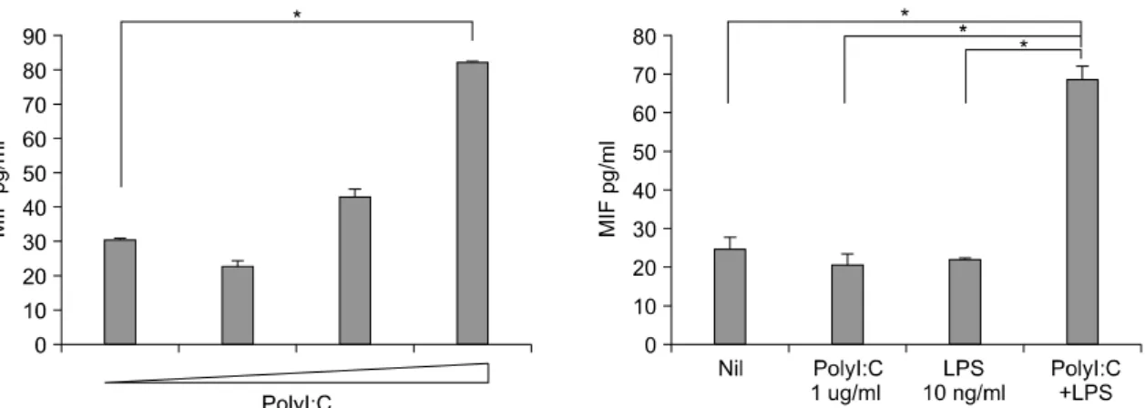
Fig. 3. Enhancement of MIF production by the stimulation of TLR3 or TLR4 engagement in FLS from patients with RA. The FLS were cultured with the TLR3 (1 μg/mL, 10 μg/mL, 50 μg/mL) and TLR4 ligands (10 ng/mL) only or combination for 72 hr. The culture supernatants were assayed for MIF by ELISA. *p<0.01.
다. 류마티스관절염 환자의 활막 세포에 PolyI:C를 처리하였을 때 농도 의존적으로 MIF와 IL-6의 생산 량이 증가하였으며 MIF는 50 ug/mL의 농도에서 최 고 생산치를 보여 통계적 유의성을 나타내었다. 저 농도의 PolyI:C와 TLR4의 리간드인 lipopolysaccharide (LPS)와 함께 자극하였을 MIF와 IL-6의 생산은 각 리간드들의 단독자극보다 그 생산량이 유의하게 증 가하였다(그림 3, 4). IL-6 이외의 TNFα, IL-1β와 같은 다른 염증성 사이토카인의 생성량은 미비하였 다(결과제시하지 않음).
3. 류마티스관절염 환자의 활막 세포에서 IL-6 중 화항체에 의한 PolyI:C에 의해 유도된 MIF 의 생산량 감소
MIF는 류마티스관절염 환자의 활막세포에서 IL-6 뿐만 아니라 IL-8, cyclooxygenase-2 (COX-2), prosta- glandin E (2)의 발현을 증가시키는 것으로 보고되었 다 (19). IL-6에 의존적인 MIF의 생성에 관여하는 알 아보고자 IL-6에 대한 중화항체를 이용하여 PolyI:C 에 의한 MIF의 생산량을 조사하였다. PolyI:C에 의 해 증가된 MIF의 생산은 IL-6에 대한 중화항체의 처 리에 의해 억제하였으나 통계적 유의성을 나타내지
Fig. 4. Enhancement of IL-6 production by the stimulation of TLR3 or TLR4 engagement in the FLS from patients with RA. FLS were cultured with TLR3 (1 μg/mL, 10 μg/mL, 50 μg/mL) and TLR4 ligands (10 ng/mL) only or a combination for 72 hr. The culture supernatants were assayed for IL-6 by ELISA. *p<0.05, **p<0.01.
Fig. 5. Blockade of the IL-6-induced decrease in MIF by the engagement of TLR3 and TLR4 in RA FLS. The RA FLS were stimulated with TLR3 (1 ug/mL) and TLR4 ligands (10 ng/mL) alone and in combination for 72 hr. The culture supernatants were assayed for MIF by ELISA. *p<0.05, **p<0.01.
않았다. 그러나 저농도의 PolyI:C와 TLR4의 리간드 인 LPS와 함께 자극하였을 때 증가된 MIF는 IL-6에 대한 중화항체의 처리에 의해 통계적으로 유의하게 감소시켰다(그림 5).
4. 류마티스관절염 환자의 혈청과 활액 내 MIF와 IL-6의 증가
류마티스관절염 환자와 대조군으로써 골관절염 환 자와 정상인의 혈청과 활액 내 존재하는 MIF와 IL-6 의 양을 조사하였다. 혈청과 활액 내 MIF와 IL-6의
양은 류마티스관절염 환자에서 골관절염과 정상인에 비해 증가되어 있었다. MIF의 경우 혈청과 활액내에 서 모두 통계적으로 유의성을 나타내었으나 IL-6의 혈청과 활액의 경우에는 통계적 유의성을 나타내진 않았다(그림 6).
고 찰
류마티스관절염의 병인연구에 있어서 IL-1과 TNF 와 같은 염증성 사이토카인에 대한 연구가 활발히 진행되어 이를 이용한 생물학적 제제가 현재 이용되
Fig. 6. Increased level of MIF and IL-6 in the synovial fluid and sera derived from patients with RA, OA and HC.
The concentrations of MIF and IL-6 in RA and OA synovial fluids and RA, OA and HC sera determined by ELISA. *p<0.05, **p<0.01.
고 있으나 류마티스관절염의 완치에 대한 접근은 아 직 미흡하여 염증병인과 관련된 다른 염증성 사이토 카인에 대한 연구가 필요하다.
MIF는 지연성 과민증(delayed-type hypersensitivity), 내독성쇼크(endotoxic shock)와 패혈쇼크(septic shock) 와 같은 질병의 선천면역과 적응면역계에서 중요한 역할을 하는 사이토카인으로 (28) 류마티스관절염의 염증반응에 관련된 병인적인 역할에 대한 많은 연구 가 진행되고 있다.
본 연구에서는 TLR3의 특이적인 리간드인 PolyI:C 의 자극에 의해 류마티스관절염의 활막세포에서의 MIF 생성능을 조사하였다. 이전 보고와 마찬가지로 류마티스관절염 환자의 활막조직, 혈청, 활액내에는 MIF를 비롯한 IL-6, TNF-α, IL-1β와 같은 염증성 사이토카인들이 과발현 되어있었다. 혈청과 활액내 존재하는 IL-6의 경우 통계적인 유의성에 도달하지
는 못하였지만 대조군과 비교 시 환자군에서 높게 증가되어 있었다. 이는 기존보고와는 다른 결과로 본 연구에서 진행된 환자군의 적은 수에 기인한 것 으로 생각되어진다.
이전연구에 따르면 류마티스관절염 환자의 활액내 에는 peptidoglycan (PGN), 괴사 세포과 같은 TLR 리 간들이 존재하므로 (5,29) 이러한 리간들에 의한 활 막세포의 활성은 과도한 면역반응에 의한 면역체계 의 불균형을 일으킨다. 따라서 활막세포에서 발현하 는 TLR의 발현과 반응성은 류마티스관절염의 병인 에 있어서 중요한 요소이다. 최근 보고에 따르면 류 마티스관절염 환자와 골관절염의 환자의 활막세포에 서 TLR1부터 TLR6에 해당하는 수용체들은 자체적 으로 발현되고 있으나 TLR7부터 TLR10은 발현되어 있지 않음이 보고되었다 (30). 특히 이러한 수용체들 중 TLR2, TLR3과 TLR4가 류마티스관절염 환자의
활막세포내 과발현이 밝혀지면서 기존 TLR2와 TLR4 에 대한 연구와 더불어 TLR3에 대한 연구의 중요성 이 부각되고 있다.
다양한 TLR 신호를 통한 활막세포의 활성은 활막 세포로부터 염증성 사이토카인의 생성을 유도할수 있는데 많은 연구를 통하여 과도한 염증반응에서의 MIF의 중추적 기능이 부각되어 후보 물질이 될 가 능성을 충분히 가지고 있다. MIF의 주요한 생산 원 천은 초기 연구에서는 활성화된 T 림프구로 알려져 있었으나 이후 많은 연구들을 통하여 LPS, IL-1과 TNF-α에 의해 T 림프구에서 보다 대식세포, 내피세 포, 활막세포에 의해 생산됨이 보고되었다 (11,18,31,32).
본 연구에서는 TLR3의 리간드인 PolyI:C에 의해 류마티스 환자의 활막세포에서 MIF 생성이 IL-6에 대한 중화항체에 의해 억제됨을 확인하였다. TLR2 와 TLR4의 리간드의 경우, 단독 자극에 의해서는 MIF 생성을 관찰하지 못하였으나 TLR4 리간드인 LPS의 경우 저농도에서 PolyI:C와의 공동 자극에 의 해 MIF생성의 상승효과를 확인하였다. 이는 TLR4 신호체계가 부분적으로 PolyI:C와 공유함으로써 MIF 생성과 관련되어 신호가 증폭되었을 것으로 생각된 다. 이러한 TLR 경로를 통한 세포내 염증성 사이토 카인 활성기전 연구는 질환병인 연구에 매우 중요하 며 이때 주요한 병인표적사이토카인 IL-6가 MIF 생 산에 관여하는 것으로 보이는 본 연구 결과는 새로 운 결과이다.
류마티스관절염을 비롯한 염증질환에서의 MIF는 단순한 염증성 사이토카인으로써 염증을 매개하는 것이 아니라 면역세포를 주변 활막세포로 유인할 수 있도록 케모카인의 생성을 유도할 뿐만 아니라 신생 혈관의 생성에도 관여하여 류마티스관절염의 병인연 구에 있어서 중추적인 사이토카인이다(11,20). 이에 TLR3의 신호를 통한 MIF의 생성은 류마티스관절염 의 염증성 병인을 근본적으로 이해하는데 큰 도움을 줄 것이다.
결론적으로 류마티스관절염의 활막세포에서 TLR3 의 신호를 통한 자극에 의해 MIF가 생성되며 이러 한 생성은 부분적으로 IL-6의 생산에 의존적으로 생 성됨을 확인하였다. 이는 TLR3을 통한 신호체계를 차단함으로써 류마티스관절염 환자의 치료에 응용될 수 있을 것으로 기대된다.
결 론
류마티스관절염 환자의 활막세포에서 TLR3의 과 발현을 확인하고 TLR3의 특이적인 리간드인 PolyI:C 에 의해 염증질환 활성 사이토카인 MIF의 생성능을 관찰함으로써 이러한 인자들을 통한 조절은 류마티 스관절염에서 보여지는 과도한 염증반응에 의한 면 역시스템 항상성의 불균형에 대한 근본적인 원인을 이해하고 극복하는데 기여할 것으로 기대한다.
참고문헌
1) Kumar A, Zhang J, Yu FS. Toll-like receptor 3 agonist poly(I:C)-induced antiviral response in human corneal epithelial cells. Immunology 2006;117:11-21.
2) Takahashi N, Yamada T, Narita N, Fujieda S. Double- stranded RNA induces production of RANTES and IL-8 by human nasal fibroblasts. Clin Immunol 2006;
118:51-8.
3) Proost P, Vynckier AK, Mahieu F, Put W, Grillet B, Struyf S, et al. Microbial Toll-like receptor ligands differentially regulate CXCL10/IP-10 expression in fibroblasts and mononuclear leukocytes in synergy with IFN-gamma and provide a mechanism for enhanced synovial chemokine levels in septic arthritis.
Eur J lmmunol 2003;33:3146-53.
4) Roelofs MF, Joosten LA, Abdollahi-Roodsaz S, van Lieshout AW, Sprong T, van den Hoogen FH, et al.
The expression of toll-like receptors 3 and 7 in rheu- matoid arthritis synovium is increased and costimu- lation of toll-like receptors 3, 4, and 7/8 results in synergistic cytokine production by dendritic cells.
Arthritis Rheum 2005;52:2313-22.
5) Brentano F, Schorr O, Gay RE, Gay S, Kyburz D.
RNA released from necrotic synovial fluid cells acti- vates rheumatoid arthritis synovial fibroblasts via Toll-like receptor 3. Arthritis Rhwum 2005;52:2656- 65.
6) Muller-Ladner U, Pap T. Pathogenesis of RA: more than just immune cells. Z Rheumatol 2005;64:396- 401.
7) Nishimoto N. Cytokine signal regulation and autoi- mmune disorders. Autoimmunity 2005;38:359-67.
8) Smolen JS, Steiner G. Therapeutic strategies for rheu- matoid arthritis. Nature Rev Drug Discov 2003;2:
473-88.
9) Nathan CF, Remold HG, David JR. Characterization of a lymphocyte factor which alters macrophage functions. J Exp Med 1973;137:275-90.
10) Burger-Kentischer A, Göbel H, Kleemann R, Zer- necke A, Bucala R, Leng L, et al. Reduction of the aortic inflammatory response in spontaneous athero- sclerosis by blockade of macrophage migration inhibi- tory factor (MIF). Atherosclerosis 2006;184:28-38.
11) Leech M, Metz C, Hall P, Hutchinson P, Gianis K, Smith M, et al. Macrophage migration inhibitory factor in rheumatoid arthritis: evidence of proinfla- mmatory function and regulation by glucocorticoids.
Arthritis Rheum 1999;42:1601-8.
12) Morand EF, Bucala R, Leech M. Macrophage migra- tion inhibitory factor: an emerging therapeutic target in rheumatoid arthritis. Arthritis Rheum 2003;48:
291-9.
13) Donnelly SC, Haslett C, Reid PT, Grant IS, Wallace WA, Metz CN, et al. Regulatory role for macrophage migration inhibitory factor in acute respiratory distress syndrome. Nat Med 1997;3:320-3
14) Mizue Y, Ghani S, Leng L, McDonald C, Kong P, Baugh J, et al. Role for macrophage migration inhibitory factor in asthma. Proc Natl Acad Sci U S A 2005;102:14410-5.
15) Magalhães ES, Mourao-Sa DS, Vieira-de-Abreu A, Figueiredo RT, Pires AL, Farias-Filho FA, et al.
Macrophage migration inhibitory factor is essential for allergic asthma but not for Th2 differentiation. Eur J Immunol 2007;37:1097-106.
16) Rossi AG, Haslett C, Hirani N, Greening AP, Rahman I, Metz CN, et al. Human circulating eosinophils secrete macrophage migration inhibitory factor (MIF).
Potential role in asthma. J Clin Invest 1998;101:
2869-74.
17) Bernhagen J, Calandra T, Mitchell RA, Martin SB, Tracey KJ, Voelter W, et al. MIF is a pituitaryderived cytokine that potentiates lethal endotoxaemia. Nature 1993;365:756-9.
18) Calandra T, Bernhagen J, Mitchell RA, Bucala R. The macrophage is an important and previously unreco- gnized source of macrophage migration inhibitory factor. J Exp Med 1994;179:1895-902.
19) Santos LL, Lacey D, Yang Y, Leech M, Morand EF.
Activation of synovial cell p38 MAP kinase by macrophage migration inhibitory factor. J Rheumatol 2004;31:1038-43.
20) Onodera S, Nishihira J, Koyama Y, Majima T, Aoki
Y, Ichiyama H, et al. Macrophage migration inhibi- tory factor up-regulates the expression of interleukin-8 messenger RNA in synovial fibroblasts of rheumatoid arthritis patients: common transcriptional regulatory mechanism between interleukin-8 and interleukin- 1beta. Arthritis Rheum 2004;50:1437-47.
21) Cunha FQ, Weiser WY, David JR, Moss DW, Mon- cada S, Liew FY. Recombinant migration inhibitory factor induces nitric oxide synthase in murine macro- phages. J Immunol 1993;150:1908-12.
22) Sampey AV, Hall PH, Mitchell RA, Metz CN, Morand EF. Regulation of synoviocyte phospholipase A2 and cyclooxygenase 2 by macrophage migration inhibitory factor. Arthritis Rheum 2001;44:1273-80.
23) Leech M, Metz C, Hall P, Hutchinson P, Gianis K, Smith M, et al. Macrophage migration inhibitory factor in rheumatoid arthritis: evidence of proin- flammatory function and regulation by glucocorti- coids. Arthritis Rheum 1999;42:1601-8.
24) Kim HR, Park MK, Cho ML, Yoon CH, Lee SH, Park SH, et al. Macrophage migration inhibitory factor upregulates angiogenic factors and correlates with clinical measures in rheumatoid arthritis. J Rheumatol 2007;34:927-36.
25) Chesney J, Metz C, Bacher M, Peng T, Meinhardt A, Bucala R. An essential role for macrophage migration inhibitory factor (MIF) in angiogenesis and the growth of a murine lymphoma. Molecular Medicine 1999;5:
181-91.
26) Amin MA, Volpert OV, Woods JM, Kumar P, Harlow LA, Koch AE. Migration inhibitory factor mediates angiogenesis via mitogen-activated protein kinase and phosphatidylinositol kinase. Circ Res 2003;93:321-9.
27) Roger T, David J, Glauser MP, Calandra T. MIF regulates innate immune responses through modu- lation of Toll-like receptor 4. Nature 2001;414:920-4.
28) Bozza M, Satoskar AR, Lin G, Lu B, Humbles AA, Gerard C, et al. Targeted disruption of migration inhibitory factor gene reveals its critical role in sepsis.
J Exp Med 1999;189:341-6.
29) Kyburz D, Rethage J, Seibl R, Lauener R, Gay RE, Carson DA, et al. Bacterial peptidoglycans but not CpG oligodeoxynucleotides activate synovial fibro- blasts by Toll-like receptor signaling. Arthritis Rheum 2003;48:642-50.
30) Ospelt C, Brentano F, Rengel Y, Stanczyk J, Kolling C, Tak PP, et al. Overexpression of toll-like receptors 3 and 4 in synovial tissue from patients with early rheumatoid arthritis: toll-like receptor expression in
early and longstanding arthritis. Arthritis Rheum 2008;58:3684-92.
31) Onodera S, Tanji H, Suzuki K, Kaneda K, Mizue Y, Sagawa A, et al. High expression of macrophage migration inhibitory factor in the synovial tissues of
rheumatoid joints. Cytokine 1999;11:163-7.
32) Nishihira J, Koyama Y, Mizue Y. Identification of macrophage migration inhibitory factor (MIF) in hu- man vascular endothelial cells and its induction by lipopolysaccharide. Cytokine 1998;10:199-205.