에스트로젠 수용체알파에 의한 Hypoxia Inducible Factor-1 의 전사 촬성조절
유광희 • 이 영 주 ^ 세중대학교 생명공학과
(Received September 24, 2009: Accepted December 3, 2009)
Activation of Hypoxia Inducible Factor-1 Alpha by Estrogen Receptor Alpha
KwangHee Ryu and YoungJoo Lee^
College of Life Science, Institute of Biotechnology, Department of Bioscknce and Biotechnology, Sejong University, Seoul 143-747, Korea
Abstract — Our previous results showed that hypoxia inducible factor-1 (HIF-1) activated estrogen receptor (ER) in the absence of ligand. In this study, we have studied the effect ER overexpression on the activation of HIF-1. ER overexpression induced transcription activation of hypoxia response element driven luciferase and vascular endothelial growth factor. As a negative control, the effect of ER on androgen receptor response element was used. Our result indicate that the two ERa and HIF-1 signaling pathways shares part of the activation pathway.
Keywords □ hypoxia, ERa, HIF-1, VEGF
저산소 상태는 혈관신생, 에너지대사, 적혈구생성 등 세포 내 다양한 반융을 조절한다.^' 저산소 상태에서의 대표적인 조절인 자는
hypoxia-inducible factor-la(HIF-la)로 정상 상태에서는 분 해되어 전사활성이 없는 반면 저 산소 상태에서는 안정화 되어 산소 항상성 유지에 필요한 당 대사, 혈관신생에 관련된 유견자 돌의 발현을 조절하는 중요한 역할을 수행한다.®
HIF-la는
Aryl Hydrocarbon Receptor Nuclear Translocator(ARNT)와
dimer로 작용을 한 다 ®
ARNT단백질은
HIF-la의 파트너로서의 역할 이외에도 에스트로젠 수용체의
coactivatoi로서도
2]용 하 :!
ARNT의 또 다른 파트너인
AhR역시 에스트로젠 수용체와의 결합을 통하여 작용을 조절함이 발 표 되 었 다 ;*이 러 한 일련의 연구결과 를 통하여 저산소의 신호 견달 체계가 에스트로젠 수용체 및 기 타 핵 호르몬 수용체의 전사 활성에 영향을 머친다는 주요 연구 결과돌이 측적되고 있다. 하지만 역으로 핵호르몬 수용체가 저 산소의 신호 견달에 영향을 미친다는 연구 걸과는 아직 거의 없다.
에스트로젠 수용체는 핵수용체
superfamily의 한 멤버이며, 에 스트로젠에 의해 활성화되는 전사인자의다.® 에스트로젠의 작용 모델은 세포 내에 있는 에스트로젠 수용체와 호르몬이 결합허여
•"본 논문에 관한 문의는 저자세게로 ( 전화) 02-3408-3640 («!스) 02-3408-4334 (E-mail) [email protected]
특정 유전자의 거울상의 염 기 서 열 을 가진
15bp의 인핸서
(hormone responsive element)®!!결합하여
basal transcription machinery와 각종
coregulator와 결합하여 전사 활성을 조절한 다고 알려져 있다.® 여성 호르몬인 에스트로젠은 난소에서 생성 되어 혈관을 통해 표적 세포에 도달하여 단순확산에 의해 표적 세포 내로 둘어가, 표적세포의 핵 내에 존재하는 전사인자인 에 스트로젠 수용체를 통하쉬 표적 유전자의 견사 활성도에 영향을 머첨으로써 그 기능을 나타낸다.® 에스트로젠 수용체는 성장인 자 경로와의
cross-talk에 의한 활성,
DNA결합 없이
AP-1파의
interaction에 의한 활 성 , 세포막에 존 재 하 는 수 용 체 를 통한
nongenomic경로 등에 의해서도 세포 내 여러 작용에 영향을 머 친다고 알려져 있 다 많 은 연구결과들에 의해 에스트로젠의 신호전달 체계에 관여하는 약
300종류의 단백질둘과 복잡한 조 절 기전이 밝혀져 있다.
에스트로젠 수용체는 유방암 세포 성장에 관여하는 많은 유전 자와 신호를 조절함으로써 유방암•의 발달과 진행에 매우 중요한 역할을 한다.^^> 에스트로젠 수용체는 유방암의 치료와 진행 추 이룰 결정하는데 매우 중요한 요소이다.^^> 최근 저산소에 의해 에스트로젠 수용체 단백질이 조절된다고 보고된바 었으며, 에스 트로젠 수용체 양성 유방암•에서
HIF-1알파의 양이 높다는 관찰 이 보고된 바도 있다.^^'
데 그 리 고 HIF-la양성 유방종양에서 에 스트로젠 수용체의 농도가 적옴도 관찰되었다.^* 이러한 일련의
102
에스트로젠 수용■체알파에 의한 Hypoxia Inducible Factor-1 의 견사 활성조절 103
관찰은 저산소와 에스트로젠 수용체가 상호 조절 함으로서 tumorigenesis에 영향을 미칠 수 있다•는 가능성을 의머한다. 본 논문에서는 에스트로젠 수용체 알파가 저산소 신호에 영향을 머 치는 가능성에 대해 고찰해보았다.
실험방법
실험재료
E; 2는 Sigma(St. Louis, MO, USA)에서 구입하였고, 100% 에 틴을에 녹였다. RPMI 역시 Sigma(St. Louis, MO, USA)에서 구 입하였고, Fetal Bovine Serum(FBS), Trizol Reagent, 그리고 penicillin/streptomycin은 GIBCO Invitxogen(Grand Island, NY, USA)에서 구입하였다. 또한 HRE-Luc DNA는 서울대학교 김규 원 박사님께 제공 받았으며, ARE-Luc DNA는 Rochester 대학 의 Dr. Chawnshang Chang에게 제공 받았다.
세포배양 및 저산소 처리
본 실험에서는 유 방 "세 포 주 인 MCF-7 세포주를 사용하였다.
MCF-7 세포주는 10% FBS을 함유하는 RPMI에서 배양하였다.
폴라스미드의 트랜스팩션에는 PEI(Polysciences)를 사용하였다.
배지는 1~2일 간격으로 교환하고 시약을 처리할 때 새 배지로 갈아주었으며 , 익물을 처리시에는 최소 24시간 이상 석탄으로 처 리한 혈청을 사용한 배지에서 배양한 후 약물을 처리하였다. 대 조군에는 에틴을 용매를 처리하였다. 저산소 상태에서의 세포는 5%의 C02 level과 1% 미만의 O2 그러고 Ng로 balance가 맞춰 진 Hypoxia chamber(Forma)률 사용하여 배양하였다.
루시퍼라OWI 활성 측정
폴라스미드룰 트랜스팩션한 세포를 PBS로 세척하고 lysis buffer(125 mM Tris pH 7.8, 10 mM CDTA, 10 mM DTT, 50% glycerol, 5% Triton X-100>t 넣고 세포률 파괴한 후 상층 액을 얻었다. 루시퍼라아제 활성측정은 20 cell extract에 20 p/
assay buffer(Promega)# 가한 루 light emmision을 Luminometer (LUMAT LB 9501/16)룰 사용하식 20초간 측정하였다.
RNA 분S| 및 Reverse Transcription-Polymerase Chain Reaction(RT-PCR)
Total RNA 주출은 Trizol Reagent(Invitrogen)률 이용하였다.
cDNA 합성 은5 |ig의 total RNA와 Random primer(Promega), 그리고 8 DEPC 처러된 물을 넣고 70°C에서 5분 동안 둔다.
그리고 40 unit의 M-MuLV 역전사효소(Promega), 5x reaction buffer(250 mM Tris-HCl; pH 8.3 at 25°C, 375 mM KCl, 15 mM MgCl2, 50 mM DTT)와 1 mM dNTP mixture, 그리고 ribonuclease inhibitor 20 unit을 넣고 37°C 에서 1시간 반응시
키고 70X에서 10S- ■동안 반응시킨다. 만들어진 cDNA 2 n/, lOx bufferdOO mM Tris-HCl; pH 8.3 at 25°C, 500 mM KCl, 15 mM MgCl) 5 jjJ, 25 unit의 rTaq polymerase(Takara, Japan), 4 |j/의 2.5 mM dNTP mixture, 100 pmoles의 primer(Geno Tech), total volume 50 |j/로 PCR을 하였다.
P-actin, VEGR E R a의 oligonucleotide 서열은 p-actin:
sense: 5-CCTGACCCTGAAGTACCCCA-3', antisense; 5'- CGTCATGCAGCTCATAGCTC-3', VEGF: sense; 5'-ATGAA- CTTTCTGCTCTCTCTGG-3', antisense; 5'-TCATCTCTCCT- ATGTGCTGGC-3', ERa: sense; 5'-CATAACGACTATATGT- GTCCAGCC-3', antisense: 5'-AACCGAGATGATGTAGCCAG- CAGC-3미다. 그리고 VEGF와 ERa의 PCR조건은 다옴과 같다.
94°C에서 3분, 94°C 45초, 55°C 45초, 72X 45초(24 cycle), 마 지막으로 72°C에서 10분간 반응시킨다. 그러고 p-actin은 20 cycle로 조건을 잡았다. 이는 P-actin을 20 cycle 이상으로 할 경 우 포화상태에 이르러 정량 분석을 하기가 힘들기 때문이다. 정 량적 분석은 각 실험 sample의 cDNA률 희석하는 방법을 사용 하였다. PCR산물은 2% gel로 전기영동 하쉬 분석하였다. bands 의 상대적 강도는 Gel Doc 2000(BIO-RAD, USA)을 통해 정량 화되었다.
실험결과 및 고찰
에스트로전] 수용체 알퍼에 의한 Hypoxia Response Elem ent (HRE)-LuciferaseSil 증가
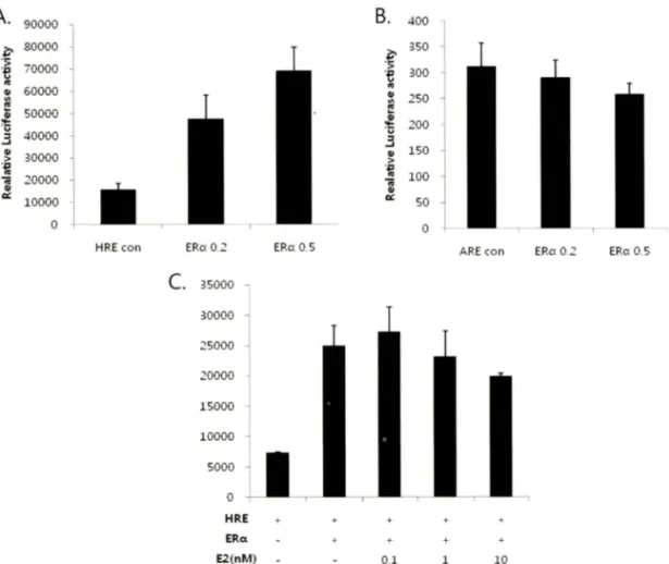
HRE(Hypoxia Response ElementO-Luciferase와 에스트로젠 수용체 알파 폴리스미드를 293 세포에 농도별로 24 h transfection 하여 HRE-luciferase 활성을 측정한 결과 luciferase의 활성이 증가하였다(Fig. 1A). 하지만 Androgen Response Element (ARE)-Ludferase와 에스트로젠 수용체 알파 폴라스미드룔 함께 transfection하여 negative control로 실험한 ARE-luciferase 측 정 결과에서는 luciferase의 활성이 보이지 않았으며 (Fig. IB), 에스트로젠 수용체 알파의 ligand로 알려진 E2률 같이 처리하 여 HRE-luciferase를 죽정하였을 때 synergy한 활성효과는 관 찰 할 수 없었다(Fig. 1C). 이 결과들은 에스트로젠 수용체의 과 발현이 HIF-1 에 의한 견사에 영향을 미칠 수 있다는 것을 제시 한다.
에스트로젠 수용체 알파에 의한 Vascular Endothelial Growth Factor(VEGF) 발현
VEGF(Vascular Endothelial Growth Factor)는 성체에서 혈관 생성뿐만 아니라 배아발달과정에서 혈관발달을 조절하는 중요한 인자이며, HIF-la와 ARNT가 interactioni: 이뤄 HRE서열부위 에 결합하여 발현되는 것으로 알려져 있 다 .^에 스 트 로 젠 수용
Vol. 54, No. 2, 2010
90000 80000 70000 60000 50000 40000 30000 20000
aoooo
400 350 300 250 200
150 100 50
H R Econ ERa 0.2 ERa 0.5
C. 35000 30000
25000
20000
15000
10000
5000
ARE con ERa 0.2 ERa 0.5
. l l l l
H R E E R a
E 2 ( n M )
0.1 10
Fig. 1 - HRE-luciferase activation was assessed by cotransfection of the ERa (0.2, 0.5 ng) with either HRE-luciferase (A) or ARE-luciferase (B) expression plasmids in HEK 293 cells, using the PEI method. At 24 h post-transfection, cells were treated with 0.1,1 and 10 nM E2 (C). Cell extracts were prepared by reporter lysis buffer and analyzed using a luciferase assay.
CON ERa
ERa
VEGF
p-actin
Fig. 2 - MCF-7 cells were transiently transfected with ERa using the PEI method. Total RNA was prepared and analyzed for ERa, VEGF and (3-actin mRNA expression by RT-PCR.
체 알파가 발현되는 cell line이며, 유방암 세포인 MCF-7세포에 에스트로젠 수용체 알파 폴라스미드를 l( ig 의 농도로 24h tmasfection하여 과발현 시켰으며, 저산소 상태에서 HIF-1 에 의
해 발현되는 VEGF의 mRNA 발현 정도를 RT-PCR로 측정하였
Fig. 3 - HEK 293 cells were transiently transfected with ERa (0.2, 0.5 (Ig) and HRE-luciferase using the PEI method. At 24 h post-txansfection, cells were incubated for 24 h hypoxic condition. Cell extracts were prepared by repoter lysis buffer and analyzed using a luciferase assay.
다. 그 결과 에스트로젠 수용체 알파의 파발현에 의해서도 VEGF 의 mRNA가 발현되는 것을 볼 수 있었다(Fig. 2).
J. Pharm. Soc. Korea
104 유광희 • 이영주
A .
>^!A
경
™
에스트로젠 수용체알파에 의한 Hypoxia Inducible Factor-1 의 전사 활성조절 105
저산소 상태에서 에스트로센 수용체 알파의 활성 변화 저산소 상태에서 HRE와 에스트로젠 수용체 알파를 293 cell 에 농도 별로 24 h transfection하늬 HRE-IuciferaseS- 측정한 결 파 normoxia 상태와는 다르게 HRE-ludferase의 활성이 약하게 저하되는 것을 관찰 할 수 있었다(Fig. 3). 이러한 결과는 에스트 로젠 수용체 알파가 정상상태와 저산소 상태에서 HIF-1 에 머치 는 영향이 다름을 의미한다.
결 론
우 리는 Normoxia 상태에서 Hypoxia Response Element (HRE) 부위에 에스트로젠 수용체 알파가 관여률 하여 HRE- luciferase의 활성을 증 가시 키 는 것을 보 았 다 . 다른 Response Element 부위에서는 에스트로젠 수용체 알파가 어떠한 관여도 하지 않는 것으로 보 이며, ligand인 E2 역시 HRE-luciferase의 활성을 추가적^ 증가시키지 못하는 것으로 밝혀졌다. 또한 에 스트로젠 수용체 알파의 과발현은 저산소 상태에서 HIF-1 에 의 해 조절되는 VEGF와 같은 유전자를 발현시켰다. 그러나 저산소 상태에서 에스트로젠 수용체 알파에 의한 HRE-luciferase 활성 증가는 Normoxia 상태와는 달러 약한 감소를 보였다. 이러한 결 과는 에스트로젠 수용체와 HIF-la가 서로 작용이 있움을 의미 하며 그 상호 작용이 정상상태와 저산소 상태에서 동일하지 않 옴을 암시한다. 에스트로젠 수용체와 HIF-la의 상호 작용에 대 해서는 향후 추가 연구가 요구된다.
감사의 말씀
이 논문은 2008년도 세종대학교 교내연구비 지원에 의한 논문임 .
참고문헌
1) Taylor, C. T. and Colgan, S. R : Therapeutic targets for hypoxia- elicited pathways. Pharm. Res. 16, 1498 (1999).
2) Ohtake, E, Takeyama, K., Matsumoto, X, Kitagawa, H,, Yamamoto, Y., Nohara, K., Tohyama, C., Krust, A., Mimura, J., Chambon, R, Yanagisawa, J., Fujii-Kuriyama, Y. and Kato, S. : Modulation of oestrogen receptor signalling by association with the activated dioxin receptor. Nature 423, 545 (2003).
3) Wang, G. L, Jiang Bing-Hua., Rue, E. A. and Semenza, G. L, : Hypoxia-inducible factor 1 is a basic-helix-loop-helix-PAS
heterodimer regulated by cellular O2 tension. Proc. N atl Acad.
Sci USA 92, 5510 (1995).
4) Brunnberg, S., Pettersson, K., Rydin, E., Matthews, J., Hanberg, A. and Pongratz, I. : The basic helix-loop-helix-PAS protein ARNT functions as a potent coactivator of estrogen receptor-dependent transcription. Proc. Natl. Acad. Sci USA 100, 6517 (2003).
5) Gu, Y.-Z., Hogenesch, J. B. and Bradfield, C. A. : The PAS superfamily: sensors of environmental and developmental signals. Annu. Rev. Pharmacol Toxicol. 40, 519 (2000).
6) Nilsson, S., Makela, S., Treuter, E., Tujague, M., Thomsen, J., Andersson, G., Enmark, E., Pettersson, K., Warner; M. and Gustafsson, J. A. : Mechanisms of estrogen action. Physiol Rev. 81, 1535 (2001).
7) Kato, S. : Estrogen receptor-mediated cross-talk with growth factor signaling pathways. Breast Cancer 8, 3 (2001).
8) Weatherman, R. V and Scanlan, T. S. : Unique protein determinants of the subtype-selective ligand responses of the estrogen receptors (ERalpha and ERbeta) at AP-1 sites./. Biol Chem. 276, 3827 (2001).
9) Kelly, M. J. and Levin, E. R. : Rapid actions of plasma membrane estrogen receptors. Trends Endocrinol Metab. 12, 152 (2001).
10) Lonard, D. M. and O'Malley, B. W : Nuclear receptor coregulators: judges, juries, and executioners of cellular regulation. M ol Cell 27, 691 (2007).
11) Shao, W and Brown, M, : Advances in estrogen receptor biology: prospects for improvements in targeted breast cancer therapy. Breast Cancer Res. 6, 39 (2004).
12) Stoner, M., Saville, B., Wormke, M., Dean, D., Burghardt, R. and Safe, S. : Hypoxia induces proteasome-dependent degradation of estrogen receptor alpha in ZR-75 breast cancer cells. MoL Endocrinol 16, 2231 (2002).
13) Bos, R., Zhong, H., Hanrahan, C. E, Mommers, E. C., Semenza, G. L., Pinedo, H. M., Abeloff, M. D., Simons, J. W, van Diest, R J. and van der Wall, E. : Levels of hypoxia- inducible factor-1 alpha during breast carcinogenesis. J. N atl Cancer Inst 93, 309 (2001).
14) Kazi, A. A. and Koos, R. D. : Estrogen-induced activation of hypoxia-inducible factor-1 alpha, vascular endothelial growth factor expression, and edema in the uterus are mediated by the phosphatidylinositol 3-kinase/Akt pathway. Endocrinology 148, 2363 (2007).
Vol. 54, No. 2,2010