<접수일:2008년 7월 14일, 심사통과일:2008년 7월 30일>
※통신저자:강 영 모
대구시 중구 삼덕2가 50번지 경북대학교병원 류마티스내과
Tel:053) 420-5495, Fax:053) 426-2046, E-mail:[email protected] 이 논문은 2005년도 경북대학교학술진흥연구비에 의하여 연구되었음.
류마티스관절염에서 TGF-β-inducible Gene-h3 (βig-h3)에 의한 활막세포 부착기전
경북대학교 의과대학 내과학교실1, 정형외과학교실2, 생화학교실3, 세포기질연구소4
남언정1ㆍ송은주1ㆍ김지민1ㆍ서재석1ㆍ사금희1ㆍ조현정1
박재용1,3ㆍ경희수2ㆍ김인산3,4ㆍ강영모1,3,4
= Abstract =
βig-h3-Mediated Adhesion of Fibroblast-Like Synoviocytes in Rheumatoid Arthritis
Eon Jeong Nam1, Eun Joo Song1, Ji Min Kim1, Jae Seok Seo1, Keum Hee Sa1, Hyung Jung Cho1, Jae Yong Park1,3, Hee-Soo Kyung2, In San Kim3,4, Young Mo Kang1,3,4
Departments of Internal Medicine1, Orthopedic Surgery2, Biochemistry and Cellular Biology3, Cell and Matrix Research Institute4, Kyungpook National University
School of Medicine, Daegu, Korea
Objective: βig-h3 is an extracellular matrix protein, which is overexpressed in synovial tissues of rheumatoid arthritis (RA) similar to adhesive glycoproteins. We sought to evaluate the compensatory role of βig-h3 with adhesive glycoproteins in mediating the adhesion of fibroblast- like synoviocytes (FLS) and to confirm the inhibitory effect of YH18 peptide of the 2nd fas-1 domain in βig-h3-mediated adhesion.
Methods: The adhesion of FLS isolated from synovial tissues of RA, was evaluated in 96 well microtiter plate coated with matrix proteins. Inhibitory effect of YH18 peptides from the 2nd and 4th fas-1 domains was estimated in βig-h3-mediated adhesion of FLS.
Results: The adhesion of FLS on βig-h3 was weaker than that of fibronectin and vitronectin.
The βig-h3-mediated adhesion was enhanced by the stimulation with phorbol myristate acetate (PMA), but not by cytokines and growth factors. Combination of fibronectin with βig-h3 synergistically enhanced the adhesion of FLS, in contrast to the additive effect of vitronectin combined with βig-h3. YH18 peptide of the 2nd fas-1 domain did not block the βig-h3-mediated
adhesion of FLS.
Conclusion: Our results reveal that βig-h3 may regulate the adhesion of FLS through the interaction with adhesive glycoproteins and confirm that the essential motifs mediating adhesion on βig-h3 are different according to the type of cells.
Key Words: βig-h3, Fas-1 domain, YH motif, Fibroblast-like synoviocyte, Rheumatoid arthritis
서 론
류마티스관절염은 활막조직의 과증식과 관절파괴 를 특징으로 하는 만성 염증성 관절질환으로서, 활 막조직에서는 염증세포의 침윤, 혈관신생, 그리고 섬 유아세포양 활막세포(이하 활막세포)의 증식 및 활 성화 등이 관찰된다. 활막조직 내의 거주세포인 활 막세포는 활막염증과 관절파괴 과정에서 중요한 역 할을 담당하며 (1), 사이토카인 및 기질 금속단백분 해효소과 함께 세포외기질단백도 생산한다 (1-5). 세 포외기질단백은 류마티스 활막조직에서 발현이 증가 되어 있으며, 염증으로 인하여 손상된 조직을 치유 하는데 중요한 것으로 밝혀져 있으나, 활막세포, 혈 관내피세포 및 염증세포의 이주, 증식 및 활성화, 연 골세포의 세포자멸사 유도 등을 통하여 활막염증의 진행에도 관여한다 (2-7).
TGF-β-inducible gene-h3 (βig-h3)는 TGF-β에 의 해 유도되는 세포외기질단백으로서, 상동 도메인인 4개의 fas-1 도메인과 Arg-Gly-Asp (RGD) motif로 구 성되어 있으며, 세포표면에 발현되어 있는 인테그린 을 통하여 세포와 결합한다 (8-15). βig-h3는 각질세 포 (8), 각막 상피세포 (9), 근위 신세뇨관 상피세포 (10), 혈관 내피세포 (11), 인간 폐 섬유아세포 (12), 그리고 활막세포 (13,14) 등 다양한 세포에서 생성되 며, 세포의 부착 (8-15), 분화 (8,10,15), 자멸사 (16), 그리고 증식을 조절함으로써 각막질환 (17), 혈관질 환 (18)과 신장질환 (19) 및 류마티스관절염 (13,14) 의 발병기전에 관여하는 것으로 규명되었다. 류마티 스관절염의 활막조직에는 βig-h3의 발현이 뚜렷하 게 증가되어 있으며 fibronectin과 vitronectin의 생산 도 증가되어 있다 (2-4,14). 기질공간 내에서 활막세 포는 여러 가지 종류의 부착성 당단백에 동시에 결 합한 상태로 활동할 것으로 추정되지만, βig-h3가 이들 당단백들과 함께 세포 외 공간에서 어떤 역할
을 하는지에 대해서는 규명된 바 없다.
βig-h3의 각 도메인에는 인테그린을 통한 세포와 의 결합에 필요한 최소 필수부위들이 존재하며 (20, 21), 돌연변이를 이용한 연구에서 이 부위가 tyrosine (Y)과 histidine (H)을 중심으로 양측에 leucine과 isol- eucine을 포함한 18개의 아미노산으로 이루어진 YH motif임이 밝혀졌다. 각 도메인에 있는 YH motif는 서열이 유사하며 세포부착을 차단하는 능력도 큰 차 이를 보이지 않는다 (11,12). 이전 연구에서 βig-h3 가 αvβ3 인테그린을 통하여 활막세포와 결합하며, 네 번째 fas-1 도메인에 위치한 YH motif가 활막세 포 부착에 관여함을 밝혔다 (14).
본 연구에서는 활막세포의 부착에 있어 βig-h3가 세포외 기질단백 가운데 다른 종류의 부착성 당단백 들과 상호보완적인 작용을 하는지 확인하고자 하였 다. 또한 βig-h3-매개 부착에 있어 네 번째 fas-1 도 메인의 YH18 펩타이드와 비교하여 두 번째 fas-1 도 메인의 YH18 펩타이드의 세포부착 차단효과를 검정 하고자 하였다.
대상 및 방법 1. 대상
미국 류마티스학회의 진단기준을 만족하는 류마티 스관절염 환자의 활막조직을 인공 관절치환술을 받 을 때 획득하였다. 활막세포를 분리하기 위해 활막 조직을 1 mm 크기로 자른 후 collagen 분해효소(1 mg/mL)로 37oC에서 3시간 동안 처리하고 나일론 거 즈로 거른 후, 부착된 세포를 배양하였다. 실험에는 3∼8 계대의 활막세포를 사용하였다. 활막세포는 고 농도 포도당이 포함된 Dulbecco’s modified Eagle’s med- ium을 사용하여 5% CO2, 37oC의 가습배양기에서 배 양하였으며, confluent layer를 형성하면 trypsin/EDTA (Gibco BRL, Calsbad, CA)를 처리하여 분리 후 계대 배양 하였다.
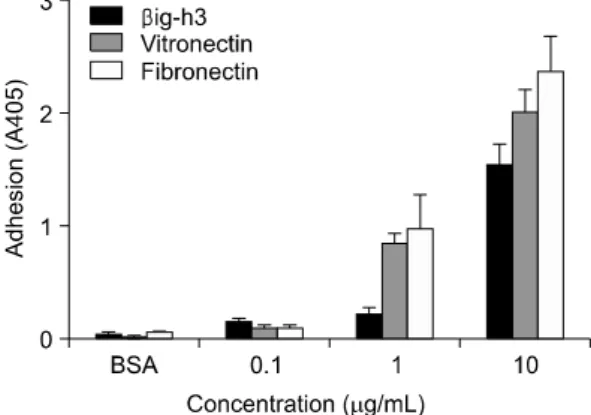
Fig. 1. Comparison of βig-h3, vitronectin, and fibronectin in adhesion of human FLS. FLS were seeded onto surfaces of 96-well culture plates which were precoated with different concentrations (0.1 ug/mL, 1 ug/mL and 10 ug/mL) of βig-h3, vitronectin, or fibronectin and then were incubated for 2 hours at 37oC. After seeding and incubation, FLS attached to the surfaces were quantified by hexosaminidase assay. Values are presented as the mean±SEM of triplicate experiments.
2. 세포 부착 시험
재조합 인간 βig-h3 단백제조를 위해 야생형 인 간 βig-h3 유전자를 PET29b 발현 벡터에 삽입시킨 후, BL21DE3 대장균에 형질전환 시켜 배양한 후 재 조합 단백을 순분리 하였다. 바닥이 편평한 96 well microtitre plate (Costar, Corning, NY)에 재조합 인간 βig-h3, vitronectin 혹은 fibronectin (Sigma)을 넣고 4oC에서 하룻밤 동안 코팅한 후, 실온에서 2% bovine serum albumin (BSA)이 함유된 phosphate buffered saline (PBS)을 이용하여 비특이적 결합을 차단시켰 다. 활막세포를 배지에서 3×10⁵ cells/mL의 농도로 부유시킨 후, 세포 부유액 0.1 mL을 재조합 인간 β ig-h3, vitronectin 혹은 fibronectin이 코팅된 well에 넣 고 5% CO2, 37oC에서 2시간 동안 배양하였으며, 부 착되지 않은 세포는 PBS로 씻어 제거하였다. 각 well 에 3.75 mM p-nitrophenyl-N-acetyl-D-glycosaminide (hexo- saminidase substrate)와 0.25% Triton X-100이 포함된 50 mM citrate buffer (pH 5.0)를 넣은 후, 37oC에서 2 시간 동안 배양하였다. 효소활성을 중지시키기 위해 5 mM EDTA (pH 10.4)가 포함된 50 mM glycine buffer 를 넣고 Multiscan MCC/340 microplate reader (Titertek Instruments, Huntsville, AL)를 이용하여 405 nm에서 흡광도를 측정하였다.
βig-h3-매개 활막세포 부착을 자극하기 위해 inter- feron (IFN)-γ, tumor necrosis factor (TNF)-α, interleukin (IL)-1β, TGF-β1, IL-4, IL-6, IL-10 등의 사이토카 인, basic fibroblast growth factor (bFGF), platelet- derived growth factor type BB (PDGF-BB), vascular endothelial growth factor (VEGF), insulin-like growth factor (IGF)-1, epidermal growth factor (EGF) (이상, R&D Systems, Minneapolis, MN) 등의 성장인자, 그 리고 비특이적 세포자극제인 phorbol 12-myristate 13-acetate (PMA) 및 lipopolysaccharide (LPS) 등을 30 분간 전처치한 후 부착 실험을 실시하였다.
세포부착 저해 실험에서 두 번째와 네 번째 fas-1 도메인의 YH motif에 포함된 18개의 아미노산으로 이루어진 YH18 펩타이드를 합성하여 이용하였으며, 대조군으로 18개의 아미노산을 무작위로 배열한 YH18 control 펩타이드(Petron, Daejeon, Korea)를 사 용하였다. YH18 펩타이드의 서열은 다음과 같다. 두 번째 도메인 YH18 펩타이드 EALRDLLNNHILK-
SAMCA, 네 번째 도메인 YH18 펩타이드 KELAN- ILKYHIGDEILVS, YH18 control 펩타이드 KELANIH- GIKLYDEILVS. 활막세포는 YH18 펩타이드들과 함 께 37oC에서 30분간 배양한 후 세포부착 실험에 사 용되었다.
3. 통계분석
실험결과는 평균과 표준오차(standard error of the mean)로 표기하였다. 평균의 차이는 Mann-Whitney U test를 이용하여 분석하였으며, 통계처리는 SPSS (statistical package for the social science) 10.0 for Windows를 사용하였고, 통계적 유의성은 p 값이 0.05 이하인 경우로 하였다.
결 과
1. 활막세포 부착에 있어 βig-h3와 부착성 당단백의 비교
염증이 있는 활막조직에 과발현된 부착성 당단백 에 의한 활막세포의 부착효율성을 비교하기 위하여, βig-h3, vitronectin 및 fibronectin을 농도별로 코팅한
Fig. 2. Regulation of βig-h3-mediated adhesion of FLS by cytokines, growth factors, and nonspecific stimu- lants. FLS were treated with different cytokines (recombinant human TNF-α, IL-1β, IL-6, IL-4, IL-10, IFN-γ, and TGF-β1), growth factors (VEGF, IGF-1, EGF, PDGF-BB, and FGF-basic), and nonspecific cell stimulants (PMA and LPS) for 30 minutes at 37oC. Pretreated FLS were added to the coated wells with 5 ug/mL of βig-h3 and were incubated for 2 hours at 37°C. After seeding and incubation, FLS attached to the surfaces were quantified by hexosaminidase assay. TGF-β1, 5 ng/mL; TNF-α, IL-1β, IL-4, IL-6, IL-10, VEGF, IGF-1 and EGF, 10 ng/mL; PDGF-BB, FGF-basic, and IFN-γ, 100 ng/mL; PMA, 60 ng/mL; and LPS 5 μg/mL. Values are presented as the mean±SEM of triplicate experiments.
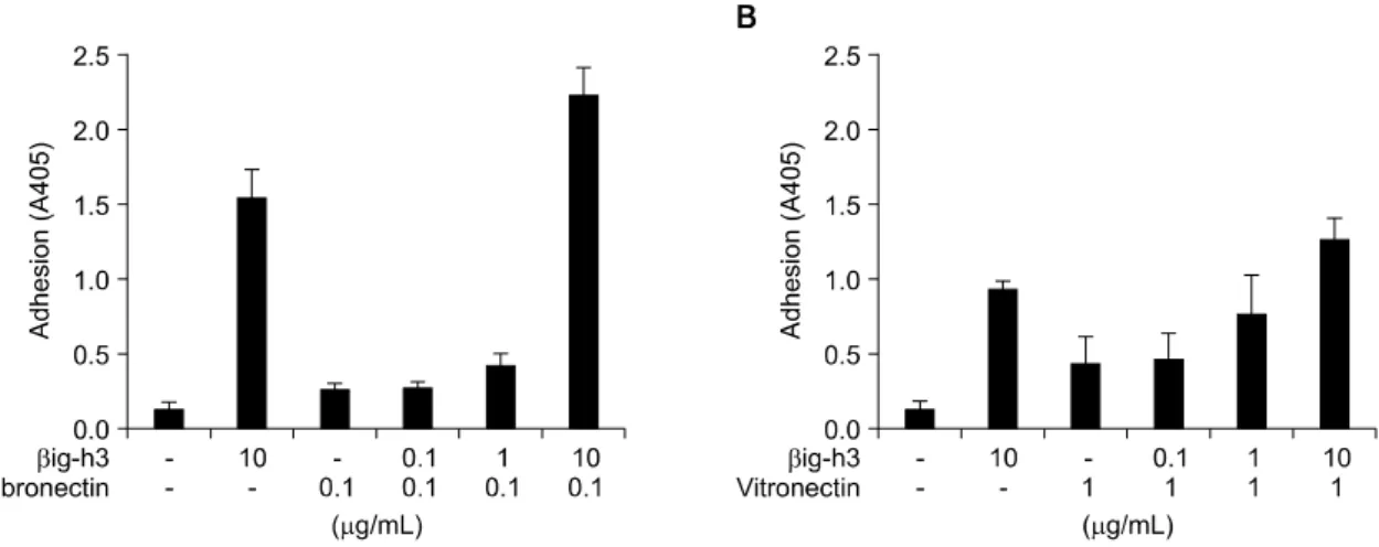
Fig. 3. Compensatory role of βig-h3 with matrix proteins in mediating adhesion of FLS. FLS were added to the precoated 96-well culture plates coated with (A) βig-h3 and fibronectin or (B) βig-h3 and vitronectin, and were incubated for 2 hours at 37oC. Low concentration (0.1 ug/mL) of fibronectin, 1 ug/mL of vitronectin, and different concen- trations (0.1 ug/mL, 1 ug/mL, and 10 ug/mL) of βig-h3 were used. After incubation, FLS attached to the surfaces were quantified. Values are presented as the mean±SEM of, at least, 3 independent experiments.
후 활막세포의 부착정도를 확인하였다. βig-h3-매개 활막세포 부착은 βig-h3에 농도 의존적으로 증가되 었으며, fibronectin과 vitronectin도 각각 농도가 증가 할수록 활막세포 부착을 증가시켰다. βig-h3와 fibro- nectin 및 vitronectin에 의한 활막세포 부착을 비교하 였을 때, 1 ug/mL 이상의 농도에서 fibronectin과 vitronectin에 의한 활막세포의 부착이 βig-h3에 의한 부착에 비해 현저히 높았으며 fibronectin에 의한 부 착이 가장 효과적으로 나타났다(그림 1).
2. βig-h3-매개 활막세포 부착에 대한 조절인자
류마티스관절염의 활막조직에는 염증촉진 및 염증 억제 사이토카인, 그리고 다양한 성장인자가 풍부하 게 존재하며 이들은 활막조직의 만성적인 염증반응 을 조절하는 역할을 담당한다 (1,22). βig-h3-매개 활막세포 부착에 미치는 영향을 조사하기 위해 염증 촉진 사이토카인들인 TNF-α (5 ng/mL), IL-1β, 그 리고 IL-6 (각각 10 ng/mL)로 전처치 하였을 때 활 막세포 부착은 거의 영향을 받지 않았으며, IFN-γ (100 ng/mL)와 염증억제 사이토카인들인 TGF-β, IL- 4, 그리고 IL-10 (각각 10 ng/mL)도 βig-h3-매개 활 막세포 부착에 거의 영향을 미치지 않았다(그림 2).
성장인자들은 성장인자 수용체와 인테그린간 cross- talk을 통해 세포부착 및 이주, 그리고 세포증식에
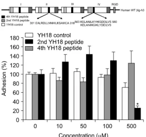
Fig. 4. Identification of the motif mediating FLS adhesion to βig-h3. The 2nd YH18 peptide (spanning amino acids 301∼318 in the 2nd fas-1 domain), 4th YH18 peptide (spanning amino acids 563∼580 in the 4th fas-1 domain), and YH18 control peptide (containing the amino acids of the 4th YH18 peptide but in a scrambled order) were prepared.
FLS were preincubated with the YH18 control, 2nd YH18, or 4th YH18 peptides at different concen- trations (0, 10, 50, 100, and 500 uM) for 30 minutes at 37oC and then added to the βig-h3-coated wells. Values are presented as the mean±SEM of at least 3 independent experiments. *p<0.05, 4th YH18 peptide versus YH18 control peptide.
관여한다 (23). 활막세포와 βig-h3의 결합에 대한 성 장인자의 영향을 조사하기 위해 PDGF-BB, bFGF (각각 100 ng/mL), VEGF, IGF-1, 그리고 EGF (각각 10 ng/mL)로 활막세포를 전처리 하였을 때, βig-h3 에 의한 세포부착은 거의 영향을 받지 않았다. 비특 이적인 세포 자극제는 인테그린의 입체적 구조 변화 를 유도하거나, cytoskeletal reorganization을 통해 세 포와 세포외기질단백과의 결합을 변화시킬 수 있다 (24,25). 비특이적인 세포자극제가 βig-h3에 의한 활 막세포 부착에 미치는 영향을 확인하기 위해 PMA 와 LPS로 전처치한 후 세포 부착 실험을 실시한 결 과, PMA를 전처리한 경우에 βig-h3-매개 활막세포 부착이 뚜렷하게 증가하였다(그림 2).
3. βig-h3와 부착성 당단백들 간의 상호작용
활막세포 부착에 있어 기질단백간의 상호작용을 조사하기 위해 βig-h3를 fibronectin 혹은 vitronectin 과 혼합하여 코팅한 후 세포부착 시험을 하였다. 저 농도(0.1μg/mL)의 fibronectin과 함께 여러 가지 농도 의 βig-h3를 혼합하여 코팅하였을 때 βig-h3에 농도 의존적으로 세포 부착이 증가되었으며, βig-h3 단독 (10μg/mL)에 비해 fibronectin (0.1μg/mL)과 βig-h3 (10μg/mL)를 혼합하여 코팅하였을 때 활막세포 부 착에 상승효과가 관찰되었다(그림 3A). 그러나 vitro- nectin과 βig-h3에 대해서는 세포부착에 있어 부가적 인 효과만 관찰되었다(그림 3B).
4. YH18 펩타이드에 의한 βig-h3-매개 세포부착 억제효과
βig-h3는 간질내에서 polymerization을 통하여 원 섬유를 형성할 수 있으며 collagen이나 부착성 당단 백과 결합하여 adapter 역할을 할 것으로 추정된다 (26). βig-h3의 4개 도메인에 존재하는 YH motif들 은 서열에 공통점이 많지만 세포에 따라 부착에 관 여하는 정도가 다소 다르다 (11,12). 이전의 연구에 서, βig-h3에 의한 활막세포의 부착과 이주에 네 번 째 도메인의 YH18 펩타이드가 차단효과를 나타내는 것이 규명되었으나 (14), 유사한 정도의 소수성를 가 지고 있는 두 번째 도메인의 YH18 펩타이드의 활막 세포 부착 억제효과에 대해서는 연구되지 않았다.
βig-h3에 의한 활막세포 부착은, 네 번째 도메인의 YH18 펩타이드로 전처치 하였을 때 100μM 하에서
는 차단효과가 없었으나 500μM에서 현저한 억제효 과를 나타내었다. 이에 비하여 두 번째 도메인의 YH18 펩타이드는 500μM까지 세포부착을 전혀 억 제하지 못하였다(그림 4).
고 찰
본 연구결과, βig-h3는 활막세포의 부착에 있어 부착성 당단백에 비해 상대적으로 약한 결합력을 보 이지만, fibronectin과는 세포부착에 대한 상승효과를 나타내는 것을 확인하였으며, βig-h3-매개 부착은 사이토카인이나 성장인자의 단기간 자극에는 거의 영향을 받지 않지만 PMA와 같은 강한 자극에 의해 서 증가되는 것을 관찰하였다. 또한, βig-h3에 의한 활막세포 부착은 네 번째 도메인의 YH18 펩타이드
와 달리, 두 번째 도메인의 YH18 펩타이드에 의해 서는 차단되지 않았다.
활막조직의 세포외기질은 I형과 II형 collagen과 같 은 미세 섬유를 구성하는 단백, 수분함량을 결정하 는 히알루로난과 같은 glycosaminoglycan (GAG)과 함께 fibronectin과 같은 부착성 당단백으로 구성되어 있다. 부착성 당단백들은 collagen에 결합된 상태로, 기질공간 내에서 이동하는 세포 표면의 인테그린들 과 결합하는 adapter 역할을 한다. βig-h3는 fibronectin 에 비하여 상대적으로 작은 크기의 단백(683개 아미 노산, 66kDa)이며 측면결합을 통해 중합되면서 원섬 유를 형성할 수 있을 뿐 아니라, collagen이나 fibro- nectin, laminin 등과 결합한 형태로 기질을 구성할 수 있기 때문에 (26), collagen과 같은 큰 구조적 단 백이 세포와 결합할 때 adapter 역할을 할 가능성이 있으며, fibronectin이나 vitronectin과 상호작용을 통해 세포의 부착과 이주를 조절할 가능성도 있다. 본 연 구결과 βig-h3는 fibronectin이나 vitronectin에 비하여 상대적으로 부착력은 약하였지만 활막세포의 부착을 촉진할 수 있으며, 저농도의 fibronectin과는 활막세 포 부착에 상승효과를 가진 것으로 확인되었다. 이 는 βig-h3와 fibronectin이 서로 다른 종류의 인테그 린과 결합하면서 세포부착의 효율이 증가되었거나, 한가지 기질단백과 인테그린의 결합이 세포내로의 신호전달을 통해 다른 인테그린의 입체형태적 변화 를 유도하여 다른 기질단백과의 결합을 촉진한 결과 로 추정할 수 있다 (2,27). βig-h3가 vitronectin과는 활막세포에 상승효과를 나타내지 못하였는데, 이는 두 기질단백이 적어도 αvβ3 인테그린을 공통으로 사용하기 때문에 상호 보완적인 결합이 일어날 여지 가 작았을 가능성이 있다 (28). 이에 대해서는 향후 좀 더 정확한 기전규명이 필요할 것이다.
세포외기질과 세포 표면에 발현되어 있는 인테그 린의 결합은 세포의 종류와 활성화 정도에 따라 다 르다. 이는 인테그린이 세포 표면을 통해 양방향의 신호전달이 가능하기 때문으로 생각된다. 인테그린 은 기질과 결합한 후 세포내로 신호전달을 하지만 (outside-in signaling) (29), 성장인자는 성장인자 수용 체를 통해 phosphoinositide-3-OH kinase (PI3-kinase)를 활성화시키고 인테그린의 세포질 도메인에 결합된 talin을 활성화시킴으로써 인테그린의 ligand binding
head를 활성화시킬 수 있다(inside-out signaling) (25, 29). 따라서, 세포의 부착과 이동은 인테그린과 성장 인자의 교차작용을 통해 촉진될 수 있으며 (23), 그 가운데 αvβ3 인테그린은 PDGF 수용체나 VEGF 수 용체 등의 신호전달과 연관되어 있다는 것이 coimm- unoprecipitation 실험으로 규명되었다 (23,29,30). 이에 비해 사이토카인과 인테그린의 상호작용은 세포의 종류와 사이토카인 및 주변여건에 따라 결과가 달라 질 수 있으며 (31,32), TNF-α와 IFN-γ로 혈관내피 세포를 자극하면 β3 인테그린의 합성이 감소되어 표면에 αvβ3 인테그린 발현율이 70% 가량 감소된 다는 보고도 있다 (32). 본 연구에서 활막세포는 VEGF 를 포함한 성장인자와 사이토카인들에 의한 βig-h3- 매개 부착의 변화가 뚜렷하지 않았으며 PDGF에 의 해서만 다소 증가되는 경향을 보였다. 이전의 연구 에서, βig-h3-매개 활막세포 부착과 PDGF에 의해 뚜렷하게 증가된 βig-h3-매개 활막세포의 이주는 αvβ3 인테그린에 의해 매개된다는 사실이 규명되었는데 (14), 이 결과들을 종합하여 보면 PDGF는 PDGF 수용 체를 통하여 αvβ3 인테그린과 cross-talk을 하는 것 으로 추정할 수 있다. PMA로 자극하였을 때 활막세 포의 βig-h3-매개 부착이 뚜렷하게 증가되었는데, 이는 PMA 자극에 의해 세포표면에 있는 인테그린 이 활성형으로 바뀌거나 (33), 세포가 활성화되면서 lipid raft의 유동성이 증가되어 monomer로 있던 인테 그린의 heterodimer 형성이 용이해지거나 (27), 전반 적으로 세포가 활성화되면서 신호전달 과정이 촉진 되어 다른 종류의 부착기전이 추가로 활성화되거나, 세포 내 cytoskeletal reorganization이 일어나면서 부착 이 촉진되기 때문 (24)일 가능성들이 존재한다. 이에 대해서는 향후 좀 더 많은 연구를 통한 규명이 필요 할 것이다.
세포의 종류에 따라 βig-h3와의 결합에 이용되는 인테그린의 종류는 다르며, 각막 상피세포가 α3β1 인테그린을 통해 결합 (9)하는데 반해, MRC-5 섬유 아세포주는 αvβ5 인테그린, 그리고 혈관내피세포 및 활막세포는 주로 αvβ3 인테그린를 이용한다 (11,12,14). βig-h3는 4개의 상동 도메인으로 구성되 어 있으며, 인테그린과의 결합에 있어 αv 인테그린 을 사용하는 세포 부착은 tyrosine과 histidine을 중심 으로 최소 18개의 아미노산으로 구성된 YH motif에
의해 매개되는 것으로 규명되었다 (11,12,14). 각각의 fas-1 도메인에 있는 YH motif의 아미노산 서열은 네 도메인 모두가 서로 유사하지만 약간의 차이가 있다. 두 번째 fas-1 도메인에 있는 YH motif의 서열 은 tyrosine (Y)이 asparagine (N)으로 대체되어 있어 다소 구조적 차이가 있으나, 네 번째 fas-1 도메인의 YH motif와 유사한 정도의 소수성을 가지고 있다.
이전의 연구에서 4개의 도메인에 있는 각 YH18 펩 타이드는 정도의 차이는 있으나 폐 섬유아세포, 그 리고 혈관내피세포의 βig-h3-매개 부착을 모두 차단 하는 것으로 보고하였다 (11,12). 하지만 본 연구에 서는 네 번째 도메인의 YH18 펩타이드와는 달리 두 번째 도메인의 YH18 펩타이드는 βig-h3와 활막세포 의 결합을 전혀 차단하지 못하였다. 이는 네 번째 fas-1 도메인은 NIF3T3 세포의 βig-h3-매개 부착을 차단할 수 있었지만 두 번째 fas-1 도메인은 차단하 지 못한다는 결과와 함께(비출간 자료) 활막세포의 부착에 두 번째 도메인의 역할이 거의 없을 가능성 을 제시하며, 향후 βig-h3를 이용한 치료제 개발에 있어 중요한 기반 정보로 사용될 수 있을 것이다.
결 론
류마티스관절염의 진행에 중요한 역할을 담당하는 활막세포는 세포외 기질단백과 결합하여 활막조직내 에서의 이주 및 활성화가 일어난다. βig-h3는 부착성 당단백들과 함께 관절염 조직에 과발현되어 있으며 활막세포의 부착을 매개한다. 본 연구의 목적은 활 막세포의 부착에 있어 βig-h3가 다른 당단백과 상 호보완적인 작용을 하는지 확인하고자 하여, β ig-h3-매개 부착에 있어 두 번째 fas-1 도메인의 YH18 펩타이드의 차단효과를 검증하고자 하였다.
활막세포를 분리한 후 기질 단백이 코팅된 microtitre plate에 부착시험을 수행하였으며 두 번째 및 네 번 째 fas-1 도메인의 YH18 펩타이드를 이용하여 차단 효과를 확인하였다. 활막세포의 부착은 fibronectin이 나 vitronectin에 비하여 βig-h3에 의해 상대적으로 약하게 일어났다. βig-h3-매개 부착은 TNF-α를 비 롯한 사이토카인이나 PDGF-BB를 비롯한 성장인자 에 의해 뚜렷한 영향을 받지 않았지만 PMA에 의해 서는 증가되었다. 활막세포의 부착에 있어 βig-h3는 fibronectin과 상승효과를 나타내었다. 네 번째 fas-1
도메인의 YH18 펩타이드와 달리 두 번째 YH18 펩 타이드는 βig-h3-매개 부착을 차단하지 못하였다.
본 연구결과로 βig-h3가 부착성 당단백들과 상호작 용을 통해 활막세포의 부착을 조절할 수 있으며 부 착에 이용되는 βig-h3의 핵심 motif가 세포의 종류 에 따라 서로 다른 것을 확인하였다.
참고문헌
1) Knedla A, Neumann E, Muller-Ladner U. Developments in the synovial biology field 2006. Arthritis Res Ther 2007;9:209.
2) Barilla ML, Carsons SE. Fibronectin fragments and their role in inflammatory arthritis. Semin Arthritis Rheum 2000;29:252-65.
3) Tomasini-Johansson BR, Milbrink J, Pejler G. Vitr- onectin expression in rheumatoid arthritic synovia- inhibition of plasmin generation by vitronectin produced in vitro. Br J Rheumatol 1998;37:620-9.
4) Lavietes BB, Carsons S, Diamond HS, Laskin RS.
Synthesis, secretion, and deposition of fibronectin in cultured human synovium. Arthritis Rheum 1985;
28:1016-26.
5) Petrow PK, Hummel KM, Schedel J, Franz JK, Klein CL, Muller-Ladner U, et al. Expression of osteopontin messenger RNA and protein in rheumatoid arthritis:
effects of osteopontin on the release of collagenase 1 from articular chondrocytes and synovial fibroblasts.
Arthritis Rheum 2000;43:1597-605.
6) Liao HX, Haynes BF. Role of adhesion molecules in the pathogenesis of rheumatoid arthritis. Rheum Dis Clin North Am 1995;21:715-40.
7) Liaw L, Almeida M, Hart CE, Schwartz SM, Giachelli CM. Osteopontin promotes vascular cell adhesion and spreading and is chemotactic for smooth muscle cells in vitro. Circ Res 1994;74:214-24.
8) Bae JS, Lee SH, Kim JE, Choi JY, Park RW, YongPark J, et al. Betaig-h3 supports keratinocyte adhesion, migration, and proliferation through alpha3- beta1 integrin. Biochem Biophys Res Commun 2002;
294:940-8.
9) Kim JE, Kim SJ, Lee BH, Park RW, Kim KS, Kim IS, et al. Identification of motifs for cell adhesion within the repeated domains of transforming growth factor-beta-induced gene, betaig-h3. J Biol Chem 2000;275:30907-15.
10) Park SW, Bae JS, Kim KS, Park SH, Lee BH, Choi
JY, et al. Beta ig-h3 promotes renal proximal tubular epithelial cell adhesion, migration and proliferation through the interaction with alpha3beta1 integrin. Exp Mol Med 2004;36:211-9.
11) Nam JO, Kim JE, Jeong HW, Lee SJ, Lee BH, Choi JY, et al. Identification of the alphavbeta3 integrin- interacting motif of betaig-h3 and its anti-angiogenic effect. J Biol Chem 2003;278:25902-9.
12) Kim JE, Jeong HW, Nam JO, Lee BH, Choi JY, Park RW, et al. Identification of motifs in the fasciclin domains of the transforming growth factor-beta- indu- ced matrix protein betaig-h3 that interact with the alphavbeta5 integrin. J Biol Chem 2002;277:46159-65.
13) Kang YM, Kim SI, Kim JS, You DW, Sa KH, Park EJ, et al. Regulation of βig-h3 Production in Rheumatoid Synovitis by Inflammatory Mediators. J Korean Rheum Assoc 2005;12:73-82.
14) Nam EJ, Sa KH, You DW, Cho JH, Seo JS, Han SW, et al. Up-regulated transforming growth factor beta- inducible gene h3 in rheumatoid arthritis mediates adhesion and migration of synoviocytes through alpha v beta3 integrin: Regulation by cytokines. Arthritis Rheum 2006;54:2734-44.
15) Thapa N, Kang KB, Kim IS. Betaig-h3 mediates osteoblast adhesion and inhibits differentiation. Bone 2005;36:232-42.
16) Kim JE, Kim SJ, Jeong HW, Lee BH, Choi JY, Park RW, et al. RGD peptides released from beta ig-h3, a TGF-beta-induced cell-adhesive molecule, mediate apoptosis. Oncogene 2003;22:2045-53.
17) Munier FL, Korvatska E, Djemai A, LePaslier D, Zografos L, Pescia G, et al. Kerato-epithelin mutations in four 5q31-linked corneal dystrophies. Nat Genet 1997;15:247-51.
18) O'Brien ER, Bennett KL, Garvin MR, Zderic TW, Hinohara T, Simpson JB, et al. Beta ig-h3, a transforming growth factor-beta-inducible gene, is overexpressed in atherosclerotic and restenotic human vascular lesions. Arterioscler Thromb Vasc Biol 1996;
16:576-84.
19) Lee SH, Bae JS, Park SH, Lee BH, Park RW, Choi JY, et al. Expression of TGF-beta-induced matrix protein betaig-h3 is up-regulated in the diabetic rat kidney and human proximal tubular epithelial cells treated with high glucose. Kidney Int 2003;64:1012-21.
20) Ferguson JW, Mikesh MF, Wheeler EF, LeBaron RG.
Developmental expression patterns of Beta-ig (betaIG- H3) and its function as a cell adhesion protein. Mech Dev 2003;120:851-64.
21) Skonier J, Neubauer M, Madisen L, Bennett K, Plowman GD, Purchio AF, et al. cDNA cloning and sequence analysis of beta ig-h3, a novel gene induced in a human adenocarcinoma cell line after treatment with transforming growth factor-beta. DNA Cell Biol 1992;11:511-22.
22) Andreakos ET, Foxwell BM, Brennan FM, Maini RN, Feldmann M. Cytokines and anti-cytokine biologicals in autoimmunity: present and future. Cytokine Growth Factor Rev 2002;13:299-313.
23) Eliceiri BP. Integrin and growth factor receptor crosstalk. Circ Res 2001;89:1104-10.
24) Kucik DF, Dustin ML, Miller JM, Brown EJ.
Adhesion-activating phorbol ester increases the mobility of leukocyte integrin LFA-1 in cultured lymphocytes. J Clin Invest 1996;97:2139-44.
25) Hynes RO. Integrins: bidirectional, allosteric signaling machines. Cell 2002;110:673-87.
26) Kim JE, Park RW, Choi JY, Bae YC, Kim KS, Joo CK, et al. Molecular properties of wild-type and mutant betaIG-H3 proteins. Invest Ophthalmol Vis Sci 2002;43:656-61.
27) Hogg N, Henderson R, Leitinger B, McDowall A, Porter J, Stanley P, et al. Mechanisms contributing to the activity of integrins on leukocytes. Immunol Rev 2002;186:164-71.
28) Plow EF, Haas TA, Zhang L, Loftus J, Smith JW.
Ligand binding to integrins. J Biol Chem 2000;275:
21785-8.
29) Giancotti FG, Ruoslahti E. Integrin signaling. Science 1999;285:1028-32.
30) Borges E, Jan Y, Ruoslahti E. Platelet-derived growth factor receptor beta and vascular endothelial growth factor receptor 2 bind to the beta 3 integrin through its extracellular domain. J Biol Chem 2000;275:
39867-73.
31) Rinaldi N, Barth T, Henne C, Mechterscheimer G, Moller P. Synoviocytes in chronic synovitis in situ and cytokine stimulated synovial cells in vitro neo- express alpha 1, alpha 3 and alpha 5 chains of beta 1 integrins. Virchows Arch 1994;425:171-80.
32) Defilippi P, Truffa G, Stefanuto G, Altruda F, Silengo L, Tarone G, et al. Tumor necrosis factor alpha and interferon gamma modulate the expression of the vitronectin receptor (integrin beta 3) in human endothelial cells. J Biol Chem 1991;266:7638-45.
33) Shimizu Y, Van Seventer GA, Horgan KJ, Shaw S.
Regulated expression and binding of three VLA (beta 1) integrin receptors on T cells. Nature 1990;345:250-3.