대한소화기학회지 2010;56:129-134 □ REVIEW □ DOI: 10.4166/kjg.2010.56.3.129
연락처: 백순구, 220-701, 강원도 원주시 일산동 162번지 연세대학교 원주의과대학 원주기독병원 소화기내과 Tel: (033) 741-1229, Fax: (033) 741-1228
E-mail: [email protected]
Correspondence to: Soon Koo Baik, M.D.
Division of Gastroenterology & Hepatology, Department of Internal Medicine, Yonsei University Wonju College of Medicine, 162, Ilsan-dong, Wonju 220-701, Korea
Tel: +82-33-741-1229, Fax: +82-33-741-1228 E-mail: [email protected]
문맥압항진증 병태 생리의 최신지견
연세대학교 원주의과대학 내과학교실
김문영ㆍ백순구
Pathophysiology of Portal Hypertension, What’s New?
Moon Young Kim, M.D., Ph.D. and Soon Koo Baik, M.D., Ph.D.
Department of Internal Medicine, Yonsei University Wonju College of Medicine, Wonju, Korea
Portal hypertension (PHT) is associated with changes in the intrahepatic, systemic and portosystemic collateral circulations. Alteration in vasoreactivity (vasodilation and vasoconstriction) plays a central role in the pathogenesis of PHT by contributing to increased intrahepatic resistance, hyperdynamic circulation and the expansion of the collateral circulation. PHT is also importantly characterized by changes in vascular structure; termed vascular re- modeling, which is an adaptive response of the vessel wall that occurs in response to chronic changes in the en- vironment such as shear stress. Angiogenesis, the sprouting of new blood vessels, also occurs in PHT, especially in the expansion of the portosystemic collateral circulation. These complementary processes of vasoreactivity, vas- cular remodeling and angiogenesis represent important targets in the research for the treatment of portal hypertension. (Korean J Gastroenterol 2010;56:129-134)
Key Words: Portal hypertension; Hepatic stellate cell; Endothelial cell; Intrahepatic vascular resistance
서 론
정상적인 간에서는 문맥 혈류가 변화되면 이에 따라 간내 혈관 저항(intrahepatic vascular resistance)이 변화하여 문맥압 이 일정하게 유지된다. 그러나 만성 간질환 때는 이러한 변 화에 대한 반응이 정상적으로 나타나지 못하여 간내 혈관 저항의 증가나 내장혈류(splanchnic circulation)의 증가 또는 이 두 가지 모두에 의해 문맥압항진증(portal hypertension)이 나타나게 되며, 보통 문맥(portal vein)과 하대정맥(inferior vena cava)간의 압력차(portal pressure gradient)가 6 mmHg 이 상인 경우를 말한다.1-4 따라서 문맥압의 변화는 수학적으로 간내 혈관을 통한 저항과 혈류 간의 함수관계에 있으며,
Ohm의 법칙(ΔP=Q×R)에 따라 문맥의 혈류와 저항의 곱으 로 표시된다.
간은 독특한 구조인 동양혈관의 구조(sinusoidal network) 로 이루어져 있으며, 동양혈관에서는 내피세포(endothelial cell)와 간성상세포(hepatic stellate cell, HSC)가 있어 서로에 게 영향을 미치며 밀접하게 연관되어 있고, 특히 간성상세 포가 혈류 조절에 주된 역할을 한다.5-7 문맥압항진증의 발 생은 혈관의 변화와 밀접한 관련성을 갖는데 이는 주로 혈 관 반응성의 변화 즉, 간내 순환(intrahepatic circulation)에 있 어서 혈관수축(vasoconstriction)과 체순환에 있어서의 혈관확 장(vasodilation)으로 대변된다. 그러나 최근에는 문맥압항진 증의 발생에 있어서 이러한 혈관 반응성의 변화 외에 혈관
130 대한소화기학회지: 제56권 제3호, 2010
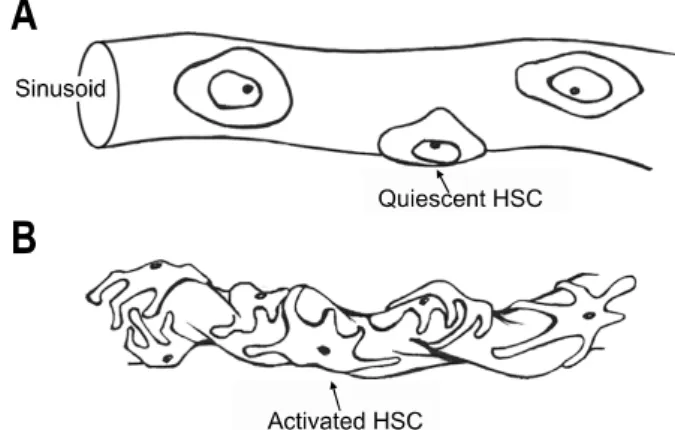
Fig. 1. Hepatic stellate cell (HSC) activation. In quiescent state, HSC do not contract (A), however in activated state, the number and contractility of HSC increase which induce the change of si- nusoidal structure and intrahepatic resistance.
의 중요한 구조적 변화 즉, vascular remodeling과 angiogene- sis가 중요한 기전으로 관심을 받고 있다.4 Vascular remodel- ing은 혈관에 대한 shear stress와 같은 환경의 지속적인 변화 및 자극에 대해 적응하는 반응이며,8 angiogenesis는 혈관 내 피세포 및 평활근육세포(smooth muscle cell)의 증식에 따른 신생 혈관의 생성뿐만 아니라, 골수로부터 혈관벽을 만드는 endothelial precursor cell을 불러와 이를 통해 신생 혈관을 생 성하는 소위 vasculogenesis를 포함한다.4 본문에서는 문맥압 항진증의 병태생리와 관련하여, 주로 이러한 혈관의 구조적 적응반응에 대한 간내 순환, 체순환 및 측부 순환의 변화에 관해 살펴 보고자 한다.
간내 순환(intrahepatic circulation)
대부분의 간질환에서 문맥압항진증의 시작은 간의 구조 적인 변화에 따라 증가된 간내 혈관 저항에 의해 발생되며, 이러한 간의 구조적인 변화는 오랫동안 정적이며 비가역적 인 변화(static and irreversible)로 인식되어 왔다. 비가역적인 요인으로는 간경변의 특징인 광범위한 세포외기질 침착과 섬유화에 의한 동양혈관의 모세혈관화(sinusoidal capillari- zation), 재생 결절의 형성, 혈관 내 혈전 형성 등을 들 수 있 으며, 주로 이들에 의해 간내 혈관 저항은 증가한다. 그러나 최근 여러 연구를 통해 간의 구조적 변화 외에도 역동적이 고 가역적인(dynamic and reversible) 부분이 20-30% 존재함 이 알려지면서 문맥압항진증 연구에 새로운 계기가 마련되 고 있다.9-13
Vasoregulatory imbalance: 동양혈관에서는 내피세포와 간성상세포가 서로에게 주변분비영향(paracrine effect)을 끼 쳐 혈류 조절의 균형을 이루고 있으며, 그 중에서 특히 간성 상세포가 혈류조절의 주된 역할을 한다.5,6 그리고 이러한 혈류조절의 중심에 혈관활성 매개체인 endothelin-1 (ET-1)과 nitric oxide (NO)가 있다. 정상적으로 ET-1은 내피세포에서 분비되고, 주로 간성상세포의 ETA 수용체에 작용하여 간성 상세포의 수축을 유발한다. 이에 대한 반대 작용으로 역시 내피세포에서 endothelial NO synthase (eNOS)에 의해 분비되 는 NO가 gluanylyl catalase pathway를 통해 주변의 간성상세 포를 이완시키게 되며, 이와 같이 ET-1과 NO 사이의 균형 에 의해 동양혈관내의 혈류는 조절된다. 그러나 간경변과 같은 간손상이 있는 상태에서는 대부분의 ET-1이 간성상세 포에서 분비되며, 활성화된 간성상세포의 ET-1에 대한 민감 도가 증가하게 되어 간성상세포의 수축이 활발해진다.14 이 와 더불어 내피세포에서 eNOS에 의한 NO의 생성에 장애가 발생하며, 이로 인해 동양혈관의 이완장애 및 활성화된 간 성상세포의 수축이 지속되어 결국 간내 혈관 저항이 증가하 게 된다.15-17 이 과정에서 실제 eNOS의 양은 변함이 없어 보
이지만, 동양혈관의 내피세포에서는 eNOS에 결합하여 이의 활동을 억제하는 caveolin-1이 증가되고, 이와 동시에 이에 반대 작용을 하는 calmodulin이 감소하여 eNOS의 기능부전 이 유발된다.15,18 그 외에 G-protein-coupled receptor kinase 2 (GRK2)에 의한 내피세포 내에서의 Akt에 의한 eNOS 효소 인산화 억제와 그에 따른 NO 생성의 감소도 또 다른 기전 으로 알려져 있다.19,20 최근에는 고지혈증치료제인 simva- statin이 eNOS를 활성화시킴으로써 간내 NO의 양을 증가시 키고, 체순환계의 혈관에 대한 부정적 영향 없이 간내 혈관 저항을 감소를 유발하여 내피세포의 기능을 호전시킨다는 보고가 있어,21,22 향후 간내 혈관 저항 완화치료로서 많은 관심을 끌고 있다.
동양혈관 remodeling (sinusoidal remodeling): 간경변이 진 행함에 따라 간섬유화에 의한 비가역적 변화 및 동양혈관 혈류 조절 장애에 따른 간내 저항의 증가와 함께 동양혈관 을 감싸는 간성상세포의 수와 면적이 증가하게 된다(Fig. 1).
또한, 이들의 수축하는 성질 등에 의해 동양혈관 remodeling 이 유발되어 동양혈관의 저항증가를 가져오며,4 이러한 동 양혈관 remodeling은 간성상세포의 collagen 침착 및 간섬유 화 작용에 의해 더욱 현저히 나타나게 된다.23 이와 같은 동 양혈관 remodeling 과정에는 간성상세포의 이동성(migration) 과 운동성(motility)이 필수적이며, 간성상세포막 구조의 변 화가 이러한 이동성과 운동성의 유발에 중요한 역할을 한 다.24 일반적으로 transforming growth factor-β (TGF-β)는 간 성상세포에 의한 collagen의 침착에 중요한 역할을 하는 것 으로 잘 알려져 있으나, TGF-β는 platelet derived growth factor (PDGF)와의 상호작용에 의해 간성상세포의 운동성에 도 영향을 미쳐 동양혈관 remodeling에 중요한 역할을 한
다.25,26 또한 endothelin과 같은 혈관활성 매개체들이 간성상
세포의 수축력의 변화와 함께 간섬유화 등에도 영향을 미친
김문영 외 1인. 문맥압항진증 병태 생리의 최신지견 131
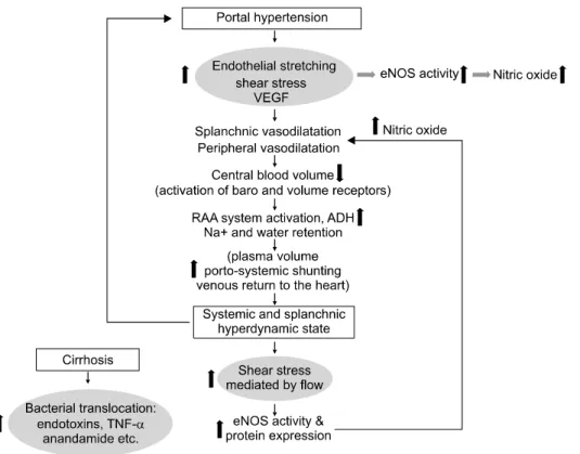
Fig. 2. Pathogenesis of hyperdyna- mic circulation.
VEGF, vascular endothelial growth factor; eNOS, endothelial nitric ox- ide synthase; RAA system, renin- angiotensin-aldosterone system; ADH, antidiuretic hormone; TNF-α, tu- mor necrosis factor-alpha.
다는 연구 결과들을 통해서, 간내 저항의 증가는 간섬유화 와 동양혈관 remodeling의 서로 공통된 인자들을 통해서 상 보적으로 발생함을 알 수 있다.5
최근에는 간내 저항의 증가에 있어서 angiogenesis의 역할 에 대한 관심이 높아지고 있다. Angiogenesis는 간섬유화의 진행에 있어서 필수적인 요소이며,27 동시에 문맥압항진증 의 유발에도 중요한 역할을 한다. 많은 신호전달체계와 vas- cular endothelial growth factor (VEGF), PDGF, TGF-β, angio- poietins, NO 등이 sinusoidal remodeling과 angiogenesis 과정 에서 간성상세포를 혈관 주변으로 불러들이는 역할을 하는 것으로 알려져 있다. 특히 PDGF는 간성상세포를 새로 형성 된 혈관들 주변으로 유도하는데 있어서 가장 중요한 역할을 하는 것으로 알려져 있으며,28 angiogenic cytokines인 leptin이 간성상세포에서 proangiogenic cytokines의 분비를 증가시킨 다는 연구 결과가 있다.27 따라서, 최근에는 간성상세포 뿐 만 아니라, 혈관 내피 세포 및 여러 혈관 활성 매개체에 대 한 연구가 문맥압항진증의 병인 및 치료에 있어 중요한 주 제가 되고 있다.
체순환 및 내장혈류(systemic and splanchnic circulation)
Vasoregulatory imbalance: 간 내에서의 NO의 생체이용률 의 저하와는 반대로 내장혈류 및 체순환에서는 상대적으로
증가된 NO의 생성이 나타나게 된다.29 이러한 NO의 생성은 주로 eNOS에 의한 혈관내피세포에서의 생성증가에 의해서 이루어지는데,30 여기에는 몇 가지 기전들이 제시되고 있다.
eNOS의 활성을 증가시키는 매개체 중 heat shock protein 90 (Hsp90)과 Akt가 이 과정에서 주로 관여하며, 이들은 VEGF, inflammatory cytokines, shear stress 등에 의해 자극되어 내장 혈관 내에서의 NO의 생성을 증가시키게 된다. 그 중에서도 angiogenic growth factor인 VEGF에 의한 eNOS의 활성화가 vasodilating angiogenesis 및 vascular remodeling 유발과 관련 이 있음이 알려져 최근 관심을 끌고 있다.31 또한, 간경변 환 자에서 흔히 발생하는 bacterial translocation이 tumor necrosis factor-α (TNF-α)의 증가를 통해 NO 분비를 증가시킨다.32 이와 같이 증가된 체순환 및 내장혈류내의 NO는 체순환 혈 관 저항의 감소로 인해 유효순환혈액량(effective circulating volume)을 감소시키고, 이는 동맥의 압력 수용체를 자극하 여 교감신경계의 활성화, renin-angiotensin-aldosteron system 과 antidiuretic hormone (ADH)의 분비 증가 등을 초래해 과 역동적 혈류흐름(hyperdynamic circulation)과 염분과 수분의 저류 및 복수를 유발하게 된다(Fig. 2).31
Vascular remodeling: 만성적인 혈류의 변화는 장기간에 걸친 적응반응으로서 체순환 특히 내장혈류계의 vascular re- modeling을 유발하게 된다. 만성적으로 증가된 혈류와 이로 인한 혈관의 확장은 내피세포에 의한 혈관의 구조변화를 유 발하고 만성적으로 혈관 직경의 확장과 많은 혈류를 담을
132 The Korean Journal of Gastroenterology: Vol. 56, No. 3, 2010
수 있는 능력의 증가를 가져와 만성적인 유효혈류량의 감소 현상을 유발한다. 이러한 현상 또한 eNOS의 활성과 연관이 있는 것으로 알려져 있다.8,33
측부순환계(collateral circulation)
Vasoregulatory imbalance: 체-문맥간 측부순환(portosyste- mic collateral circulation)의 발생은 문맥압항진증의 중요한 합병증 중 하나이다. 식도 및 항문 주위에 기존의 측부혈관 의 확장 및 새로운 발생은 증가된 문맥압에 대한 보상 기전 으로 발생하며 문맥압을 낮추는 역할을 하나, 불행히도 이 들은 간경변 및 문맥압항진증에 의한 여러 질환 및 사망의 주요 원인이 되고 있다. 따라서 이들 측부혈관 및 혈류에 대 한 조절 기전은 향후 문맥압항진증으로 인한 합병증의 실질 적인 예방 및 치료법 개발과 관련하여 많은 관심을 받고 있 고 또한 연구가 진행 중에 있다.
Angiogenesis and vascular remodeling: 측부혈관 및 혈류 의 변화는 단순한 혈관의 확장 외에도 angiogenesis와 깊은 연관이 있다. 이러한 내장혈관 및 측부혈관의 angiogenesis에 있어서 VEGF가 중요한 역할을 하는 것으로 알려져 있는데, VEGF는 NO 의존적 또는 비의존적으로 혈관의 확장과 vascular remodeling, 내피세포 증식의 활성화를 통한 angio- genesis 등에서 여러 역할을 한다.34 또한 일단 혈관이 형성 화되면 이의 성숙과 안정화는 주로 PDGF에 의해 유도된 pericyte들에 의해 이루어지게 된다.34 이와 관련하여 최근에 VEGF receptor-2에 대한 중화항체 또는 multikinase inhibitor 인 sorafenib 등은 동물실험에서 문맥-체순환계 단락이 저하 되고 문맥압항진증이 호전됨을 보여줌으로써 문맥압항진증 치료의 새로운 가능성으로 주목받고 있다.35-37 특히, sora- fenib을 이용한 연구에서는 현저한 내장 및 측부혈관 신생 및 단락 형성의 억제와 함께 간내 섬유화와 염증의 호전도 함께 보여 본 약제가 angiogensis의 과정뿐 아니라 간성상세 포의 활성의 억제에도 영향을 미침을 보여 주어, 간내 저항 과 체순환 및 측부순환의 증가까지 함께 조절할 수 있는 가 능성을 보여 주었다. 그러나 angiogensis가 간의 재생에 있어 서도 필수적인 요소임을 감안할 때 과연 이러한 anti-angio- genic therapy가 문맥압항진증 및 간섬유화의 치료에 있어서 실제 임상에서도 도움이 될 지는 더 많은 연구가 필요하다.
결 론
만성간질환의 진행과 그에 따른 간성상세포의 활성화 및 간섬유화는 간내 혈관의 저항 증가와 문맥압의 상승을 초래 하고, 상승된 문맥압은 문맥과 전신 혈관 사이에 단락(porto- systemic shunt)을 유발시킨다. 이 단락을 통해 심장으로의
혈류량 유입이 많아지고, 심박출량이 증가하게 되는데, 이 러한 심박출량의 증가는 동맥 혈관에 압력을 증가시켜 동맥 혈관 내피세포에서 NO 생성 증가를 가져 오게 된다. 증가 된 NO는 내장혈관의 확장과 그에 따른 혈류량의 증가를 유 발하여 궁극적으로 문맥으로의 혈류 유입을 증가시킴으로 써, 간내 혈관 저항의 상승에 의해 처음 유발된 문맥압항진 증이 지속적으로 유지되도록 한다.1 또한 말초혈관에서의 이러한 혈관확장 물질의 증가는 혈관의 확장과 혈압의 강하 를 유발하며, 이는 유효혈장량(effective circulatory volume)의 감소로 나타나 교감신경계를 항진시키고 renin-angiotensin- aldosteron system 활성화를 유발시킨다. 이는 전반적인 체내 혈장량의 증가를 야기하는 과역동적 혈류흐름을 발생시키 고 결국, 복수 및 정맥류와 같은 합병증이 생기게 된다.38 최 근, 문맥압항진증의 발생 및 유지에 있어서 vacular remodel- ing 및 angiogenesis의 역할에 새로운 해석이 이루어지고 있 어, 이를 통한 새로운 문맥압항진증 치료 모델 연구에 많은 관심이 모아지고 있다.
참고문헌
1. Baik SK. Assessment and current treatment of portal hyper- tension. Korean J Hepatol 2005;11:211-217.
2. Lebrec D. Methods to evaluate portal hypertension. Gastroen- terol Clin North Am 1992;21:41-59.
3. Baik SK, Jeong PH, Ji SW, et al. Acute hemodynamic effects of octreotide and terlipressin in patients with cirrhosis: a randomized comparison. Am J Gastroenterol 2005;100:631- 635.
4. Lee JS, Semela D, Iredale J, Shah VH. Sinusoidal remodeling and angiogenesis: a new function for the liver-specific peri- cyte? Hepatology 2007;45:817-825.
5. Rockey DC. Hepatic blood flow regulation by stellate cells in normal and injured liver. Semin Liver Dis 2001;21:337-349.
6. Reynaert H, Thompson MG, Thomas T, Geerts A. Hepatic stellate cells: role in microcirculation and pathophysiology of portal hypertension. Gut 2002;50:571-581.
7. Baik SK, Park DH, Kim MY, et al. Captopril reduces portal pressure effectively in portal hypertensive patients with low portal venous velocity. J Gastroenterol 2003;38:1150-1154.
8. Fernández-Varo G, Ros J, Morales-Ruiz M, et al. Nitric ox- ide synthase 3-dependent vascular remodeling and circulatory dysfunction in cirrhosis. Am J Pathol 2003;162:1985-1993.
9. Rockey DC. Vascular mediators in the injured liver. Hepato- logy 2003;37:4-12.
10. Baik SK. Pharmacological therapy of portal hypertension--fo- cused on Korean data. Korean J Gastroenterol 2005;45:381-
Kim MY, et al. Pathophysiology of Portal Hypertension, What’s New? 133
386.
11. Bhathal PS, Grossman HJ. Reduction of the increased portal vascular resistance of the isolated perfused cirrhotic rat liver by vasodilators. J Hepatol 1985;1:325-337.
12. Park DH, Baik SK, Choi YH, et al. Inhibitory effect of an- giotensin blockade on hepatic fibrosis in common bile duct-li- gated rats. Korean J Hepatol 2007;13:61-69.
13. Rockey D. The cellular pathogenesis of portal hypertension:
stellate cell contractility, endothelin, and nitric oxide.
Hepatology 1997;25:2-5.
14. Reinehr RM, Kubitz R, Peters-Regehr T, Bode JG, Häussinger D. Activation of rat hepatic stellate cells in cul- ture is associated with increased sensitivity to endothelin 1.
Hepatology 1998;28:1566-1577.
15. Shah V, Toruner M, Haddad F, et al. Impaired endothelial ni- tric oxide synthase activity associated with enhanced caveolin binding in experimental cirrhosis in the rat. Gastroenterology 1999;117:1222-1228.
16. Rockey DC, Chung JJ. Reduced nitric oxide production by endothelial cells in cirrhotic rat liver: endothelial dysfunction in portal hypertension. Gastroenterology 1998;114:344-351.
17. Gupta TK, Toruner M, Chung MK, Groszmann RJ. Endothe- lial dysfunction and decreased production of nitric oxide in the intrahepatic microcirculation of cirrhotic rats. Hepatology 1998;28:926-931.
18. Yokomori H, Oda M, Yoshimura K, et al. Elevated ex- pression of caveolin-1 at protein and mRNA level in human cirrhotic liver: relation with nitric oxide. J Gastroenterol 2003;38:854-860.
19. Morales-Ruiz M, Cejudo-Martín P, Fernández-Varo G, et al.
Transduction of the liver with activated Akt normalizes portal pressure in cirrhotic rats. Gastroenterology 2003;125:522-531.
20. Liu S, Premont RT, Kontos CD, Zhu S, Rockey DC. A cru- cial role for GRK2 in regulation of endothelial cell nitric ox- ide synthase function in portal hypertension. Nat Med 2005;
11:952-958.
21. Abraldes JG, Rodriguez-Vilarrupla A, Graupera M, et al.
Simvastatin treatment improves liver sinusoidal endothelial dysfunction in CCl4 cirrhotic rats. J Hepatol 2007;46:1040- 1046.
22. Zafra C, Abraldes JG, Turnes J, et al. Simvastatin enhances hepatic nitric oxide production and decreases the hepatic vas- cular tone in patients with cirrhosis. Gastroenterology 2004;
126:749-755.
23. Friedman SL, Rockey DC, McGuire RF, Maher JJ, Boyles JK, Yamasaki G. Isolated hepatic lipocytes and Kupffer cells from normal human liver: morphological and functional char-
acteristics in primary culture. Hepatology 1992;15:234-243.
24. Lee JS, Kang Decker N, Chatterjee S, Yao J, Friedman S, Shah V. Mechanisms of nitric oxide interplay with Rho GTPase family members in modulation of actin membrane dynamics in pericytes and fibroblasts. Am J Pathol 2005;166:
1861-1870.
25. Daniels CE, Wilkes MC, Edens M, et al. Imatinib mesylate inhibits the profibrogenic activity of TGF-beta and prevents bleomycin-mediated lung fibrosis. J Clin Invest 2004;114:
1308-1316.
26. Ceni E, Crabb DW, Foschi M, et al. Acetaldehyde inhibits PPARgamma via H2O2-mediated c-Abl activation in human hepatic stellate cells. Gastroenterology 2006;131:1235-1252.
27. Aleffi S, Petrai I, Bertolani C, et al. Upregulation of proin- flammatory and proangiogenic cytokines by leptin in human hepatic stellate cells. Hepatology 2005;42:1339-1348.
28. Hellström M, Kalén M, Lindahl P, Abramsson A, Betsholtz C. Role of PDGF-B and PDGFR-beta in recruitment of vas- cular smooth muscle cells and pericytes during embryonic blood vessel formation in the mouse. Development 1999;126:
3047-3055.
29. Langer DA, Shah VH. Nitric oxide and portal hypertension:
interface of vasoreactivity and angiogenesis. J Hepatol 2006;44:209-216.
30. Wiest R, Shah V, Sessa WC, Groszmann RJ. NO over- production by eNOS precedes hyperdynamic splanchnic circu- lation in portal hypertensive rats. Am J Physiol 1999;276:
G1043-1051.
31. Iwakiri Y, Groszmann RJ. The hyperdynamic circulation of chronic liver diseases: from the patient to the molecule.
Hepatology 2006;43(2 suppl 1):S121-S131.
32. Wiest R, Das S, Cadelina G, Garcia-Tsao G, Milstien S, Groszmann RJ. Bacterial translocation in cirrhotic rats stim- ulates eNOS-derived NO production and impairs mesenteric vascular contractility. J Clin Invest 1999;104:1223-1233.
33. Rudic RD, Shesely EG, Maeda N, Smithies O, Segal SS, Sessa WC. Direct evidence for the importance of endothe- lium-derived nitric oxide in vascular remodeling. J Clin Invest 1998;101:731-736.
34. Folkman J, D'Amore PA. Blood vessel formation: what is its molecular basis? Cell 1996;87:1153-1155.
35. Fernandez M, Mejias M, Angermayr B, Garcia-Pagan JC, Rodés J, Bosch J. Inhibition of VEGF receptor-2 decreases the development of hyperdynamic splanchnic circulation and portal-systemic collateral vessels in portal hypertensive rats. J Hepatol 2005;43:98-103.
36. Fernandez M, Vizzutti F, Garcia-Pagan JC, Rodes J, Bosch J.
134 대한소화기학회지: 제56권 제3호, 2010
Anti-VEGF receptor-2 monoclonal antibody prevents por- tal-systemic collateral vessel formation in portal hypertensive mice. Gastroenterology 2004;126:886-894.
37. Mejias M, Garcia-Pras E, Tiani C, Miquel R, Bosch J, Fernandez M. Beneficial effects of sorafenib on splanchnic, intrahepatic, and portocollateral circulations in portal hyper-
tensive and cirrhotic rats. Hepatology 2009;49:1245-1256.
38. Baik SK, Kim JW, Kim HS, et al. Recent variceal bleeding:
doppler US hepatic vein waveform in assessment of severity of portal hypertension and vasoactive drug response.
Radiology 2006;240:574-580.