© 2017 The Korean Ophthalmological Society
This is an Open Access article distributed under the terms of the Creative Commons Attribution Non-Commercial License (http://creativecommons.org/licenses /by-nc/3.0/) which permits unrestricted non-commercial use, distribution, and reproduction in any medium, provided the original work is properly cited.
Original Article
Efficacy and Safety of Intravitreal Dexamethasone Implants for Treatment of Refractory Diabetic Macular Edema
Erkan Unsal, Kadir Eltutar, Pınar Sultan, Sezin Ozdogan Erkul, Ozen Ayranci Osmanbasoglu
Department of Ophthalmology, Istanbul Research and Training Hospital, Istanbul, Turkey
Purpose: To evaluate the safety and efficacy of intravitreal dexamethasone (IVD) implants in eyes with diabetic macular edema that did not respond to previous treatment.
Methods: We included 46 eyes of 46 patients in this retrospective study. Each month, we recorded patient vi- sual acuity with logarithm of the minimum angle of resolution using the Early Treatment Diabetic Retinopathy Study chart, central macular thickness measurements with optical coherence tomography, intraocular pres- sure (IOP), and posttreatment complication occurrence.
Results: The mean follow-up time was 8.95 ± 1.33 months (range, 6 to 12). Best-corrected visual acuity improved significantly in the first 4 months after IVD, but no statistically significant change was observed over the follow- ing 2 months. Although a statistically significant decrease in central macular thickness was observed in the first 3 months, the change was not statistically significant in the following 3 months. There was a statistically significant increase in IOP in the first 2 months, but no statistically significant change was observed in the fol- lowing months. IOP was controlled with medication in all patients with elevated IOP. Of the 26 phakic patients, two had cataracts requiring surgery.
Conclusions: Cases of refractory diabetic macular edema that did not respond to previous treatment, such as anti-vascular endothelial growth factor injections and laser photocoagulation, exhibited improvements in visual acuity and decreases in retinal thickness after IVD implantation. Both functional and anatomical effects were observed in the first 3 months after injection. Repeat injections and frequent examination might be required for continued improvement. Side effects, such as cataracts and elevation of IOP, may require medical or surgical treatment.
Key Words: Diabetic macular edema, Intravitreal dexamethasone implants
The most common cause of vision loss in 20 to 74 years old in developed nations is diabetic retinopathy (DR), and 75% of this loss is due to diabetic macular edema (DME)
[1]. The results of the Wisconsin DR Epidemiological Study showed that 20.1% of type 1 diabetes mellitus (DM) cases and 25.4% of type 2 insulin-dependent DM cases ex- perienced vision loss within 10 years [2]. According to data from the Early Treatment Diabetic Retinopathy Study (ETDRS), DME causes severe and moderate vision loss in 6.4% and 24% of cases, respectively [3].
The pathogenesis of DME, which can emerge alongside both preproliferative and proliferative DR, is not well un-
Received: October 5, 2015 Accepted: December 1, 2015
Corresponding Author: Erkan Unsal, MD. Department of Ophthalmol- ogy, Istanbul Research and Training Hospital, Gurman Cd, PK 34098, Fatih, Istanbul, Turkey. Tel: 90-505-238-2250, Fax: 90-212-459-6145, E-mail: [email protected]
derstood. This disorder is thought to arise as a result of several types of changes, including leakage associated with retinal vascular anomalies, localized ischemia, chronic in- flammation, or vascular cell degeneration and death [4].
Earlier studies demonstrated that several cytokines play a role in this process, including vascular endothelial growth factor (VEGF), intercellular adhesion molecule-1 (an im- portant mediator in leukostasis), tumor necrosis factor-al- pha and interleukin-1 (which increase vasodilatation and vascular permeability), interleukin-6, angiopoietin-2, and fibroblastic growth factor-2 [5,6].
Corticosteroids block arachidonic acid secretion from cell membranes, reduce prostaglandin synthesis and block leukotriene formation. Corticosteroids in human vascular smooth muscle cells have been shown to suppress VEGF and decrease VEGF expression and vascular permeability in rats [7,8]. Moreover, they prevent the release of media- tors that lead to inflammation, such as VEGF, tumor ne- crosis factor-alpha and leucocyte migration. Corticoste- roids also reduce the release of molecules such as intercellular adhesion molecule-1, which is responsible for vascular endothelial cell activity, vascular cell adhesion molecule 1 and major histocompatibility complexes 1 and 2. Moreover, they suppress acute processes and inflamma- tion processes [9].
The anti-inflammatory effects of corticosteroids are di- rect and fast when applied intravitreally. Corticosteroids stabilize tight bonds between endothelial cells in retinal veins and strengthen them by increasing their number and decreasing edema. Because of these properties, corticoste- roids are considered to have anti-inflammatory, antiapop- totic, antiedematous, and antiangiogenic effects [9-13].
Devices that deliver long-release intravitreal steroids were developed after promising results from intravitreal steroid injections. Compared to intravitreal steroid injec- tions, long-release delivery advantageously offers long- term effects and consistent drug levels.
Ozurdex (Allergan, Irvine, CA, USA) is a biodegradable intravitreal implant that slowly releases dexamethasone, which has potent anti-inflammatory and anti-VEGF ef- fects. Ozurdex is inserted into the vitreous cavity through the pars plana using a single use 22-gauge applicator de- vice [14]. Direct intravitreal dexamethasone (IVD) admin- istration results in high intraocular concentration with minimal systemic absorption [14,15]. The implant consists of a biodegradable copolymer matrix of lactic acid and
glycolic acid that enables the slow release of dexametha- sone [14]. Dexamethasone implantation resulted in sus- tained levels of dexamethasone and biological activity for 6 months, with the drug at peak levels during the first 2 months [16].
The aim of this study was to evaluate the results of dexa- methasone (Ozurdex) implantation for treating eyes with DME that did not respond to previous treatment.
Materials and Methods
We retrospectively searched the medical records of 46 DME patients who received IVD implants and were fol- lowed up for 12 months between January 2013 and Decem- ber 2014 at Istanbul Research and Training Hospital.
The local ethics committee approved this study, which was conducted in accordance with the Declaration of Hel- sinki. Before IVD application, we informed all patients about the potential adverse effects of treatment and ob- tained their consent.
Our patients underwent complete ophthalmoscopic ex- amination before treatment. These examinations involved determination of best-corrected visual acuity (BCVA; log- arithm of the minimum angle of resolution [logMAR]) us- ing the ETDRS scale, slit-lamp examination and intraocular pressure (IOP) measurement with Goldmann applanation tonometry. Patients’ pupils were dilated with 2.5% phenyl- ephrine and 1% tropicamide, and the fundus was examined using a 90-D lens. Central macular thickness (CMT, mm) was measured via fundus photography, fundus fluorescein angiography, and spectral domain optic coherence tomog- raphy (Optovue OCT V 5.1, RTVue 100-2; Optovue, Fre- mont, CA, USA). Gonioscopic examination was conducted before and after injection. Hemoglobin A1c levels in the 3 and 6 months were evaluated before and after IVD appli- cation.
DME diagnosis, defined as clinically significant macu- lar edema, was determined based on criteria developed in the ETDRS [3]. Despite at least three administrations of an intravitreal anti-VEGF drug (ranibizumab [Lucentis] or bevacizumab [Avastin]; Genentech Inc., South San Fran- cisco, CA, USA), DME persisted beyond 6 months in many patients; those with CMT thickness ≥300 µm were includ- ed in this study. Performance in a focal photocoagulation test was assessed 3 months before the first injection. If
CMT thickness remained >300 µm, or if a fall in BCVA was detected after 6 months following the first injection, a second injection was given.
Patients were excluded from this study if they ever un- derwent eye surgery (except cataract surgery), had a trau- ma history, epiretinal membrane or vitreomacular traction in optic coherence tomography, macular ischemia in fluo- rescein angiography, glaucoma, IOP over 21 mmHg or used topical and/or systemic steroids within the last 3 months. Also, patients who had a hemorrhage disorder, ac- tive ocular infections, a recent history of myocardial in- farction, uncontrolled hypertension or who were pregnant at the time of treatment were excluded. Finally, patients who underwent eye surgery or who received DME treat- ment other than a dexamethasone implant, such as intrav- itreal anti-VEGF, or who underwent photocoagulation during their follow-up were not included in the study.
During the 6-month study period, patients were only in- jected with a dexamethasone implant and did not receive intravitreal anti-VEGF or photocoagulation.
A single vitreoretinal surgeon (EU) conducted all appli- cations under sterile conditions after the pupils were dilat- ed. After applying topical proparacaine HCL 0.5% (Al- caine; Alcon, Mississauga, ON, Canada), eyelids and the supraorbital area were cleaned with 10% povidone iodine.
Povidone iodine (5%) was applied to the conjunctiva after eyelids were opened with a blepharostat. Intravitreal injec- tions were performed in the upper temporal quadrant and positioned 3.0 to 4.0 mm from the limbus. The patients re- mained seated immediately after injection and the fundus
was examined with indirect ophthalmoscopy. Optical nerve perfusion was controlled. The patients were cau- tioned not to go to bed for at least 1 hour. Following injec- tion, patients were prescribed lomefloxacin HCL (Okacin;
Novartis, Basel, Switzerland) four times a day for a week.
IOP was regarded as high when IOP ≥22 mmHg. Timo- lol, dorzolamide, brinzolamide, or a dorzolamide-timolol combination were the first options for patients who had IOP ≥25 mmHg. When necessary, brimonidine was added.
Statistical analysis
The relationships between BCVA, IOP, and CMT, which were measured prior to injection and during initial exam- inations, were analyzed using paired t-tests with SPSS ver.
15.0 for Windows (SPSS Inc., Chicago, IL, USA). A p <
0.05 was considered statistically significant.
Results
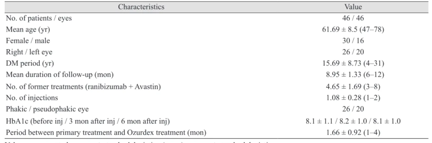
In total, 46 eyes of 46 patients (30 females [65.2%] and 16 males [34.8%]) with a mean age of 61.69 ± 8.5 years (range, 47 to 78) were included in the study (Table 1). The mean duration of diabetes was 15.69 ± 8.73 years (range, 4 to 31), the average follow-up period was 8.95 ± 1.33 months (range, 6 to 12), and the average injection number was 1.08
± 0.28 (range, 1 to 2) (Table 1). The mean period between primary treatments and Ozurdex treatment was 1.66 ± 0.92 months (range, 1 to 4).
Table 1. Characteristics of patients
Characteristics Value
No. of patients / eyes 46 / 46
Mean age (yr) 61.69 ± 8.5 (47–78)
Female / male 30 / 16
Right / left eye 26 / 20
DM period (yr) 15.69 ± 8.73 (4–31)
Mean duration of follow-up (mon) 8.95 ± 1.33 (6–12)
No. of former treatments (ranibizumab + Avastin) 4.65 ± 1.69 (3–8)
No. of injections 1.08 ± 0.28 (1–2)
Phakic / pseudophakic eye 26 / 20
HbA1c (before inj / 3 mon after inj / 6 mon after inj) 8.1 ± 1.1 / 8.2 ± 1.0 / 8.1 ± 1.0 Period between primary treatment and Ozurdex treatment (mon) 1.66 ± 0.92 (1–4) Values are presented as mean ± standard deviation (range) or mean ± standard deviation.
DM = diabetes mellitus; HbA1c = hemoglobin A1c; inj = injection.
Although a second injection was recommended for all patients, only four received the second injection within 6 months. Other patients refused the second injection due to financial or other reasons.
Efficacy
Although a statistically significant increase was detected (p < 0.05) during months 1 to 4 of the follow-up period when compared to patient condition before BCVA treat- Table 2. Comparison of VA, CMT, and IOP at study onset and during follow-up Parameter
Initial (n = 46) 1 mon (n = 46)
p-value
2 mon (n = 46)
p-value
3 mon (n = 46)
p-value
4 mon (n = 46)
p-value
5 mon (n = 46)
p-value
6 mon (n = 46)
p-value BCVA (logMAR)
0.88 ± 0.3 (0.5–1.3) 0.55 ± 0.29 (0.2–1)
0.0000.60 ± 0.32 (0.2–1)0.000
0.87 ± 0.23 (0.5–1.3)
0.000
0.80 ± 0.27 (0.5–1.3)
0.027
0.83 ± 0.29 (0.4–1.3)
0.066
0.85 ± 0.28 (0.5–1.3)
0.063 CMT (μm)498.1 ± 90.7 (366–620)370.5 ± 84.45 (195 ± 534)0.000
380.6 ± 68.7 (236–487)
0.000
486.0 ± 87.9 (355–647)
0.039
499.7 ± 93.2 (309–630)
0.951
484.7 ± 79.2 (359–607)
0.075489.0 ± 82.3 (370–636)0.760 IOP (mmHg) 16.47 ± 3.1 (10–21)18.91 ± 4.63 (11–29)0.07518.43 ± 2.44 (12–22) 0.00317.13 ± 2.26 (10–21)0.37816.65 ± 2.3 (12–21)0.96716.52 ± 1.97 (16–21)0.87716.56 ± 2.10 (15–20)0.676 Values are presented as mean ± standard deviation (range). VA = visual acuity; CMT = central macular thickness; IOP = intraocular pressure; BCVA = best-corrected visual acuity; logMAR = logarithm of the minimum angle of resolution. Paired t-test (p < 0.05 indicates statistical significance). BCVA (logMAR)
Preop 1 mon 2 mon 3 mon 4 mon 5 mon 6 mon 1.0
0.8 0.6 0.4 0.2 0
CMT (μm)
Preop 1 mon 2 mon 3 mon 4 mon 5 mon 6 mon 600
400 200 0
IOP (mmHg)
Preop 1 mon 2 mon 3 mon 4 mon 5 mon 6 mon 20
19 18 17 16 15
Fig. 1. Mean BCVA, CMT, and IOP at study onset and during follow-up. (A) Mean BCVA (logMAR), (B) mean CMT, (C) mean IOP before and after treatment. BCVA = best-corrected visual acuity; logMAR = logarithm of the minimum angle of resolution;
Preop = preoperative; CMT = central macular thickness; IOP = intraocular pressure.
A
B
C
ment, no statistically significant changes were detected during months 5 and 6 (p > 0.05) (Table 2 and Fig. 1A).
Macular thickness
Although macular thickness was found (p < 0.05) to de- crease at a statistically significant rate during months 1, 2 and, 3 when compared to pretreatment conditions, the rate of decrease was not statistically significant during months 4 to 6 (p > 0.05) (Table 2 and Fig. 1B).
Decreases in CMT were greatest (370 mm) after the first injection during month 1 (Fig. 1B).
Safety
Although IOP increased at a statistically significant rate during the follow-up month 2 (p < 0.05) compared to pre- IVD treatment conditions, rate increases were not statisti- cally significant after that point (p > 0.05) (Table 2 and Fig. 1C).
The first day after injection, one eye had an IOP of 27 mmHg; two eyes had an IOP of 26 mmHg; three eyes had an IOP of 25 mmHg; and one eye had an IOP of 29 mmHg.
In total, eight eyes had an IOP >25 mmHg in the month following injection. All of these patients’ IOP values de- creased to within normal limits (<22 mmHg) after admin- istration of topical antiglaucomatous drugs and did not re- quire surgery. Twelve patients (26%) experienced an IOP increase >5 mmHg after injection.
With the exception of subconjunctival hemorrhage, which occurred after 6 of the 50 injections (12%) given to patients, no associated complications, such as endophthal- mitis, retinal detachment or any IVD-dependent systemic adverse effects, were observed.
During follow-up, four patients were diagnosed with cataracts in month 11 and two of these patients underwent cataract surgery.
Discussion
A major cause of DR-associated visual loss is DME.
Capillary leakage resulting from destruction of the blood-retina barrier, fluid, increases in macular thickness, inflammatory mediators and VEGF are the main causes of DME pathogenesis [17,18].
The number of treatment modalities that exist parallels the number of pathogenic etiologies. Visual acuity loss can be prevented by application of focal/grid laser photocoagu- lation [19] in microaneurysms and diffuse capillary layer leakage. However, sufficient visual acuity cannot be achieved through this method alone [3].
Repeat injections of anti-VEGF are required and deter- mining which patients are partial responders or non-re- sponders is a major challenge. Detection of ranibizumab and BCVA below 20 / 40 in 40% of the patients in RISE and RIDE (a study of Ranibizumab Injection in Subjects with Clinically Significant Macular Edema with Center Involvement Secondary to Diabetes Mellitus) studies re- vealed that add-on therapies are required to achieve suffi- cient improvement [20].
Intravitreal steroids have positive effects on DME through VEGF inhibition, anti-inflammatory effects and vascular disorder reduction via tight bonds among endo- thelial cells. The most frequent complications from ste- roids are IOP increase and cataract formation. Infectious endophthalmitis and retinal detachment have been report- ed as well [21,22].
Dexamethasone is a potent water-soluble corticosteroid.
Because its intravitreal half-life is short, it is applied to the vitreous through a drug release system. Ozurdex (Aller- gan) is a biodegradable dexamethasone implant that con- tains lactic acid and glycolic acid and can be injected through a 22-gauge applicator. The implant undergoes hy- drolysis over time and decomposes into carbon dioxide and water components [14] and was designed to reduce the number of repeat injections, which last for 6 months at most [16]. Dexamethasone is less lipophilic than triamcin- olone and fluocinolone acetonide, so it decreases IOP and prevents cataract formation by reducing accumulation in the trabecular meshwork and lens [23].
In a randomized, sham-controlled 3-year study (n = 1,048), Boyer et al. [24] observed an average decrease in CMT of 111.6 µ after a mean of 4.1 injections and 22.2% of the patients had a visual acuity gain of more than 15 let- ters. Meanwhile, 59.2% of the patients underwent cataract operation, and almost one-third exhibited a significant in- crease in IOP. Only one patient required surgery to control IOP, whereas other patients only needed medical therapy.
Intravitreal hemorrhage, which does not require surgery, was observed in 6.9% of the patients and endophthalmitis was observed in one patient [24].
In our retrospective study, a mean CMT decrease of 128 µ after an average of 1.08 ± 0.28 injections was achieved;
78.2% and 21.7% of patients demonstrated visual gains of more than 10 and 15 letters, respectively. Two patients un- derwent cataract operations and significant IOP increases were detected in 10 patients. Neither hemorrhage nor en- dophthalmitis was observed among our patients. Further- more, none of our patients experienced neovascularization.
The low incidence of both cataract surgery and significant IOP increases might be due to the short-term follow-up pe- riod and small sample size.
In a retrospective study of nine patients, Zucchiatti et al.
[25] observed CMT decreases in refractory DME patients who had treatments at an average of 13.4 months that did not include Ozurdex injections. On day 3, by applying a single Ozurdex injection, Zucchiatti et al. found that BCVA improvements started in month 1 and continued for 4 months. They reported that 88.8% of their patients re- quired a second injection in month 6. In this study, the av- erage hemoglobin A1c value was 8.2 [25]. In our study, the average hemoglobin A1c level was 8.1. Because the last treatment before Ozurdex injection was performed 1.66 months prior, 100% of the patients required a second injec- tion in month 6, but only 9.09% of the patients underwent a second injection.
Similar to our study, Scaramuzzi et al. [26] determined that the effect of dexamethasone implantation on BCVA and CMT continued for 4 months. In contrast, in a retro- spective study of 58 patients, Dutra Medeiros et al. [27]
found that patients with DME refractory to other treat- ments, were under the effect of dexamethasone implants during 6 months of follow-up and the maximum effect was observed in month 3. CMT decreases of 37% CMT and 0.44 ± 0.27 logMAR BCVA improvement were detect- ed during the period of maximum effect after a single in- jection of Ozurdex. In our study, the maximum effect was observed in month 1 and it continued for 4 months. CMT decreases of 25% and 0.33 logMAR BCVA improvement were detected during the period of maximum effect.
In the PLACID study of patients with diffuse macular edema, laser photocoagulation treatment was compared to a combination treatment of laser photocoagulation and dexamethasone; no significant differences were observed among groups at the end of the first year, although vision improvements were greater among the combination group [28]. However, DME patients who were refractory to focal
laser photocoagulation and anti-VEGF treatment were sus- ceptible to the dexamethasone implant in our study and a significant increase in visual acuity was observed in the first 4 months.
In their randomized control trial, Haller et al. [29] deter- mined that the percentage of eyes with DME that achieved a BCVA improvement of 10 letters or more by day 90 after receiving 700 μg of dexamethasone was similar to the overall study population, which also included eyes with macular edema due to uveitis, Irvine-Gass syndrome and retinal vein occlusion. Rishi et al. [30], however, concluded that the effect of Ozurdex implantation lasted for a shorter period in patients with refractory DME (4 months) than in patients with retinal vein occlusion dependent-macular edema (6 months). Furthermore, they suggested that CMT decreases in month 1 did not affect BCVA increases, which could be due to the chronic effect of DM on the macula. Even though CMT changed in month 4 among pa- tients who had been diagnosed with DM at least 15 years prior in our study, continued BCVA increases do not sup- port this theory.
The effect of a single anti-VEGF injection appears to last 6 to 8 weeks [31]; in our study, the average period be- tween the treatments before Ozurdex and receipt of Ozur- dex treatment was 1.66 ± 0.92 months (range, 1 to 4), indi- cating that anti-VEGF might affect the early follow-up period after switching from anti-VEGF to Ozurdex.
The limitations of our study are its retrospective nature and the absence of a control group. In future studies, ran- domization of patients not responding adequately to an- ti-VEGF either to a continued anti-VEGF treatment group or to a dexamethasone implantation group could help ad- dress the question of whether dexamethasone implantation is superior to anti-VEGF treatment.
The advantages of our study are that we were able to de- termine the clinical effect of dexamethasone implantation on DME refractory to anti-VEGF treatment and focal laser photocoagulation.
In summary, treatment of DME patients who do not re- spond to anti-VEGF and laser photocoagulation treatment and who have prolonged duration DME can be a challenge.
Herein, we analyzed the effect of Ozurdex implantation in this difficult patient group. Consequently, for patients who require DM-dependent refractory macular edema treat- ment, IVD implantation is effective and well-tolerated; it increases visual acuity and decreases macular edema. Al-
though its effective period has been observed to be longer than that of anti-VEGF treatments, repeat injections may still be needed if the effective period is <6 months. The ef- fective period and reliability of Ozurdex intravitreal injec- tion should be determined in future randomized studies.
Conflict of Interest
No potential conflict of interest relevant to this article was reported.
References
1. Williams GA. Diabetic macular edema. In: Bandello F, Querques G, editors. ESASO course series. Vol. 1. Medical retina. Basel: Karger; 2012. p. 99-104.
2. Klein R, Klein BE, Moss SE, Cruickshanks KJ. The Wis- consin Epidemiologic Study of Diabetic Retinopathy. XV:
the long-term incidence of macular edema. Ophthalmology 1995;102:7-16.
3. Photocoagulation for diabetic macular edema: Early Treat- ment Diabetic Retinopathy Study report number 1. Early Treatment Diabetic Retinopathy Study research group.
Arch Ophthalmol 1985;103:1796-806.
4. Adamis AP. Is diabetic retinopathy an inflammatory dis- ease? Br J Ophthalmol 2002;86:363-5.
5. Grant MB, Afzal A, Spoerri P, et al. The role of growth factors in the pathogenesis of diabetic retinopathy. Expert Opin Investig Drugs 2004;13:1275-93.
6. Nguyen QD, Shah SM, Van Anden E, et al. Supplemental oxygen improves diabetic macular edema: a pilot study. In- vest Ophthalmol Vis Sci 2004;45:617-24.
7. Nauck M, Karakiulakis G, Perruchoud AP, et al. Cortico- steroids inhibit the expression of the vascular endothelial growth factor gene in human vascular smooth muscle cells.
Eur J Pharmacol 1998;341:309-15.
8. Heiss JD, Papavassiliou E, Merrill MJ, et al. Mechanism of dexamethasone suppression of brain tumor-associated vas- cular permeability in rats: involvement of the glucocorti- coid receptor and vascular permeability factor. J Clin In- vest 1996;98:1400-8.
9. McGhee CN. Pharmacokinetics of ophthalmic corticoste- roids. Br J Ophthalmol 1992;76:681-4.
10. Parke DW 3rd, Sisk RA, Houston SK, Murray TG. Ocular
hypertension after intravitreal triamcinolone with vitrecto- my and phacoemulsification. Clin Ophthalmol 2012;6:925- 31.
11. Massin P, Audren F, Haouchine B, et al. Intravitreal triam- cinolone acetonide for diabetic diffuse macular edema:
preliminary results of a prospective controlled trial. Oph- thalmology 2004;111:218-24.
12. Park CH, Jaffe GJ, Fekrat S. Intravitreal triamcinolone ace- tonide in eyes with cystoid macular edema associated with central retinal vein occlusion. Am J Ophthalmol 2003;
136:419-25.
13. Edelman JL, Lutz D, Castro MR. Corticosteroids inhibit VEGF-induced vascular leakage in a rabbit model of blood-retinal and blood-aqueous barrier breakdown. Exp Eye Res 2005;80:249-58.
14. Haller JA, Bandello F, Belfort R Jr, et al. Randomized, sh- am-controlled trial of dexamethasone intravitreal implant in patients with macular edema due to retinal vein occlu- sion. Ophthalmology 2010;117:1134-46.
15. Goldfein A. Adrenocorticosteroids and adrenocortical an- tagonists. In: Katzung BG, editor. Basic and clinical phar- macology. 6th ed. London: Prentice Hall; 1995. p. 592-607.
16. Chang-Lin JE, Attar M, Acheampong AA, et al. Pharma- cokinetics and pharmacodynamics of a sustained-release dexamethasone intravitreal implant. Invest Ophthalmol Vis Sci 2011;52:80-6.
17. Yau JW, Rogers SL, Kawasaki R, et al. Global prevalence and major risk factors of diabetic retinopathy. Diabetes Care 2012;35:556-64.
18. Bhagat N, Grigorian RA, Tutela A, Zarbin MA. Diabetic macular edema: pathogenesis and treatment. Surv Ophthal- mol 2009;54:1-32.
19. American Diabetes Association. Standards of medical care in diabetes--2009. Diabetes Care 2009;32 Suppl 1:S13-61.
20. Brown DM, Nguyen QD, Marcus DM, et al. Long-term outcomes of ranibizumab therapy for diabetic macular edema: the 36-month results from two phase III trials.
RISE and RIDE. Ophthalmology 2013;120:2013-22.
21. Gillies MC, Sutter FK, Simpson JM, et al. Intravitreal tri- amcinolone for refractory diabetic macular edema: two- year results of a double-masked, placebo-controlled, ran- domized clinical trial. Ophthalmology 2006;113:1533-8.
22. Jonas JB, Degenring R, Kreissig I, Akkoyun I. Safety of intravitreal high-dose reinjections of triamcinolone aceton- ide. Am J Ophthalmol 2004;138:1054-5.
23. Thakur A, Kadam R, Kompella UB. Trabecular meshwork
and lens partitioning of corticosteroids: implications for el- evated intraocular pressure and cataracts. Arch Ophthalmol 2011;129:914-20.
24. Boyer DS, Yoon YH, Belfort R Jr, et al. Three-year, ran- domized, sham-controlled trial of dexamethasone intravit- real implant in patients with diabetic macular edema. Oph- thalmology 2014;121:1904-14.
25. Zucchiatti I, Lattanzio R, Querques G, et al. Intravitreal dexamethasone implant in patients with persistent diabetic macular edema. Ophthalmologica 2012;228:117-22.
26. Scaramuzzi M, Querques G, Spina CL, et al. Repeated in- travitreal dexamethasone implant (Ozurdex) for diabetic macular edema. Retina 2015;35:1216-22.
27. Dutra Medeiros M, Postorino M, Navarro R, et al. Dexa- methasone intravitreal implant for treatment of patients with persistent diabetic macular edema. Ophthalmologica
2014;231:141-6.
28. Callanan DG, Gupta S, Boyer DS, et al. Dexamethasone intravitreal implant in combination with laser photocoagu- lation for the treatment of diffuse diabetic macular edema.
Ophthalmology 2013;120:1843-51.
29. Haller JA, Kuppermann BD, Blumenkranz MS, et al. Ran- domized controlled trial of an intravitreous dexamethasone drug delivery system in patients with diabetic macular ede- ma. Arch Ophthalmol 2010;128:289-96.
30. Rishi P, Rishi E, Kuniyal L, Mathur G. Short-term results of intravitreal dexamethasone implant (OZURDEX(®)) in treatment of recalcitrant diabetic macular edema: a case se- ries. Oman J Ophthalmol 2012;5:79-82.
31. Badala F. The treatment of branch retinal vein occlusion with bevacizumab. Curr Opin Ophthalmol 2008;19:234-8.