J Lung Cancer 2010;9(1):24-25
24
Fig. 1. Initial Brain MRI shows necrotic tumors in Lt cerebral hemisphere, with severe peritumoral edema.
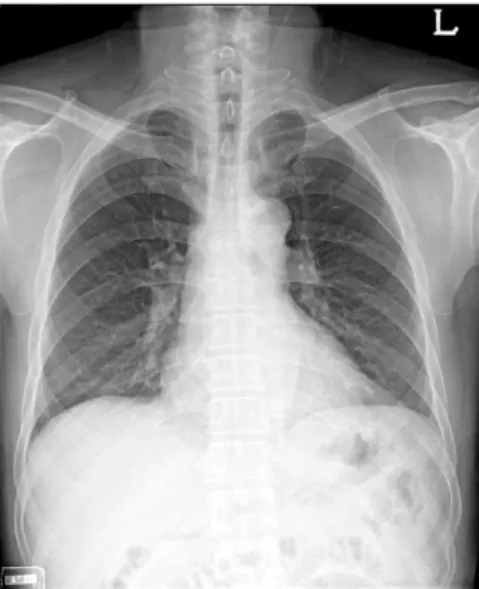
Fig. 2. Initial chest PA shows no active lung lesion.
Small Cell Lung Cancer at Subcarina Presenting as a Metastatic Brain Tumor
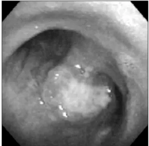
A 59-year-old man was rushed to the emergency room. The patient complained of headache with impaired memory function. Brain MRI showed a necrotic tumor in Lt cerebral hemisphere, with severe peritumoral edema (Fig. 1). Pathologic examination of the brain lesion confirmed that the tumor was a small cell lung cancer (SCLC). Chest computed tomography revealed a large soft tissue mass with central necrosis at subcarinal area in spite of an initial normal chest X-ray (Fig. 2). Bronchoscopic biopsy of the polypoid mass at subcarina revealed that the mass was a SCLC (Fig. 3). This is the case of SCLC only with an extrapulmonary symptoms despite of a normal chest X-ray. When metastatic brain tumor was found, appropriate chest evaluation should be performed even though chest X-ray was normal because brain is a common site of invasion of lung cancer. (J Lung Cancer 2010;9(1):24 25)
Key Words: Small cell lung cancer, Metastatic brain tumor
Mi-Ae Kim, M.D.
Eun-Kyung Kim, M.D.
Ji-Hyun Lee, M.D. and Hye-Cheol Jeong, M.D.
Department of Internal Medicine, Bundang CHA Hospital, CHA Univer- sity College of Medicine, Seongnam, Korea
Received: April 21, 2010 Accepted: May 10, 2010 Address for correspondence Hye-Cheol Jeong, M.D.
Division of Pulmonary and Critical Care Medicine, Department of Internal Medicine, CHA Bundang Medical Center, College of Medicine, CHA Uni- versity, 351, Yatap-dong, Bundang- gu, Sungnam 463-712, Korea Tel: 82-31-780-6142
Fax: 82-31-780-4800 E-mail: [email protected]
Small Cell Lung Cancer Presenting as a Metastatic Brain Tumor 25
Fig. 3. Bronchoscopy shows the polypoid mass at subcarina.