This is an open-access article distributed under the terms of the Creative Commons Attribution Non-Commercial License (http://creativecommons.org/
licenses/by-nc/4.0/), which permits unrestricted non-commercial use, distribution, and reproduction in any medium, provided the original work is properly cited.
CC
An idiopathic delayed maxillary hemorrhage after orthognathic surgery with Le Fort I osteotomy: a case report
Byungho Park, Wan-Hee Jang, Bu-Kyu Lee
Department of Oral and Maxillofacial Surgery, Asan Medical Center, University of Ulsan College of Medicine, Seoul, Korea
Abstract(J Korean Assoc Oral Maxillofac Surg 2019;45:364-368)
A Le Fort I osteotomy is a common procedure for correcting dental and facial deformities in orthognathic surgery. In rare cases, a delayed hemorrhage can occur as early as several hours or up to 12 weeks, postoperatively. The most frequently involved blood vessels in a delayed hemorrhage are the de- scending palatine artery, the internal maxillary artery, and the pterygoid venous plexus of veins. Intraoral bleeding accompanied by severe epistaxis in these cases makes it difficult to locate the precise bleeding focus. Eventual uncontrolled bleeding would require Merocel packing or surgical interven- tion. In general, a severe late postoperative hemorrhage is most effectively managed by angiography and embolization. Herein we describe a delayed hemorrhage case in which the cause was not evident on angiography. We were able to detect the bleeding point through an endoscopic nasal approach and treat it using direct cauterization.
Key words: Epistaxis, Postoperative complication, Le Fort I osteotomy, Pseudoaneurysm
[paper submitted 2019. 11. 5 / revised 2019. 11. 14 / accepted 2019. 11. 18]
Copyright © 2019 The Korean Association of Oral and Maxillofacial Surgeons. All rights reserved.
I. Introduction
Orthognathic surgery is an option for obtaining proper oc- clusion and profile in patients with dento-facial deformities1,2. The Le Fort I osteotomy procedure is the most frequently used method in orthognathic surgery and is regarded as a safe and predictable approach. However, diverse complications have been reported in these surgeries, including neurosensory disorder, maxillary sinusitis, internal fixation loosening, in- fection, hemolacria, unfavorable fractures, and vascular com- plications3,4. Panula et al.5 reported that intraoperative and postoperative bleeding are the most serious complications of orthognathic surgery.
Bleeding that occurs after Le Fort I osteotomy appears
mainly in the form of an epistaxis. An isolated epistaxis is caused by trauma from the nasal intubation process or by separation of the nasal mucosa from the nasal floor or septal area. Injury to the posterior vessels should be suspected in cases of epistaxis caused by either of these events. Most ex- cess bleeding that arises during orthognathic surgery is due to incorrect instrument manipulation, especially when an in- strument is positioned high toward the pterygopalatine fossa.
In the Le Fort I osteotomy procedure, the internal maxillary, sphenopalatine, and ascending palatine arteries are suscep- tible to damage. However, such acute hemorrhage events can be detected easily and managed during the operation5.
Delayed hemorrhage in the form of epistaxis can occur as early as several hours after Le Fort I osteotomy. This can be easily controlled, however, by bed rest and anterior nasal packing. The bleeding is not typically as extensive when it occurs this early after surgery and usually ceases within 1-2 days. However, if hemostasis fails and repeated bleeding oc- curs, definitive therapy and diagnosis are required. Although the risk is low, pseudoaneurysms can cause life-threatening bleeding6.
Pseudoaneurysms, also known as false aneurysms, are caused by incomplete damage of the blood vessels. Their progression affects the degree of vascular injury, blood flow, and elasticity of blood vessels and surrounding tissues. In- Bu-Kyu Lee
Department of Oral and Maxillofacial Surgery, Biomedical Engineering Research Center, Asan Institute for Life Sciences, Asan Medical Center, University of Ulsan College of Medicine, 88 Olympic-ro 43-gil, Songpa-gu, Seoul 05505, Korea
TEL: +82-2-3010-5907 FAX: +82-2-2045-4068 E-mail: [email protected]
ORCID: https://orcid.org/0000-0001-8888-1719
complete injury to one or more vessel wall layers results in gradual expansion of all vessel walls, resulting in hematoma.
Once a hematoma has developed, it continues to progress due to arterial pressure as long as blood flow is maintained; this leads to increased leakage and rupture3,7.
In this case report, we discuss onset of delayed epistaxis from the posterior lateral nasal artery, which is a branch of the sphenopalatine artery, after Le Fort I osteotomy. We contend that high clinical suspicion of a pseudoaneurysm should be maintained in patients with delayed postoperative bleeding.
Pseudoaneurysms of the sphenopalatine artery caused by Le Fort I osteotomy are rare. However, our report supplements the small existing case pool in the literature by describing a patient with a delayed but massive, life-threatening epistaxis that occurred at three weeks after Le Fort I osteotomy due to perioperative trauma of a branch of the sphenopalatine ar- tery. Maxillofacial surgeons need to be aware of the potential severity of delayed epistaxis and be able to rapidly diagnose and treat such cases.
II. Case Report
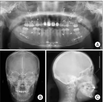
A 30-year-old female underwent Le Fort I osteotomy and bilateral sagittal split ramus osteotomy at a local dental clinic.
(Fig. 1) Three weeks later, she presented at our hospital due to intermittent and significant bleeding in both nostrils and in the oral cavity. Although there was no bleeding at presenta- tion, she was trembling and appeared pale and confused on physical examination. A routine complete blood count found hemoglobin level of 9.0 g/dL, hematocrit of 26.7%, and a platelet count of 180×103/μL. Prior to visiting our clinic, she had been transfused with two red blood cell (RBC) packs and received nasal packing using Merocel (Merocel 2000;
Medtronic Xomed, Heerlen, The Netherlands). A computed tomography (CT) angiogram with contrast media was used but revealed no definite active bleeding focus in the major vessels of the maxillary areas and no hemorrhage or en- hanced lesion in the brain parenchyma.(Fig. 2) At 3 days after admission, on December 19, a second episode of mas- sive epistaxis and oropharyngeal bleeding occurred, totaling 1 L. The patient’s hemoglobin level dropped to 7.7 g/dL, and she was transfused with two packs of RBC. Anterior nasal packing was again conducted, and the bleeding eventually stopped.
Before reopening the surgical area, the patient was referred to our department of otolaryngology for a nasal cavity ex- amination because anterior nasal packing had been effective when the second massive hemorrhage occurred. Using an en- doscopic nasal approach, slight oozing was observed on the right superior turbinate lateral side. The wound was irritated by a curved suction tip and showed massive arterial bleeding, strongly suggesting that it was the bleeding focus. The right inferior turbinate was outfractured via an endoscopic assistant using a Freer dissector. The right posterior lateral nasal artery, a branch of the sphenopalatine artery that was suspected to be the bleeding focus, was then electrocauterized.(Fig. 3) No
A B
Fig. 2. Images produced three weeks after surgery in the study patient. A. Computed tomography with contrast. B. Angiogram.
Byungho Park et al: An idiopathic delayed maxillary hemorrhage after orthognathic surgery with Le Fort I osteotomy: a case report. J Korean Assoc Oral Maxillofac Surg A
B C
Fig. 1. Images produced three weeks after surgery in the study patient. A. Panoramic x-ray. B. Posteroanterior cephalogram. C.
Lateral cephalograms.
Byungho Park et al: An idiopathic delayed maxillary hemorrhage after orthognathic surgery with Le Fort I osteotomy: a case report. J Korean Assoc Oral Maxillofac Surg
further bleeding episodes occurred after this intervention. The patient was discharged four days after electrocauterization and remained asymptomatic during the three-month follow- up, with no further episodes of bleeding.
III. Discussion
The maxillary osteotomy approach in orthognathic surgery can cause serious bleeding complications. These adverse events may arise immediately during surgery or may occur postoperatively. Bleeding after Le Fort I osteotomy manifests mainly as an epistaxis that can occur anywhere from several hours to 12 weeks after the procedure. Unilateral epistaxis that appears a few days after surgery may occur occasionally but is generally self-limiting or manageable with conservative treatment. Traditionally, most instances of epistaxis can be al- leviated and stopped by simple use of anterior-posterior nasal packing for 3-5 days6,8. In contrast, recurrent uncontrolled epistaxis after Le Fort I surgery is a very rare complication, with a frequency less than 1%6. The number of reported cases of this more serious complication is therefore not sufficient to establish optimal treatment guidelines. Here, we describe the diagnosis and treatment of a rare patient with massive epi- staxis after Le Fort I osteotomy, which may shed further light on management of such cases.
If postoperative epistaxis persists, radiographic imaging is recommended to achieve an accurate differential diagnosis.
In this regard, enhanced CT is a useful diagnostic tool when vascular complications occur as it can reveal anatomical details with high definition3. However, a metal mini-plate is used for maxillary fixation in Le Fort I osteotomy and can
lead to generation of metal artifacts in the imaging results and complicate proper diagnosis. Furthermore, because the diam- eter of the branch of the sphenopalatine artery is very small, enhanced CT is limited in its ability to discriminate postero- anterior from vascular injury7.
As an alternative approach, radiographic examination via angiography is also very useful for detecting vascular abnor- malities in a specific part of the body. This method detects a radioactive isotope flowing along the blood vessel and thereby pinpoints any deformation, injury, or bleeding3. Angi- ography is the preferred technique for diagnostic examination if severe bleeding occurs after Le Fort I osteotomy. In our current case, intracranial CT angiography was performed us- ing an external carotid artery protocol. However, no vascular abnormality was observed on either the internal maxillary or sphenopalatine artery, and no active bleeding focus was found. The exact bleeding focus in our study patient was not detected on this angiogram because the image was produced after the bleeding had been stopped, and the change was not large enough to detect prior to re-bleeding. Nevertheless, in terms of the clinical aspects of this case, a pseudoaneurysm can be considered the cause of delayed and substantial hem- orrhage. This patient had a recurrent and persistent oral and nasal bleeding pattern, followed by massive epistaxis that resulted in severe hemodynamic instability8.
There are several factors underlying the occurrence of a pseudoaneurysm. The main causes include infection associ- ated with the arterial wall, iatrogenic injury, or trauma. Many prior studies have reported trauma as the leading cause of an- eurysms in the head and neck3,5-9. For example, a downfrac- ture or repositioning of the maxilla after Le Fort I osteotomy
A B C
Fig. 3. The right posterior lateral nasal artery, a branch of the sphenopalatine artery that was suspected to be the bleeding focus, was electrocauterized. A. The bleeding focus in the study case was observed at the right posterior lateral nasal artery site. B. Bleeding oc- curred at the focus site when irritated by a suction tip. C. Bleeding was successfully stopped by electrocauterization.
Byungho Park et al: An idiopathic delayed maxillary hemorrhage after orthognathic surgery with Le Fort I osteotomy: a case report. J Korean Assoc Oral Maxillofac Surg 2019
can result in considerable bone fragmentation and thereby lead to vascular damage9. The most frequently involved blood vessels in these cases are the internal maxillary artery, the descending palatine artery, and the pterygoid venous plexus of veins. Arterial pseudoaneurysms, on the other hand, are caused by blunt trauma to the vessel wall, rather than by sharp damage from incorrect instrumentation5. Progression of pseudoaneurysm involves incomplete destruction of the arte- rial wall, causing the artery to expand abnormally and enlarge the lesion between the injured artery and surrounding tissue.
After the arterial wall has been destroyed, hemorrhaging oc- curs in the partially incised vessel and adjacent tissue until in- hibited by the pressure of the formed hematoma. Such cases usually involve the internal maxillary artery system, which has an increased probability of damage in these circumstance due to the proximity of the osteotome3,5,10.
Angiographic embolization is a commonly used method of treating bleeding and vascular damage in the head and neck areas. The purpose of an embolization is not to block the blood vessels around the injury in the presence of a pseudoa- neurysm, but to stimulate the formation of ancillary blood vessel networks. This procedure is less invasive than surgical intervention10-12. For selective embolization, a micro-catheter method is mainly used. The catheter is inserted through the femoral artery to the external carotid artery or its branch.
New techniques and various materials (i.e., micro-coils, gel- form, detachable balloons, N-butyl cyanoacrylate, autologous clots, and polyvinyl alcohol) have been introduced to achieve vascular embolization, and successful examples and draw- backs have been reported. However, the materials best suited to oral maxillofacial area embolization remain controversial, and further research is needed3,9,10,13.
There is a method of arterial ligation to treat persistent epistaxis that does not respond to the traditional method.
The external carotid artery has previously been used for liga- tion. Although greater anatomical understanding and better surgical procedures made it possible to suggest a method of ligating the internal carotid artery through the transantral approach, there were shortcomings with this technique. The anatomy of the antrum changes after Le Fort I osteotomy, and the space filled with blood limits detection of arteries and branches. Hence, ligation through a transantral approach has a significantly lower success rate13,14.
An endoscopic nasal approach is a widely accepted method for treatment of massive epistaxis. Recent studies have re- ported that the endoscopic approach is superior to nasal pack-
epistaxis are the sphenopalatine artery and the posterior nasal artery, the terminal branches of the internal maxillary artery.
They are easily accessible through the endoscopic nasal ap- proach, and ligation or direct cauterization can be performed.
This method is also less painful, more cost-effective, mini- mally invasive, and more clinically effective than others14-16. In our current case, endoscopy revealed bleeding in the pos- terior nasal artery, a branch of the sphenopalatine artery, and direct cauterization was performed.
In summary, sudden bleeding may occur in the nasal or oral cavity after Le Fort I osteotomy for orthognathic surgery.
This adverse event is caused by overlooked vascular injury during the operation. Hemostasis of the resulting pseudoa- neurysm appears to be achieved easily by nasal packing but may recur within a short time and can cause a life-threatening massive hemorrhage. If massive epistaxis occurs, angiogra- phy may be necessary for an accurate diagnosis12 and is often performed to treat ligation or embolization based on radio- graphic information. However, if the result of the examina- tion is unclear or a diagnosis is not performed in emergency cases, a less invasive and immediate endoscopic approach is a good alternative.
ORCID
Byungho Park, https://orcid.org/0000-0001-6085-1745 Wan-Hee Jang, https://orcid.org/0000-0002-8941-2313 Bu-Kyu Lee, https://orcid.org/0000-0001-8888-1719
Authors’ Contributions
B.P. participated in data collection and wrote the manu- script. W.H.J. and B.K.L. participated in the design and per- formed the draft. B.K.L. participated in the coordination and helped to draft the manuscript.
Conflict of Interest
No potential conflict of interest relevant to this article was reported.
References
1. Lee WY, Park YW, Kwon KJ, Kim SG. Change of the airway space in mandibular prognathism after bimaxillary surgery involv- ing maxillary posterior impaction. Maxillofac Plast Reconstr Surg 2016;38:23.
temporomandibular joint symptoms. Maxillofac Plast Reconstr Surg 2015;37:14.
3. Avelar RL, Goelzer JG, Becker OE, de Oliveira RB, Raupp EF, de Magalhães PS. Embolization of pseudoaneurysm of the inter- nal maxillary artery after orthognathic surgery. J Craniofac Surg 2010;21:1764-8.
4. Shin YM, Lee ST, Kwon TG. Surgical correction of septal devia- tion after Le Fort I osteotomy. Maxillofac Plast Reconstr Surg 2016;38:21.
5. Panula K, Finne K, Oikarinen K. Incidence of complications and problems related to orthognathic surgery: a review of 655 patients.
J Oral Maxillofac Surg 2001;59:1128-36; discussion 1137.
6. Garg S, Kaur S. Evaluation of post-operative complication rate of Le Fort I osteotomy: a retrospective and prospective study. J Max- illofac Oral Surg 2014;13:120-7.
7. Bykowski MR, Hill A, Garland C, Tobler W, Losee JE, Goldstein JA. Ruptured pseudoaneurysm of the maxillary artery and its branches following Le Fort I osteotomy: evidence-based guide- lines. J Craniofac Surg 2018;29:998-1001.
8. Politis C. Life-threatening haemorrhage after 750 Le Fort I oste- otomies and 376 SARPE procedures. Int J Oral Maxillofac Surg 2012;41:702-8.
9. Procopio O, Fusetti S, Liessi G, Ferronato G. False aneurysm of the sphenopalatine artery after a Le Fort I osteotomy: report of 2 cases. J Oral Maxillofac Surg 2003;61:520-4; discussion 524-5.
10. Zachariades N, Rallis G, Papademetriou G, Papakosta V, Spanomi- chos G, Souelem M. Embolization for the treatment of pseudoan- eurysm and transection of facial vessels. Oral Surg Oral Med Oral Pathol Oral Radiol Endod 2001;92:491-4.
11. Krempl GA, Noorily AD. Pseudoaneurysm of the descending pala- tine artery presenting as epistaxis. Otolaryngol Head Neck Surg 1996;114:453-6.
12. Silva AC, O'Ryan F, Beckley ML, Young HY, Poor D. Pseudoaneu- rysm of a branch of the maxillary artery following mandibular sag- ittal split ramus osteotomy: case report and review of the literature.
J Oral Maxillofac Surg 2007;65:1807-16.
13. Fernández-Prieto A, García-Raya P, Burgueño M, Muñoz-Caro J, Frutos R. Endovascular treatment of a pseudoaneurysm of the de- scending palatine artery after orthognathic surgery: technical note.
Int J Oral Maxillofac Surg 2005;34:321-3.
14. Viehweg TL, Roberson JB, Hudson JW. Epistaxis: diagnosis and treatment. J Oral Maxillofac Surg 2006;64:511-8.
15. Ellinas A, Jervis P, Kenyon G, Flood LM. Endoscopic sphenopala- tine artery ligation for acute idiopathic epistaxis. Do anatomical variation and a limited evidence base raise questions regarding its place in management? J Laryngol Otol 2017;131:290-7.
16. McClurg SW, Carrau R. Endoscopic management of posterior epi- staxis: a review. Acta Otorhinolaryngol Ital 2014;34:1-8.
How to cite this article: Park B, Jang WH, Lee BK. An idiopathic delayed maxillary hemorrhage after orthognathic surgery with Le Fort I osteotomy: a case report. J Korean Assoc Oral Maxil- lofac Surg 2019;45:364-368. https://doi.org/10.5125/jkaoms.
2019.45.6.364

![OPEN BID INVITATION [SECURITY SERVICE]](data:image/gif;base64,R0lGODlhAQABAIAAAP///wAAACH5BAEAAAAALAAAAAABAAEAAAICRAEAOw==)