Background and PurposezzBrain lesions involving the cerebral cortex are rarely described in patients with neuromyelitis optica spectrum disorder (NMOSD), in contrast to multiple scle- rosis. We investigated cerebral cortex involvement using conventional brain magnetic resonance imaging (MRI) in anti-aquaporin-4 (AQP4)-antibody-positive NMOSD patients.
MethodszzThe study enrolled 215 NMOSD patients who were seropositive for the anti-AQP4 antibody from 5 referral hospitals, and retrospectively analyzed their demographic, clinical, and MRI findings. Abnormal cerebral cortex lesions on brain MRI were identified by a neuroradi- ologist and two neurologists using consensus.
ResultszzMost of the 215 enrolled patients (87%) were female. The median age at onset was 22.5 years (range: 15–36 years) and the mean follow-up duration was 123 months. Brain lesions were found in 143 of 194 patients (74%) in whom MRI was performed during follow-up. Brain lesions involving the cerebral cortex were identified in 6 of these 194 patients (3.1%). Five of the patients were female, and the six patients together had a median age of 29 years (range: 15–36 years) at the time of lesion presentation. Three of them showed leptomeningeal enhancement in the lesions. At presentation of the cortex-involving lesions, five of these patients were not be- ing treated at the time of presentation, while the sixth was being treated with interferon-beta.
ConclusionszzAlthough rare, cortical involvement occurs in NMOSD and is commonly com- bined with leptomeningeal enhancement. We speculate that this occurs only in patients who are not treated appropriately with immunosuppressant drugs.
Key Wordszz neuromyelitis optica, neuromyelitis optica spectrum disorder, magnetic resonance imaging, cerebral cortex.
Cerebral Cortex Involvement in Neuromyelitis Optica Spectrum Disorder
INTRODUCTION
Neuromyelitis optica (NMO) is an inflammatory disorder of the central nervous system (CNS) that is distinct from multiple sclerosis (MS).1 The discovery of a specific antibody against aquaporin-4 (AQP4) in the sera of NMO patients resulted in the concept of NMO being changed.2 The broadened term “neuromyelitis optica spectrum disorder” (NMOSD) includes anti-AQP4-antibody-positive patients with limited or inaugural forms of NMO, and also the cerebral, diencephalic, and brainstem lesions, as well as anti-AQP4-antibody- positive patients with coexisting autoimmune disorders [e.g., systemic lupus erythematosus (SLE) or Sjögren syndrome (SS)].1
The presence of brain lesions with characteristic locations and configurations is helpful in the diagnosis of NMOSD.3 The 2015 diagnostic criteria for NMOSD classified typical NMOSD brain lesion patterns as follows: 1) lesions involving the dorsal medulla, especially the area postrema, either small and localized, often bilateral, or contiguous with an upper cervical spinal cord lesion; 2) lesions involving the periependymal surfaces of the fourth Woojun Kima
Jee Eun Leea Su-Hyun Kimb So-Young Huhc Jae-Won Hyunb In Hye Jeongb Min-Su Parkd Joong Yang Choe Sang-Hyun Leef Kwang Soo Leea Ho Jin Kimb
a Department of Neurology, The Catholic University of Korea College of Medicine,
Seoul, Korea
bDepartments of Neurology and
f Radiolgoy, Research Institute and
Hospital of National Cancer Center, Goyang, Korea
c Departments of Neurology and
Kosin University College of Medicine, Busan, Korea
d Department of Neurology,
Yeungnam University College of Medicine, Daegu, Korea
e Department of Neurology, Inje University Ilsan Paik Hospital, Goyang, Korea
pISSN 1738-6586 / eISSN 2005-5013 / J Clin Neurol 2016;12(2):188-193 / http://dx.doi.org/10.3988/jcn.2016.12.2.188
Received September 15, 2015 Revised September 20, 2015 Accepted September 24, 2015 Correspondence Woojun Kim, MD, PhD Department of Neurology, The Catholic University of Korea College of Medicine,
222 Banpo-daero, Seocho-gu, Seoul 06591, Korea
Tel +82-2-3779-2077 Fax +82-2-782-8654 E-mail [email protected]
cc This is an Open Access article distributed under the terms of the Creative Commons Attribution Non-Com- mercial License (http://creativecommons.org/licenses/by-nc/3.0) which permits unrestricted non-commercial use, distribution, and reproduction in any medium, provided the original work is properly cited.
JCN
Open Access ORIGINAL ARTICLEKim W et al.
JCN
ventricle in the brainstem/cerebellum; 3) lesions involving the hypothalamus, thalamus, or periependymal surfaces of the third ventricle; 4) large, confluent, unilateral, or bilateral sub- cortical or deep white-matter lesions; 5) long (at least half the length of the corpus callosum), diffuse, heterogeneous, or edematous corpus callosum lesions; 6) long corticospinal- tract lesions, unilateral or bilateral, contiguously involving the internal capsule and cerebral peduncle; and 7) extensive peri- ependymal brain lesions, often with gadolinium enhancement.1 Cortical lesions have been considered as “red flags” against the diagnosis of NMOSD.1 However, brain lesions involving the cerebral cortex are found in clinical practice, albeit very rarely. Lesions involving the cerebral cortex in patients with NMOSD have also been reported.4,5
We examined the involvement of the cerebral cortex using conventional brain magnetic resonance imaging (MRI) in NMOSD patients who were seropositive for the anti-AQP4 antibody, and described their imaging and clinical charac- teristics.
METHODS
This study enrolled 215 consecutive NMOSD patients who were seropositive for the anti-AQP4 antibody from 5 refer- ral hospitals, from May 2005 to April 2014. The diagnosis of NMOSD was based on anti-AQP4-antibody seropositivity and the 2007 NMOSD description.6 The presence of NMO- IgG or the anti-AQP4 antibody was tested according to a previously reported tissue-based indirect immunofluores- cence assay,7 cell-based indirect immunofluorescence assay,8 and enzyme-linked immunosorbent assay.9 We retrospec- tively reviewed the demographic, clinical, and MRI findings of the enrolled patients, including age, sex, dates of the first neurological symptom presentation and last follow-up, at- tack history, and dates of MRI scans. All MRI scans were ob- tained using either a 1.5- or 3.0-T machine. Abnormal lesions involving the cerebral cortex on brain MRI were identified by three experienced observers using consensus: a neurora- diologist (S.H. Lee) and two neurologists (W. Kim and H.J.
Kim).
We collected more-detailed clinical information on pa- tients in whom cortical lesions were identified, including the relapse history of optic neuritis, myelitis, and brain symp- toms, and treatments received for NMOSD. Cerebrospinal fluid (CSF) findings at the time of presentation of the cortical lesions were reviewed. The presence of comorbid systemic autoimmune diseases was reviewed, including SLE, SS, pso- riasis, and autoimmune thyroiditis.
This study was approved by the Institutional Review Board of the National Cancer Center, Korea.
RESULTS
Most of the 215 enrolled patients (87%) were female. The me- dian age at onset was 22.5 years (range: 15–36 years) and the mean follow-up duration was 123 months. Brain lesions were identified in 143 of 194 patients (74%) in whom MRI was performed during follow-up. Brain lesions involving the ce- rebral cortex were identified in 6 of these 194 patients (3.1%) (Fig. 1). All of the MRI scans were obtained during the acute stage of the correlated neurological symptoms. During the follow-up period, all six patients presented more than one of the “core clinical characteristics” of NMOSD as defined by Wingerchuk et al.1 in recently published international con- sensus diagnostic criteria, such as optic neuritis, acute my- elitis, area postrema syndrome, acute brainstem syndrome, symptoms correlated with NMOSD-typical diencephalic MRI lesions, or symptomatic cerebral syndrome with NMOSD- typical brain lesions. None of these patients had a history of vaccination during the few months preceding when the cor- tex-involving lesions first appeared.
Five of the six patients (83.3%) exhibiting cortex-involving brain lesions were female, and the six patients together had a median age of 29 years (range: 15–36 years) at the time of lesion presentation (Table 1). Three of them (50%) had those lesions at the first presentation of their CNS inflammatory symptoms. At the time of presentation of the cortex-involv- ing lesions, five of the six patients were not being treated, while the sixth was being treated with interferon-beta-1b.
Three patients (Patients 1, 2, and 3) presented with enceph- alopathy, including seizures, confusion, decreased mental sta- tus, or hypersomnolence. One (Patient 4) experienced an intermittent myoclonic seizure of the contralateral arm two to five times a day, as confirmed by electroencephalography, and two (Patients 5 and 6) did not show obvious symptoms that were correlated with the cortical lesions. Patient 6 pre- sented hemiparesis on her left limbs that was correlated with lesions involving the right corticospinal tract.
The cortex-involving lesions were located most com- monly in the frontal, posterior parietal, and occipital lobes (Fig. 1). In fluid-attenuated inversion recovery (FLAIR) im- ages, many of the cortex-involving lesions showed hetero- geneous signal intensities and blurred margins, and simul- taneously involved the cortex and subcortex. At the time of presentation of the cortex-involving lesions, various other lesions were seen in the brain. Some were characteristic le- sions of NMOSD, such as periependymal lesions of the third (Patient 1) and fourth (Patient 6) ventricles, large confluent subcortical and deep white-matter lesions (Patients 3 and 6), long corpus callosum lesions (Patients 3 and 6), corticospi- nal-tract lesions (Patient 2, 3, and 6), and extensive periepen-
Cortical Involvement in NMOSD
JCN
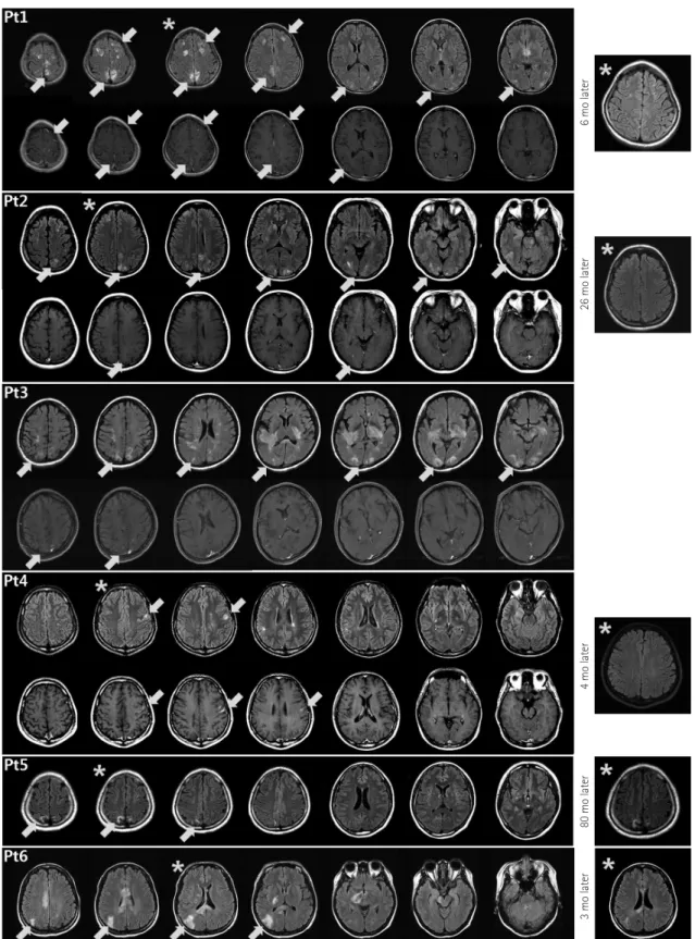
Fig. 1. Brain abnormalities in neuromyelitis optica spectrum disorder (NMOSD) patients on fluid-attenuated inversion recovery (FLAIR) (upper rows) and T1-weighted enhanced (lower rows) magnetic resonance imaging (MRI) scans obtained at the time of cortical lesion presentation. The brain MRI scans of Patient 2 have been described previously.15 The lesions involving the cerebral cortex are accompanied by leptomeningeal (Pa- tients 1, 2, and 3) or cortical (Patient 4) enhancement, or no enhancement (Patients 5 and 6). In addition to the cortex-involving lesions, other characteristic brain lesions of NMOSD are evident. In representative follow-up FLAIR images (Patients 1, 2, 4, 5, and 6), obtained 3 to 80 months after the presentation of the cortex-involving lesions, most of the lesions had disappeared or become faint—compared to the correlated slices in the previous images (*)—in all patients except Patient 5.mo: months.
6 mo later26 mo later4 mo later80 mo later3 mo later
Kim W et al.
JCN
Table 1. Clinical characteristics of neuromyelitis optica spectrum disorder patients with involvement of cerebral cortex in brain MRI Pt.SexOnset age Age & disease duration at the presentation of cortical lesion(s)
NMO-IgG or anti- aquaporin 4 antibody test
Enhancement
Symptoms at the presentation of the cortical lesion(s)
CSF findings at the presentation of the cortical lesion(s)
NMOSD symptoms presented before the presentation of cortical lesions
NMOSD symptoms presented during disease duration
Treatment proceeding the presentation of cortical lesion(s)
Treatment after presentation of cortical lesion(s)
Comorbid systemic autoimmune disease 1F1515 (onset)TBA(+) CBA(+) ELISA(+)
LMSeizure, confusion, dysarthria, decreased mental status Myelitis (confirmed) WBC 6, Protein 141, Glucose 59
-ON Myelitis Brain Sx
NoRTSjögren syndrome 2F3636 (onset)TBA(+) CBA(+) ELISA(+)
LMHeadache, confusion, hypersomnolence
N/A-Myelitis Brain SxNoMTSjögren syndrome 3F2429 (5 years)TBA(+) CBA(+) ELISA(+)
LMFever, headache, confusion, decreased mental status, paraparesis WBC 45, Protein 178, Glucose 35
ONON Brain SxNoMT- 4M2121 (onset)CBA(+) ELISA(+)CorticalDysarthria, facial numbness, myoclonic seizure of the Rt. arm
WBC 1, Protein 25.4, Glucose 61
-ON Brain SxNoAZA → MMF- 5F1929 (10 years)TBA(+) CBA(+) ELISA(-)
NoDizzinessN/AON Myelitis Brain Sx
ON Myelitis Brain Sx
NoRT- 6F3434 (6 months)TBA(+) CBA(+) ELISA(+)
NoLeft hemiparesis, dizzinessN/AON MyelitisON Myelitis Brain Sx
Interferon- beta 1bRT- AZA: azathioprine, CBA: cell-based indirect immunofluorescence assay, ELISA: enzyme-linked immunosorbent assay, LM: leptomeningeal, MMF: mycophenolate mofetil, MT: mitoxantrone, N/A: not avail- able, NMO: neuromyelitis optica, NMO-IgG: neuromyelitis optica-immunoglobulin G, NMOSD: neuromyelitis optica spectrum disorder, ON: optic neuritis, RT: rituximab, Sx: symptoms, TBA: tissue-based indirect immunofluorescence assay, WBC: white blood cells.
Cortical Involvement in NMOSD
JCN
dymal brain lesions (Patient 3). Three of the lesions showed leptomeningeal cortical enhancement (Patients 1, 2, and 3) and one showed cortical enhancement (Patient 4).
Follow-up brain MRI scans were available in five patients (Patients 1, 2, 4, 5, and 6). In the follow-up FLAIR images, obtained 3 to 80 months after the presentation of the cor- tex-involving lesions, most of the lesions had disappeared or become faint in four of these five patients (Patients 1, 2, 4, and 6) (Fig. 1).
DISCUSSION
This study found that in very rare cases cortical involvement occurs in NMOSD patients and is commonly combined with leptomeningeal enhancement. We speculate that this rare finding occurs only in patients who are not treated appropri- ately with immunosuppressant drugs.
Several studies using advanced techniques, such as double inversion recovery sequences10 or 7.0-T MRI,11 did not ob- serve cortical lesions in NMOSD; however, those studies in- volved relatively small numbers of patients (30 and 10 NMO/
NMOSD patients, respectively), and imaging was not per- formed at the acute stage of relapses. The absence of cortical demyelination in NMO was also found in a study of the pa- thology of the lesions that performed immunohistochemi- cal analyses of 19 NMO cases.12 This led to a lack of cortical demyelination in NMOSD being suggested as one of the fea- tures that could help to differentiate it from MS.13 However, our results suggest that brain lesions involving the cerebral cortex do not absolutely rule out NMOSD. It is noteworthy that the MRI characteristics of the lesions involving the ce- rebral cortex observed in our patients are quite different from the typical cortical lesions described in MS.10,11,14
Two of our patients (Patients 1 and 2) have been described in our previous report on NMOSD patients who showed brain abnormalities as an initial manifestation.15 However, the present study had a totally different focus: we examined brain MRI scans focusing on cortical involvement in this study, whereas our previous report merely described the brain lesions of Patient 2 as “diffuse subcortical lesions.”15 In fact, the brain lesions of that patient involved mainly the subcor- tex, but we also found some lesions simultaneously involv- ing the cerebral cortex since we concentrated on the corti- cal area.
Lesions involving the cerebral cortex in patients with NMOSD have been found in previous studies, although only very rarely.4,5 In a study describing brain abnormalities in 17 NMO patients, 11 patients (64.7%) had abnormal MRI findings, and 1 of them (5.8%) had lesions in the cerebral cortex.4 A case with cortical oscillopsia without nystagmus as a mani-
festation of NMOSD with lesions of the visual cortex has also been reported.5
Most of the cortex-involving lesions in our patients dis- appeared or became faint on FLAIR images after 3 to 80 months, as has also been reported for other brain lesions.3,13,15,16 Considering that brain lesions in NMOSD can disappear on subsequent imaging MRI, performing MRI evaluations during the acute phase of the clinical symptoms is important to discovering brain lesions in NMOSD.
At least three of the six patients exhibiting cortex-involv- ing brain lesions developed encephalopathy, including sei- zures, confusion, decreased mental status, or hypersomno- lence, and the symptoms and brain MRI features showed reversibility in most of our patients. The clinical and imaging features of these three patients resembled acute disseminated encephalomyelitis (ADEM) or posterior reversible encepha- lopathy syndrome (PRES). In fact, some of our patients were diagnosed with ADEM or PRES at the time of presentation of encephalopathy, as described in our previous report.15 In NMOSD, the encephalopathy associated with diffuse white- matter lesions may appear similar to ADEM.17 A pediatric case of ADEM was recently reported in which seropositivity for the anti-AQP4 antibody presented with optic neuritis and myelitis with encephalopathy at 14 days after an influen- za vaccination.18 Several cases of PRES-type lesions in NMOSD have also been described.13,19 However, none of our patients had a history of vaccination during the few months preceding when the cortex-involving lesions first appeared, and they did also not have any comorbid medical conditions or drug histories typical of PRES.
Three of our patients showed leptomeningeal enhance- ment in association with cortical lesions. Leptomeningeal en- hancement in brain MRI is not commonly observed in NMOSD patients, but this was recently found to be more frequent in patients with NMOSD than in patients with MS.20 Disrup- tion of the blood-brain barrier is normally the main expla- nation for gadolinium enhancement.20 Consistent with the location of AQP4 in the leptomeninges, which is associated with the integrity of the interface between the brain and blood, and between the CNS and CSF, the anti-AQP4 antibody binds to such sites selectively and readily.7,21 Therefore, the infiltration of inflammatory cells via the damaged BBB into the adjacent cortex is one possible explanation of the cortical involvement—especially with leptomeningeal enhancement—
in our patients.
None of our patients had been treated appropriately when they presented with the cortex-involving lesions. Three of them already had the lesions at the first presentation of their disease, while among the others, on presentation of the cor- tex-involving lesions, two of them had not been treated and
Kim W et al.
JCN
one was being treated with interferon-beta-1b. This means that cortex-involving lesions could be present in NMOSD patients when the disease activity is high, given that none of the six patients experienced relapse associated with cortex- involving lesions after being treated with appropriate im- munosuppressive drugs.
This study had some limitations, including its retrospec- tive design, the inclusion of a population with a single eth- nicity, the possibility of unintentional selection bias, and the unknown status of the anti-AQP4 antibody at the time of pre- sentation of the cortex-involving lesions. In addition, only conventional 1.5- and 3.0-T MRI machines were used.
In conclusion, cortical involvement occurs in NMOSD pa- tients, although only very rarely. Further evaluations are needed using advanced MRI techniques or pathological in- vestigations to reveal the characteristics of those cortex-in- volving lesions and leptomeningeal enhancement in NMOSD.
Conflicts of Interest
Ho Jin Kim has received honoraria for speaking or consulting from Bay- er Schering Pharma, Biogen Idec, Genzyme, Merck Serono, Novartis, MedImmune, and Teva-Handok and has received research grants from the Ministry of Science, ICT & Future Planning, Genzyme, Merck Sero- no, and Kael-GemVax. He serves on a steering committee for MedIm- mune and serves as an editor of Multiple Sclerosis Journal - Experimen- tal, Translational and Clinical. Woojun Kim, Su-Hyun Kim, So-Young Huh, Jae-Won Hyun, In Hye Jeong, Min-Su Park, Joong Yang Cho, Sang- Hyun Lee, and Kwang Soo Lee have nothing to declare.
REFERENCES
1. Wingerchuk DM, Banwell B, Bennett JL, Cabre P, Carroll W, Chitnis T, et al. International consensus diagnostic criteria for neuromyelitis optica spectrum disorders. Neurology 2015;85:177-189.
2. Kim W, Kim SH, Kim HJ. New insights into neuromyelitis optica. J Clin Neurol 2011;7:115-127.
3. Kim HJ, Paul F, Lana-Peixoto MA, Tenembaum S, Asgari N, Palace J, et al. MRI characteristics of neuromyelitis optica spectrum disorder:
an international update. Neurology 2015;84:1165-1173.
4. Kim JE, Kim SM, Ahn SW, Lim BC, Chae JH, Hong YH, et al. Brain abnormalities in neuromyelitis optica. J Neurol Sci 2011;302:43-48.
5. Kim SM, Kim JS, Heo YE, Yang HR, Park KS. Cortical oscillopsia without nystagmus, an isolated symptom of neuromyelitis optica spectrum disorder with anti-aquaporin 4 antibody. Mult Scler 2012;
18:244-247.
6. Wingerchuk DM, Lennon VA, Lucchinetti CF, Pittock SJ, Weinshen- ker BG. The spectrum of neuromyelitis optica. Lancet Neurol 2007;
6:805-815.
7. Lennon VA, Wingerchuk DM, Kryzer TJ, Pittock SJ, Lucchinetti CF, Fujihara K, et al. A serum autoantibody marker of neuromyelitis op- tica: distinction from multiple sclerosis. Lancet 2004;364:2106-2112.
8. Takahashi T, Fujihara K, Nakashima I, Misu T, Miyazawa I, Nakamura M, et al. Establishment of a new sensitive assay for anti-human aqua- porin-4 antibody in neuromyelitis optica. Tohoku J Exp Med 2006;
210:307-313.
9. Kim W, Lee JE, Li XF, Kim SH, Han BG, Lee BI, et al. Quantitative measurement of anti-aquaporin-4 antibodies by enzyme-linked im- munosorbent assay using purified recombinant human aquaporin-4.
Mult Scler 2012;18:578-586.
10. Calabrese M, Oh MS, Favaretto A, Rinaldi F, Poretto V, Alessio S, et al. No MRI evidence of cortical lesions in neuromyelitis optica. Neu- rology 2012;79:1671-1676.
11. Kister I, Herbert J, Zhou Y, Ge Y. Ultrahigh-field MR (7 T) imaging of brain lesions in neuromyelitis optica. Mult Scler Int 2013;2013:
398259.
12. Popescu BF, Parisi JE, Cabrera-Gómez JA, Newell K, Mandler RN, Pittock SJ, et al. Absence of cortical demyelination in neuromyelitis optica. Neurology 2010;75:2103-2109.
13. Cabrera-Gomez JA, Kister I. Conventional brain MRI in neuromye- litis optica. Eur J Neurol 2012;19:812-819.
14. Puthenparampil M, Poggiali D, Causin F, Rolma G, Rinaldi F, Perini P, et al. Cortical relapses in multiple sclerosis. Mult Scle 2015. [Epub ahead of print]
15. Kim W, Kim SH, Lee SH, Li XF, Kim HJ. Brain abnormalities as an initial manifestation of neuromyelitis optica spectrum disorder. Mult Scler 2011;17:1107-1112.
16. Kim SH, Huh SY, Hyun JW, Jeong IH, Lee SH, Joung A, et al. A lon- gitudinal brain magnetic resonance imaging study of neuromyelitis optica spectrum disorder. PLoS One 2014;9:e108320.
17. Wingerchuk DM, Weinshenker BG. Acute disseminated encephalo- myelitis, transverse myelitis, and neuromyelitis optica. Continuum (Minneap Minn) 2013;19(4 Multiple Sclerosis):944-967.
18. Okumura A, Nakazawa M, Igarashi A, Abe S, Ikeno M, Nakahara E, et al. Anti-aquaporin 4 antibody-positive acute disseminated encepha- lomyelitis. Brain Dev 2015;37:339-343.
19. Magaña SM, Matiello M, Pittock SJ, McKeon A, Lennon VA, Rabin- stein AA, et al. Posterior reversible encephalopathy syndrome in neu- romyelitis optica spectrum disorders. Neurology 2009;72:712-717.
20. Long Y, Chen M, Zhang B, Gao C, Zheng Y, Xie L, et al. Brain gadolin- ium enhancement along the ventricular and leptomeningeal regions in patients with aquaporin-4 antibodies in cerebral spinal fluid. J Neu- roimmunol 2014;269:62-67.
21. Pittock SJ, Lennon VA, de Seze J, Vermersch P, Homburger HA, Wing- erchuk DM, et al. Neuromyelitis optica and non organ-specific auto- immunity. Arch Neurol 2008;65:78-83.