A cervical aortic arch is a rare aortic arch anomaly as- sumed to result from persistence of the third aortic arch and regression of the normal fourth arch (1). Most pa- tients with this anomaly are asymptomatic, but symp- toms of dysphagia and respiratory distress due to com- pression by the vascular ring have been reported (1-7).
Aneurysm formation involving the cervical aortic arch is very rare (2-14). Subaortic left innominate vein have usually been associated with heart anomaly (15-17).
We report a case in which aneurysm formation involved the cervical aortic arch associated with the subaortic left innominate vein.
Case Report
A 26-year-old man who had been healthy and sympto- matic was admitted to our institution, where plain chest radiography initially indicated a mediastinal mass.
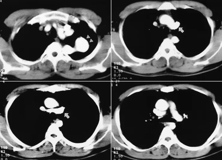
Blood pressure in the right and left arm was 180/100 mmHg and 134/93 mmHg, respectively, without med- ication. A chest radiograph depicted a superior mediasti- nal mass in the left side (Fig. 1), and the aortic knob ap- peared to be small. CT demonstrated that the mass was a left-sided cervical aortic arch (Fig. 2) that compressed the left upper lobe at lung window setting. Linear calci- fication denoted a fusiform dilated aneurysmal wall (Fig. 3), but none was present in the ascending, thoracic, or abdominal aorta. The size of the ascending aorta was normal (30 mm), but the descending aorta was narrower than usual, only 15 mm. The left innominate vein drained into the superior vena cava behind the aorta, which was in the anomalous subaortic position of the brachiocephalic vein (Fig. 2).
Routine laboratory data were normal. MR angiogra- phy depicted a left-side aortic arch with normal branch- ing of the innominate and left common carotid arteries,
Aneurysm Formation of Cervical Aortic Arch Combined with Subaortic Left Innominate Vein: Case Report1
Young-Min Han, M.D.1, 4, Ja-Hong Gu, M.D.2, Gong-Yong Jin, M.D., Hyo-Sung Kwak, M.D., Gyung-Ho Chung, M.D., Myoung-Ja Chung, M.D.3
An asymptomatic 26-year-old man was initially admitted with a suspicious mediasti- nal mass. On the basis of the contrast-enhanced chest CT findings, aneurysm forma- tion involving the left cervical aortic arch associated with subaortic left innominate vein was diagnosed. The aneurysm was confirmed by MR angiography and DSA. The arch aneurysm was surgically removed. We describe this case, and review the litera- ture.
Index words :Anomalies, congenital Aorta, aneurysm CT, MRI, angiography Cervical aortic arch
1Department of Radiology, Chonbuk National University Medical School
2Department of Thoracic Surgery, Chonbuk National University Medical School
3Department of Pathology, Chonbuk National University Medical School
4Institute of Cardiovascular Research, Chonbuk National University Medical School
Received June 27, 2003 ; Accepted October 20, 2003
Address reprint requests to : Young-Min Han, M.D., Department of Radiology, Institute of Cardiovascular Research, Chonbuk National University Hospital, 634-18 Keumam-dong, Chonju 560-182, Korea.
Tel. 82-63-250-1176 Fax. 82-63-272-0481 E-mail: [email protected]
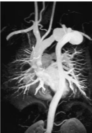
hypertrophy of the right vertebral artery and oblitera- tion of the left vertebral artery (Fig. 4). Thoracic aortog- raphy using a right axillary approach demonstrated a left-side aortic arch with normal branching of the in- nominate and left common carotid arteries (Fig. 5). The
configuration of the ascending aorta was normal, but the diameter of the descending thoracic aorta was less than usual. The aortic arch was elongated and tortuous be- tween the origin of the left common carotid and the left subclavian arteries, and inferior to the arch, a fusiform
Fig. 3. Non-contrast CT shows linear calcifications (arrows) along the dilated aneurysmal wall.
Fig. 1. A chest radiograph shows a superior mediastinal mass.
Fig. 2. Contrast-enhanced CT shows a tortuous fusiform dilated transverse arch aneurysm (arrowheads). The subaortic left innomi- nate vein (arrows) is noted.
aneurysm had formed (Fig. 5). The left vertebral artery was not identified, and the descending thoracic aorta was left-sided.
At surgery, an aneurysm was found at the site at which the trunchus arteriosum is usually present. The aneurysm was resected, and a Gore-Tex bypass graft used to replace the tortuous aortic arch between the ori- gin of the left common carotid artery and the descend- ing thoracic aorta. The left subclavian artery was su- tured to the bypass graft. Histologic examination re- vealed a true aneurysm, which had atherosclerosis of the thick intima and media of the aortic wall, with dys- trophic calcification, and a layer of adventitia (Fig. 6).
Discussion
The mechanism of development of an aortic arch anomaly is explained by the hypothetical double aortic arch system (1). Between the fifth and seventh embry- onic weeks, the persistence of the left fourth branchial arch and regression of the right fourth branchial arch produce a normal left aortic arch. Cervical aortic arch anomaly arises from the abnormal persistence of the
third arch and regression of the fourth, which normally forms the normal aortic arch (1). Haughton et al. (12) classified cervical aortic arches as one of five types ac- cording to the configuration of the aorta, sequence of brachiocephalic branching, and embryogenesis, al- though some cases were unclassifiable. The descending
Fig. 6. The microscopic findings show thickening of aortic wall, accumulation of foamy histiocytes and dystrophic calci- fication (arrows) in the media of the aneurysmal wall.
Fig. 4. MR angiography shows a tortuous dilated transverse aortic arch aneurysm. The ascending aorta reveals a normal configuration, and the size of the descending aorta revealed a small diameter.
Fig. 5. Thoracic aortography shows a tortuous dilated trans- verse aortic arch aneurysmal formation in the left anterior oblique view. The brachiocephalic and left common carotid arteries show normal branching, and the ascending aorta is not tortuous. The left vertebral artery is not identified.
thoracic aorta is left sided in the type-D cervical aortic arch of the original classification, though Akduman et al. (8) and Hirao et al (13) showed that a right-sided de- scending thoracic aorta can also be classified as type D.
In our case, the elongated tortuous transverse arch is thought to have originated from a persistent left third branchial arch.
The left brachiocephalic vein extends from the junc- tion of the left internal jugular and left subclavian vein to the junction of the right brachiocephalic and right su- perior caval vein. The normal course is obliquely down- wards and to the right, passing in front of the left subcla- vian, left common carotid, and brachiocephalic arteries, beneath the aortic arch. Very rarely, this vein follows an anomalous course, passing from left to right below the arch of the aorta, to enter the superior caval vein below the orifice of the azygos vein (15-17).
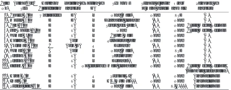
To our knowledge, this is the first documented case of aneurysm formation involving the cervical aortic arch associated with subaortic left innominate vein to appear in the literature in English (Table 1). In reported cases in which an aneurysm involved the cervical aortic arch, patient age was 6-59 (mean, 30) years, but among the 14 patients (M:F= 4:10), only two were children. Type D in the Haughton classification is the most frequently occurring type of cervical aortic arch associated with aortic aneurysm (10/14 cases: 71%). A ventricular septal defect occurred in two patients, and in ten, involved a cervical aortic arch, with characteristic symptoms which in three cases included pain around the neck,
shoulder, chest or back. An ischemic cerebrovascular at- tack occurred in two patients, including one child; dys- phagia in two, and dyspnea in three. The aneurysm rup- tured in one patient (10), and another developed aortic dissection (4). In one other, aortic coarctation and aneurysm involved the left cervical aorta (14). In con- trast, adult patients with no cervical aortic arch aneurysm were usually asymptomatic or experienced dysphagia (1, 7).
Aortic aneurysms were located in the aortic arch in 11 patients, the descending thoracic aorta in two and both the arch and descending thoracic aorta in one. Nine of ten Haughton type-D aortic arch aneurysms occurred between the left common carotid and the subclavian artery, the location in which they are least common (6, 10).
In congenital heart disease detected at echocardiogra- phy, the brachiocephalic vein has been reported to occu- py an anomalous position in 0.98% of cases (24/2457) (15). Gerlis et al. (16) reviewed the literature, finding that in 25 cases, the brachiocephalic vein occupied an anomalous subaortic position. Sixteen cases were con- firmed at necropsy; in ten, associated cardiovascular dis- ease and anomaly were present, but in six there was no such association. Nine cases were confirmed by echocardiography and angiography, and in eight, cardio- vascular disease was also present (16). Mill et al. (17) re- ported seven cases in which the course of the left bra- chiocephalic vein was found during surgery to be anom- alous; six of these involved the tetralogy of Fallot, and in
Table 1. Cervical Aortic arch Associated with Aneurysm
Case Author/Age/ Haughton Location of Position of Symptoms Stenosis of aorta Other Pathology of
No. Sex Classification aneurysm DA of major vessels anomaly aneurysm
01. Cooley, 39/F Unclassified DA L None (Mass) None VSD (-)
02. Morris, 35/F D DA L Dyaphagia, dyspnea (+) None (-)
03. Cao, 21/F D TA L Severe neck pain* (+) None Cystic Medial Necrosis
04. van Nooten, 17/F D TA L None (+) None Cystic Medial Necrosis
05. Kumar, 37/F D TA L Cough, mass None None (-)
06. Holland, 42/F E Arch L Retrosternal pain None None (-)
07. Akduman, 41/F C Arch, DA R Dyspnea (+) None (-)
08. Kumar, 19/M B Arch L None (Mass) None VSD (-)
09. Pearson, 6/F D TA L Paresthesia None None (-)
10. Pearson, 11/F D TA L Dyspnea** (+) None (-)
11. Farsak, 24/F D TA Not described Mass, dysphagia None None Cystic Medial Necrosis (basophilic degeneration)
12. Hirao, 59/M D TA R Previous TIA (+) None Atherosclerosis
13. Imai, 48/M D TA L H/T, murmur*** None None Atherosclerosis
14. Present case, 26/M D TA L None (Mass) (+) V. A**** Atherosclerosis
DA: Descending Aorta, L: Left, VSD: Ventricular Septal Defect, (-): Not obtained, TA: Transverse Arch, located between left common carotid and subclavian arteries, *: Aortic Dissection, R: Right, **: Aneurysm Rupture, TIA: Transient Ischemic Attack, H/T:
Hypertension, ***: Aortic Coarctation, V.A****: Anomalous subaortic position of innominate vein
one, the aortic arch was interrupted. Kim et al. (18) re- ported 14 cases of subarotic left innominate vein. In our case, spiral chest CT showed good delineation of the anomalous subaortic position of the brachiocephalic vein.
Pathologically, medial cystic necrosis of the wall of the aortic aneurysm was observed in three younger patients age 17-24 (mean, 21) years, and atherosclerosis in three older patients age 26-59 (mean, 44) years. Pearson et al.
(10) stated that the aortic aneurysms develop in the cer- vical aortic arch for reasons which include abnormal embryologic development, abnormal connective tissue, altered hemodynamics and aortic wall stress, and trau- ma. In our case, heavy calcification was observed only in the walls of the tortuous transverse arch and arch aneurysm, not in those of other regions of the aorta.
Akduman et al. (8) and Hirao et al. (13) also reported the same findings, which suggest that abnormal histology of the walls of the cervical aortic arch, arch aneurysm, and the upper descending aorta, as well as the tortuosity of the transverse arch, caused hemodynamic disturbance and produced a large aneurysm (3). Taneja et al. (19) re- ported pseudocoarctation of the aorta, but in our case, the pathologic findings demonstrated the presence of a true aneurysm, which was differentiated from pseudo- coarctation. The diameter of the ascending aorta was normal, but that of the descending aorta was smaller than usual. After surgery, our patient suffered hyperten- sion, which may have been caused by a discrepancy be- tween the sizes of the ascending and descending tho- racic aorta. In our case, the abnormal course of the left innominate vein behind the aorta was visible at chest CT. The presence of an aneurysm involving the cervical aortic arch associated with the subaortic left innominate vein has not previously been reported in the literature.
In conclusion, aneurysm formation involving the left cervical aortic arch, visible at plain radiography, as a left superior mediastinal mass, is very rare. Contrast en- hanced CT, MR angiography and DSA very easily differ- entiated between the aneurysm formation associated with subaortic left innominate vein and other masses.
Acknowledgements
We thank Bonnie Hami, M.A., at the Department of Radiology, University Hospitals of Cleveland, for her
editorial assistance in the preparation of this manu- script.
References
1. Acikel U, Ugurlu B, Hazan E, Salman E. Cervical aortic arch. A case report. Angiology 1997;48:659-662
2. Cooley DA, Mullins CE, Gooch JB. Aneurysm of right-sided cervi- cal aortic arch: surgical removal and graft replacement. J Thorac Cardiovasc Surg 1976;72;106-108
3. Morris T, Ruttley M. Left cervical aortic arch associated with aor- tic aneurysm. Br Heart J 1978;40:87-90
4. Cao P, Angelini P, Colonna L, Cristallo E, Cooley DA. Cervical aor- tic arch with medial cystic necrosis. Bull Texas Heart Inst 1980;7:
188-193
5. Van Nooten G, Deuvaert F, De Paepe J, Primo G. Left-sided cervi- cal aortic arch. Acta Chir Belg 1986;86:248-250
6. Kumar S, Mandalam KR, Unni M, Roy S, Gupta AK, Rao VR. Left cervical arch and associated abnormalities. Cardiovasc Intervent Radiol 1989;12:88-91
7. Holland P, Fitzpatrick JD. Case report: magnetic resonance imag- ing of a right-sided cervical aortic arch with a congenital aneurysm. Clin Radiol 1991;43:352-355
8. Akduman EI, Balkanci F, Ariyurek OM. Rare anomaly of the aor- tic arch complex with extreme degenerative changes. A case re- port. Angiology 1994;45:249-252
9. Kumar S, Bajaj R, Gujral R. Case report: MR angiography of cervi- cal aortic arch. Clin Radiol 1997;52:717-719
10. Pearson GD, Kan JS, Neill CA, Midgley FM, Gardner TJ, Hougen TJ. Cervical aortic arch with aneurysm formation. Am J Cardiol 1997;79:112-114
11. Farsak B, Yilmaz M, Kaplan D, Boke E. Cervcial aortic arch with aneurysm formation. Eur J Cardiothorac Surg 1998;14: 437-439 12. Haughton VM, Fellows KE, Rosenbaum AE. The cervical aortic
arches. Radiology 1975;114:675-681
13. Hirao K, Miyazaki A, Noguchi M, Shibata R, Hayashi K. The cervi- cal aortic arch with aneurysm formation. J Comput Assist Tomogr 1999;23:959-962
14. Imai Y, Harada T, Yamada H, et al. Left cervical aortic arch with aortic coarctation and saccular aneurysm. Jpn Circ J 2000;64:544- 546
15. Choi JY, Jung MJ, Kim YH, Noh CI, Yun YS. Anomalous subaortic position of the brachiocephalic vein (innominate vein): an echocar- diographic study. Br Heart J 1990;64:385-387
16. Gerlis LM, Ho SY. Anomalous subaortic position of the brachio- cephalic (innominate) vein: a review of published reports and re- port of three new cases. Br Heart J 1989;61:540-545
17. Mill MR, Wilcox BR, Detterbeck FC, Anderson RH. Anomalous course of the left brachiocephalic vein. Ann Thorac Surg 1993;55:
600-602
18. Kim SH, Chung JO, Im JG, Choi YW, Choe YH, Han MC.
Subarotic left innominate vein: radiologic findings and considera- tion of embryogenesis. J Thorac Imaging 1999;14:142-146
19. Taneja K, Kawlra S, Sharma S, Rajani M. Pseudocoarctation of the aorta: complementary findings on plain film radiography, CT, DSA, and MRA. Cardiovasc Intervent Radiol 1998;21:439-441
대한방사선의학회지 2004;50:27-32
경 동맥궁 동맥류에 합병된 대동맥 판막하의 왼쪽 무명 정맥: 증례 보고1
1전북대학교 의과대학 진단방사선과학교실
2전북대학교 의과대학 흉부외과학교실
3전북대학교 의과대학 병리학교실
4전북대학교 의과대학 심혈관 연구소
한영민1, 4・구자홍2・진공용・곽효성・정경호・정명자3
26세 남자가 종격동 종괴를 주소로 내원하였다. 조영 증강 전산화 단층 촬영상 왼쪽 경 동맥궁 동맥류와 합병된 대동 맥 판막하의 왼쪽 무명 정맥으로 진단 하였다. 자기공명영상 및 계수적 감산 혈관 촬영상 경 동맥궁 동맥류로 확진되었 다. 동맥류는 수술로 제거하였다. 저자들은 경 동맥궁 동맥류에 합병된 대동맥 판막하의 왼쪽 무명 정맥 1예를 문헌 고 찰과 함께 보고하고자 한다.