선척적인 대동맥궁 기형은 모든 환자에서 임상적인 문제를 일 으키지는 않지만, 기형적인 혈관이 혈관륜을 형성하여 기도와 식도를 둘러싸면서 압박을 하여 기도협착이나 연하 곤란증을 일 으킬 수 있다. 이 경우 임상적인 증상의 유무는 혈관륜의 압박 정도와 연관된다 (1, 2). 심각한 대동맥궁 기형은 대부분 영아 기나 소아기에 증상을 일으키지만 성인에서 발견되는 경우는 기 형이 심하지 않거나 압박 증상이 미미하여 대부분 증상 없이 우 연히 발견되는 경우가 많다. 이런 질환들이 선천성 심장질환에 병발하는 경우 수술 전에 정확히 진단이 되지 않으면 여러 가지 수술 후 합병증을 일으킬 수 있다. 또한 수술 후에 산소호흡기 를 뗄 수 없는 경우가 많으므로 대동맥 기형과 이에 동반된 기 도의 협착 여부는 수술 전에 정확히 진단되어야 한다 (1-3).
대동맥 기형의 진단에는 심혈관조영검사가 표준(gold stan- dard)으로 되어 있으나 이는 침습적이기 때문에 심초음파 검 사로 대체하는 경우가 많다 (4). 심초음파 검사는 정확하고 편 리해서 대부분의 선천성 심장질환의 영역에 있어서 심혈관 조 영술을 대체하고 있다. 그러나 기도의 이상을 동반한 대동맥 기형의 경우에는 심초음파의 특성상 공기를 직접 묘사할 수 없 고 상부 종격동의 초음파 창이 제한적이기 때문에 진단이 매 우 어렵다 (4). 식도 조영검사와 기관 조영검사가 간접적으로 대동맥 기형을 진단할 수는 있으나 원인이 되는 대동맥 기형 자체를 보는 것은 불가능하다. 자기공명 영상 (이하 MRI)은 이 러한 단점들이 없기 때문에 대동맥 기형의 진단에 자주 이용 되고 있으나 대동맥 기형에 동반된 기도의 정확한 묘사가 어 렵다는 단점을 가지고 있다 (3). 전산화 단층 촬영술 (이하
CT)은 MRI보다 우수한 해상도를 가지고 있으며 특히 기관을 정확히 묘사할 수 있다. 삼차원 영상은 기존의 횡단영상만으로 는 쉽게 이해하기 힘들었던 대혈관의 기형과 동반한 기도압박 을 명료하게 보여 줌으로서 방사선과 의사뿐만 아니라 임상 의 사들에게도 큰 도움이 되고 있다 (4). 본 화보는 다양한 종류 의 대동맥궁 기형의 삼차원 영상을 소개하고 영상을 얻을 수 있는 방법에 대해 기술하고자 한다.
임상 경험
1997년 7월부터 2000년 5월까지 나선형 CT를 시행한 54 명의 환자에서 삼차원 영상을 얻었다. 이중 36명은 수술로 확 진 되었다. 환자의 성별은 남자가 29명, 여자가 25명이었으며 나이범위는 8일에서 55세로 중앙값은 8개월이었다. 48명 (89%)의 환자는 5세 이하였고, 그 중 4명이 신생아, 36명이 영아였다. 대상 환자의 체중범위는 2.6-55 kg으로서 중앙값 은 7.2 kg이었다. 동반된 심장 질환은 활로우 4징 / 폐동맥 협 착증이 14명, 심실중격결손증이 8명 등이었고 동반 심기형이 없는 경우가 8명이었다.
영상의 획득
영상의 획득은 Somatom plus 4 CT scanner (Siemens, Erlangen, Germany)를 사용하여 먼저 조영전 스캔을 하여 병 변의 위치를 확인한 후, 병변을 포함하는 최소한의 영역을 머 리쪽에서부터 발쪽으로 스캔하였다. 영상의 해상도와 직결되는 절편두께를 가능한 한 줄이고 스캔하고자 하는 부위를 포함하 기 위하여 테이블 이동속도를 증가시키는 방법으로 시행을 하 였다. 절편두께는 1-3 mm로 하였는데, 작은 영아에서는 1
선천성 대동맥궁 기형의 CT 혈관조영술
1문 융・김양민・김태훈・김미영・이재영2・김형석3
대동맥궁 기형은 태생기에 대동맥궁의 형성과정에서 정상적으로 퇴화하는 혈관이 남거나, 남 아있어야 하는 혈관이 퇴화해서 발생한다. 기형적인 대동맥궁은 기도와 식도를 둘러싸서 혈관 륜을 형성할 수 있다. 대동맥궁 기형과 이에 동반된 기도 협착은 환자의 자연적인 경과나 수술 후 경과에 합병증을 일으킬 수 있으므로 정확한 진단이 매우 중요하다. CT는 혈관조영술상 보 이지 않는 기도와 식도를 잘 볼 수 있고, 이들을 압박하는 혈관을 동시에 볼 수 있다. 나선형CT 로 얻은 삼차원 영상은 복잡한 장기의 공간적인 이해를 쉽게 할 수 있으며, 이차원 영상에 익 숙하지 않은 임상의사들에게 쉬운 해부학적 정보를 제공하고 수술의 계획에 도움을 줄 수 있다.
1세종병원 방사선과
2서울대학교 의과대학 방사선과학교실
3지방공사 제주의료원
이 논문은 2000년 8월 21일 접수하여 2000년 11월 4일에 채택되었음.
mm, 큰 영아와 소아에서는 2 mm를 사용하였고, 성인에서는 3 mm를 사용하였다. 협조가 안되거나 호흡을 참을 수 없는 43명의 환자에서는 호흡 중에 스캔을 하였고 나머지 6명의 환 자에서는 한차례의 호흡정지 동안에 스캔을 하였다. 삽관이 되 어 있고 자기 호흡이 없는 5명의 환자에서는 20초까지의 무호 흡 동안에 스캔을 하였다. 모든 환자에서 얕은 호흡을 유도하 거나 숨을 참을 수 있는 시간을 길게 하기 위하여 산소를 주 면서 과호흡을 시킨 후에 스캔을 하였다.
검사에 비협조적인 46명의 환자에서는 kg당 60-80 mg의 경 구 클로랄 하이드레이트(Chloral hydrate, 포크랄, 한림제약, 한 국)를 투여해서 진정을 시킨 후에 스캔을 하였다. 경구 투여로 진정이 되지 않는 경우에는 kg당 0.1 mg의 미다졸람 (mida- zolam, Roche, Swiss)을 3 회까지 정맥내 주사를 하여 진정을 시켰다. 진정 환자는 환자의 호흡상태를 평가하기 위해서 경피 적인 산소포화도 측정과 무호흡 감시(monitoring)를 하였다. 모 든 환자에서 비이온성 조영제인 울트라비스트 (Ultravist 370, Schering, Berlin, Germany )를 기계식 주사기를 사용하여 정 맥내 주사를 하였다. 상대정맥의 조영제에 의한 인공물이 상부 종격동의 장기영상에 영향을 끼치기 때문에 가능하면 하지 정 맥으로 조영제를 주입하였으나, 8명의 환자에서는 상지 정맥이 나 경부 정맥으로 주입하였다. 조영제의 주입속도는 체중 kg당 0.052-0.093 ml/sec의 속도로 주입하였다. 조영제 주입을 시 작한 후에 하지 정맥의 경우는 15-25초, 상지 정맥의 경우는 10-18초의 지연 후에 스캔을 시작하였다.
3차원 영상의 재구성
3차원 영상의 재구성은 Magic View workstation (Magic view, Siemens, Erlangen, Germany)을 사용하였으며 필요에
따라 multiplanar reformation (이하 MPR), maximum inten- sity projection (이하 MIP)와 minimum intensity projection 및 shaded surface display (이하 SSD)를 얻었다. 3차원 영상 의 질을 높이기 위해서 절편두께의 50-20% 정도로 중첩하여 횡단면 영상을 재구성하였다. 혈관의 3차원 영상은 SSD와 MPR을 주로 사용하였으며 기도의 3차원 영상은 SSD와 curved planar reformation을 사용하였다. 혈관의 SSD영상은 170내지 220HU의 역치값 이상으로 분절조작(segmentation)하였다. 기 도의 SSD영상은 최저 -800 HU, 최고 -300 HU의 역치값으 로 다른 조직과 구분하여 삼차원영상을 얻었다. 관심부위를 가 리고 있는 혈관이나 늑골 및 척추를 제거하기 위해서 횡단면 영상에서 이들을 먼저 분절조작한 후에 3차원 영상을 얻었다.
가끔 심장혈관과 늑골, 흉골 및 척추를 함께 포함해서 이들 구 조물과 혈관과의 상호연관성을 보여주기도 하였다. 3차원 영 상을 획득하기 위하여 추가로 소요된 시간은 10분내지 60분 정도였다.
대동맥궁 기형
좌대동맥궁과 동반된 우쇄골하동맥 이상 기시 (Left aortic arch with aberrant right subclavian artery)
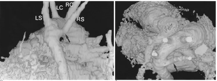
가장 흔한 대동맥궁의 기형으로 전 인구의 0.5%에서 보이 며 다운 증후군환자에서는 38%로 매우 높은 빈도를 보인다 (1, 5). 분지 방식은 대동맥 근위부에서 원위부 쪽으로 우총경 동맥, 좌총경동맥, 좌쇄골하동맥이 순서대로 기시하고 마지막 으로 우쇄골하동맥이 기시한다. 마지막으로 분지하는 우쇄골하 동맥은 대동맥궁의 원위부 후내측에서 기시하며 식도 뒤쪽으 로 주행한다 (Fig. 1). 흔히 쇄골하동맥의 기시부가 국소적 확 장을 보이며 이를 콤머렐 게실(aortic diverticulum of
A B
Fig. 1. Left aortic arch with aberrant right subclavian artery in a 2-year-old boy.
A. Posterior view, SSD image. The branching order is right common carotid artery (RC), left common carotid artery (LC), left sub- clavian artery (LS), and right subclavian artery (RS). Right subclavian artery arises aberrantly from the medial aspect of the distal part of the right-sided aortic arch. It is not dilated at its proximal portion (arrow).
B. Superior view, SSD image. The right subclavian artery (arrow) crosses the midline to reach the left side.
Kommerell) 이라 한다. 임상적으로 거의 대부분은 무증상이 나, 다만 고령환자에서나 혹은 콤머렐 게실이 동반된 경우 식 도 압박증상으로 연하곤란증을 일으킬 수 있다 (1, 2).
우대동맥궁과 동반된 좌쇄골하동맥 이상 기시 (Right aortic arch with aberrant left subclavian artery)
우측 대동맥의 가장 흔한 형으로 전체 인구의 0.1%에서 발 견되며 5-12%는 선천성 심질환을 가지고 있다. 동반된 선천
성 심질환중 활로우 4징이 70%이며 활로우 4징 환자의 약 2%
에서 이 기형이 동반된다. 분지순서는 좌총경동맥, 우총경동맥, 우쇄골하동맥이 순서대로 기시하고 마지막으로 좌쇄골하동맥 이 비정상적으로 대동맥궁 원위부에서 기시한다. 좌쇄골하동맥 의 기시부는 늘어나서 콤머렐 게실을 보일 수도 있다 (Fig. 2).
콤머렐 게실이 있을 때는 동맥관(ductus arteriosus)이 남아 있 으면 혈관륜이 성립된다. 그러나 동맥관이 막혀서 인대 형태로 남아 있는 경우는 비교적 헐거운 혈관륜이 되므로 기관 및 식
A B
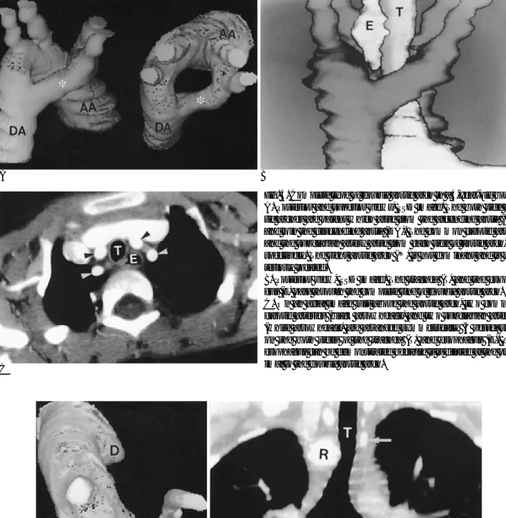
Fig. 2. Right aortic arch with aberrant left subclavian artery in a 42-year-old woman with dysphagia.
A. Anterior view, SSD image. The aberrant left subclavian artery is dilated at its proximal portion to form “diverticulum of Kommerell (D)”and it tapers abruptly in caliber to the normal subclavian artery.
B. Oblique sagittal MPR image. The proximal portion of diverticulum of Kommerell (D) is markedly dilated and compresses the esophagus (E) posteriorly. The trachea (T) is anteriorly displaced by the diverticulum.
A B
Fig. 3. Right aortic arch with left descending aorta (Circumflex retroesophageal aortic arch) in a 54-year-old man with dysphagia.
A. Anterior view, SSD image. The branching pattern is identifical to that of right aortic arch with aberrant left subclavian artery.
The aortic arch is initially located at the right side and abruptly crosses the midline to the left. The left subclavian artery (S) arises from the left sided descending aorta (DA) and its proximal portion is dilated to form diverticulum of Kommerell (D).
B. Oblique sagittal MPR image. The vessel (A) which compresses the esophagus from the posterior aspect is retroesophageal aortic arch , not the diverticulum of Kommerell.
A B
C
Fig. 4. Complete type of double aortic arch in a 4-year-old boy.
A. Posterior and superior views, SSD image. The both side aor- tic arches are patent which arise from the ascending aorta (AA) and join the descending aorta (DA). The common carotid artery and the subclavian artery arise from each side of aortic arch, re- spectively. The right aortic arch (*) is not dominant and is pos- teriorly located.
B. Posterior view, SSD image. The trachea (T) and the esopha- gus (E) pass through the complete ring of double aortic arch.
C. On an axial image just above the aortic arch, two common carotid arteries (black arrowheads) and two subclavian arteries (white arrowheads) are arranged symmetrically (4 vessel sign) on the both sides of the trachea (T) and esophagus (E). The esophagus can be demonstrated because it is dilated at the prox- imal to the double aortic arch.
A B
Fig. 5. Double aortic arch with segmental atresia of left aortic arch in a 16-year-old girl with dysphagia and shortness of breath.
A. Superior view, SSD image. The left brachiocephalic branches of the aortic arch are relatively symmetrically arranged with right bracheocephalic branches, which are not like mirror-image branching pattern of right aortic arch. The remnant of the left aortic arch is dilated to form a diverticulum of Kommerell (D).
B. Coronal reformatted image. The trachea (T) is indented at the level of aortic arch. The remnant of the left aortic arch (arrow) is smaller and higher than right aortic arch (R). In this patient, the barium esophagogram showed bilateral indentation of esophagus (not shown).
도 압박증상은 미미하거나 없을 수 있다 (1-3).
우대동맥궁에 동반된 좌하행대동맥 (Right aortic arch with left descending aorta, circumflex retroesophageal aortic arch) 대동맥궁은 우측에 위치해 있으나 하행 대동맥은 네번째 또 는 다섯번째 흉추 수준에서 식도 뒤 정중선을 넘어 좌측 동맥 관과 연결된 후 척추의 좌측을 따라 주행한다. 이렇게 좌우로 길어진 대동맥궁은 경부로 연장되어 정상 대동맥궁보다 높게 위치하여 경부 대동맥궁(cervical aortic arch)을 형성하는 경 우가 많다. 분지순서는 좌총경동맥, 우총경동맥, 우쇄골하동맥 이 순서대로 기시하고 좌쇄골하동맥은 식도 뒤를 지난 대동맥 에서 기시한다. 이때 대동맥궁은 식도 뒤 위치를 취하지만 쇄
골하동맥이나 대동맥 게실은 식도의 좌측에 위치한다 (Fig. 3).
식도는 식도를 감싸는 대동맥궁에 의해서 후측으로부터 압박 을 받는다 (1, 2).
중복 대동맥궁 (Double aortic arch)
태생기 때의 양측의 제 4 대동맥궁이 모두 남아서 생기는 것 으로 가장 흔한 완전형 혈관륜의 원인이다. 좌우는 대칭적 모 양을 보이지만 대개는 우측이 더 높고 뒤쪽에 위치한다. 양측 의 경동맥과 쇄골하동맥은 각각의 대동맥궁에서 대칭적으로 기 시하여 기관이나 식도의 양측에 위치하는 소위“4 vessel sign”
을 보인다 (Fig. 4). 하행 대동맥은 대개 왼쪽에 위치하나 오 른쪽에 위치할 수도 있다. 임상적으로 식도 및 기도 폐색의 증
A B
Fig. 6. Persistent fifth aortic arch in a 18-day-old girl.
A. Lateral view, SSD image.
B. Oblique sagittal MPR image. All of the brachiocephalic arteries (B) arise as a common trunk from ascending aorta (AA). The aor- tic arch is rather inferiorly located and shows focal stenosis (arrows).
A B
Fig. 7. Anomalous origin of innominate artery in a 7-month-old boy.
A. Axial MPR image.
B. Sagittal MPR image. Left innominate artery (I) arises from more posterior part of aortic arch and crosses the midline in front of the trachea (T). The trachea shows mild stenosis. It is flattened by the compression of the innominate artery.
상 발현정도 및 중증도는 혈관륜의 압박정도에 달려 있다. 양 측의 대동맥궁이 모두 넓게 개통된 형태부터 한쪽의 대동맥궁 이 반대쪽의 대동맥궁에 비해 작은 형태와 한쪽의 대동맥궁이 폐색(atresia)되어 인대형의 띠만 남아 있는 경우도 있다 (Fig.
5). 폐색형 중복 대동맥궁의 경우에는 인대형 폐색분절을 CT 로 직접 볼 수는 없지만 대동맥궁의 분지형태와 기관의 국소 적 협착으로 진단할 수 있다.
제5 대동맥궁 잔존 (Persistent fifth aortic arch)
네번째와 다섯번째 대동맥궁이 기관의 동측에 모두 존재하 는 기형이다. 세 개의 아군이 있는데, 첫번째 아군은 두 대동 맥궁의 내강이 모두 개통돼 있는 형태로 가장 많은 빈도를 보 인다. 이는 두 대동맥궁이 상하로 배열되어 다섯번째 대동맥궁 이 지하철 형태(subway vessel)를 보인다 (6). 다섯번째 대동 맥궁은 무명동맥 근위부에서 기시하여 동맥관 근위부로 연결 된다. 두번째 아군은 위쪽에 위치한 네번째 대동맥궁은 폐색되 고, 아래쪽에 위치한 다섯번째 대동맥궁은 개통되어 있는 형태 이다. 이 형태에서는 모든 상완두동맥이 상행 대동맥으로부터 공통기시를 갖는다 (Fig. 6). 세번째 아군은 다섯번째 대동맥 궁이 폐동맥과 연결되는 형태를 취한다 (1).
무명동맥 이상 기시(Anomalous innominate artery)
무명동맥이 대동맥으로부터 정상보다 후외측에서 기시하여 이 동맥이 정중선을 넘는 과정에서 전방으로부터 기관을 압박 할 수 있다 (Fig. 7). Janet 등 (7)에 의하면 심장병이 동반된 경우에는 흔히 보이는 현상이며 반드시 기관을 누르지는 않는 다. 또한 대부분에서는 증상이 없거나 미미하고, 증상이 심한
경우에도 나이가 듦에 따라서 증상이 약해지거나 없어진다. 그 러나 흡기시 천명이나 무호흡을 일으키는 경우에는 수술적 적 응증이 된다 (1).
대동맥 축착 (Coarctation of aorta)
대동맥 축착은 대동맥궁의 분절적 협착이나 전반적인 형성 부전(hypoplasia)을 포함한다. 대동맥궁 형성부전증은 전반적 으로 대동맥궁이나 대동맥의 협부가 형성부전되어 내강이 좁
Fig. 8. Isthmic hypoplasia in a 44-day-old boy.
Oblique lateral SSD image. The aortic arch is diffusely nar- rowed. The isthmic segment of aortic arch shows posterior in- dentation (arrow). The descending aorta (DA) is rather dilated.
A B
Fig. 9. Arch hypoplasia with coarctation in a 8-day-old boy.
A. Oblique lateral SSD image of aorta. Aortic arch is very elongated and tortuous and shows diffuse hypoplasia. The isthmic area shows focal stenosis with posterior indentation (arrow). Small vascular stump (arrowhead) at the opposite side of coarctation is the closing patent ductus arteriosus.
B. Oblique lateral SSD image of thoracic cage and cardiovascular system. Both internal mammary arteries (arrows) are dilated, which may serve as collateral pathways.
아지는 것으로 과거의 preductal type에 해당한다 (Fig. 8). 국 소형 대동맥 축착은 동맥관 주위의 내강이 국소적으로 좁아지 는 것으로, 과거의 postductal type에 해당한다. 두 형태가 혼 합된 형태도 있으며 (Fig. 9), 동맥관은 대동맥 축착의 정도에 따라 열려 있을 수 있다. 심한 대동맥 축착이 지속되면 단순 흉부촬영상 양측성으로 상부늑골의 하단에 절흔이나 상부 종 격동의“3 자”징후를 보일 수 있으나, 이른 소아기에는 이런 소견을 보기 어렵다. 심 초음파검사의 음파창의 제한 때문에 상부 종격동에 위치한 대동맥궁의 병변을 명확하게 묘사하기 어렵다. CT상 내흉동맥이나 늑간동맥등의 측부 혈행을 쉽게 발견할 수 있다 (6, 8).
대동맥궁 단절 (Interruption of the aorta)
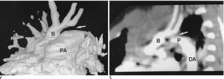
대동맥 협착의 심한 형태로 대동맥궁이 완전히 단절되는 것 으로, 해부학적으로는 근위부와 원위부 대동맥 사이의 단절된 부분을 연결하는 섬유성 잔존물이 남아 있는 경우가 있다. 단 절 위치가 세번째 분지인 좌쇄골하동맥 직후방인 A형 (Fig.
10), 두 번째 분지 (좌 총경동맥)와 세번째 분지 (좌 쇄골하동 맥) 사이인 B형 (Fig. 11), 첫 번째 분지 (무명동맥)와 두번째 분지 (좌 총경동맥) 사이인 C형의 세가지의 아군으로 나뉜다 (1, 8). 서양의 보고에 의하면 B형이 가장 빈도가 높으나 (1), 한국인에서는, 보고된 바는 없으나 A형이 흔한것으로 알려져 있다. C형, 즉 무명동맥과 좌총경동맥 사이의 단절은 드물다.
동맥관은 하행대동맥으로의 혈류를 유지하기 위해 모든 경우 에 동반되어 하행대동맥이 마치 폐동맥에서 기시하는 것처럼
A B
Fig. 10. Interruption of aortic arch, type A in a 11-day-old boy.
A. Frontal SSD image.
B. Oblique sagittal MPR image. The isthmic segment of the aortic arch is interrupted (arrows) after the origin of the left subclavian artery. The brachiocephalic artery (B) looks like the last branch of aortic arch. The pulmonary artery (PA) is connected to descend- ing aorta (DA) via the patent ductus arteriosus (P).
A B
Fig. 11. Interruption of aortic arch, type B, on anterior view of MIP images (A) and SSD image (B) in a 10-day-old boy.
A, B. The aortic arch is interrupted (arrows) between the origins of the left common carotid artery and left subclavian artery (S).
The left subclavian artery arises from the descending aorta (not shown) which is directly connected with the pulmonary artery via patent ductus arteriosus.
보인다. 심실중격결손증 (90%)이나 좌심실유출로 폐쇄 등이 흔히 동반된다 (1).
참 고 문 헌
1. Weinberg PM. Aortic arch anomalies. In Moss and Adams. Heart disease in infants, children, and adolescents. Baltimore: Willams &
Wilkins, 1995;810-837
2. Predey TA, McDonald V, Demos TC. CT of congenital anomalies of the aortic arch. Semin Roentgenol 1989;24:96-111
3. Friese KK, Dulce MC, Higgns CB. Airway obstruction by right aor- tic arch with right sided patent ductus arteriosus: demonstration by MRI. J Comput Assist Tomogr 1992;16:888-892
4. Becker C, Soppa C, Fink U, Haubner M, Englmeier KH. Spiral CT angiography and 3D reconstruction in patients with aortic coarcta- tion. Eur Radiol 1997;7:1473-1447
5. Goldstein WB. Aberrant right subclavian artery in mongolism. AJR Am J Roentgenol 1965;95:131-134
6. Gerlis LM, Dickinson DF, Wilson N, Gibbs JL. Persistent 5th aor- tic arch: a report of two news and a review of the literature. Int J Cardiol 1987;16:185-192
7. Strife JL, Baumel AS, Dunbar JS. Tracheal compression by the in- nominate artery in infancy and childhood. Radiology 1981;139:73- 75
8. Oppenheimer Dekker A, Gittenberger de groot AC, Roozendaal H.
The ductus arteriosus and associated cardiac anomalies in inter- ruption of the aortic arch. Pediatr Cardiol 1982;2:185-193
J Korean Radiol Soc 2001;44:51-58
Address reprint requests to : Yang Min Kim, M.D., Department of Radiology, Sejong General Hospital 91-121 Sosa-dong, Sosa-gu, Bucheon, Gyoungido 422-232, Korea.
Tel. 82-32-340-1175 Fax. 82-32-340-1236
Congenital Anomalies of Aortic Arch: CT Angiography1
Yung Moon, M.D., Yang-Min Kim, M.D., Tae-Hoon Kim, M.D.
Mi-Young Kim, M.D., Jae-Young Lee, M.D.2, Hyung Seok Kim, M.D.3
1Department of Radiology, Sejong General Hospital
2Department of Diagnostic Radiology, Seoul National University College of Medicine
3Department of Radiology, Cheju Medical Center
Aortic arch anomalies result from the failure of an embryonic vascular structure to persist and regress in the usual manner during formation of the aortic arch. The anomalous aortic arch may encircle and compress the trachea and esophagus as a form of a vascular ring. The diagnosis of aortic arch anomaly and the recognition of airway compression are important because they are conditions which complicate the natural and surgical course of related diseases. CT can demonstrate the nature of anatomic structures such as the trachea and esophagus not revealed by angiography, simultaneously disclosing the relationship of stenotic airways and of- fending mediastinal vessels. Volumetric data acquisition by means of spiral CT enables three dimensional re- construction, which can provide easy global understanding of the complex anatomy and spatial relationship of airway and cardiovascular structures. Three dimensional imaging is very useful for the physician and surgeon who are not accustomed to mentally reconstructing axial images, and can facilitate surgical planning.
Index words :Computed tomography (CT), three-dimensional Aorta, abnormalities
Aorta, CT