• 검색 결과가 없습니다.

Left Ventricular Pseudoaneurysm after Valve ReplacementJun Ho Lee, M.D.

N/A
N/A
Protected

Academic year: 2021

Share "Left Ventricular Pseudoaneurysm after Valve ReplacementJun Ho Lee, M.D."

Copied!
4
0
0

로드 중.... (전체 텍스트 보기)

전체 글

(1)

ISSN: 2233-601X (Print) ISSN: 2093-6516 (Online)

− 63 −

Departments of

1

Thoracic and Cardiovascular Surgery and

2

Radiology, Hanyang University Seoul Hospital, Hanyang University College of Medicine,

3

Department of Thoracic and Cardiovascular Surgery, Seoul National University Bundang Hospital, Seoul National University College of Medicine

Received: September 4, 2014, Accepted: September 23, 2014, Published online: February 5, 2015

Corresponding author: Hyuck Kim, Department of Thoracic and Cardiovascular Surgery, Hanyang University Seoul Hospital, Hanyang University College of Medicine, 222 Wangsimni-ro, Seongdong-gu, Seoul 133-791, Korea

(Tel) 82-2-2290-8467 (Fax) 82-2-2290-8467 (E-mail) khkim@hanyang.ac.kr

C

The Korean Society for Thoracic and Cardiovascular Surgery. 2015. All right reserved.

CC
This is an open access article distributed under the terms of the Creative Commons Attribution Non-Commercial License (http://creative- commons.org/licenses/by-nc/3.0) which permits unrestricted non-commercial use, distribution, and reproduction in any medium, provided the original work is properly cited.

Left Ventricular Pseudoaneurysm after Valve Replacement

Jun Ho Lee, M.D.

1

, Seok Chol Jeon, M.D.

2

, Hyo-Jun Jang, M.D.

3

, Won-Sang Chung, M.D.

1

, Young Hak Kim, M.D.

1

, Hyuck Kim, M.D.

1

We present a case of left ventricular pseudoaneurysm, which is a very rare and fatal complication of cardiac pro- cedures such as mitral valve replacement. A 55-year-old woman presented to the Department of Thoracic and Cardiovascular Surgery at Hanyang University Seoul Hospital with chest pain. Ten years prior, the patient had under- gone double valve replacement due to aortic regurgitation and mitral steno-insufficiency. Surgical repair was successfully performed using a prosthetic pericardial patch via a left lateral thoracotomy.

Key words: 1. Left ventricular pseudoaneurysm 2. Left ventricular rupture

3. Mitral valve, replacement

CASE REPORT

A 55-year-old woman visited the emergency clinic of Hanyang University Seoul Hospital with chest pain that had persisted for one month and had become aggravated two days earlier. Ten years before this admission, the patient had un- dergone double valve replacement with aortic and mitral me- chanical valves due to aortic regurgitation and mitral steno-insufficiency.

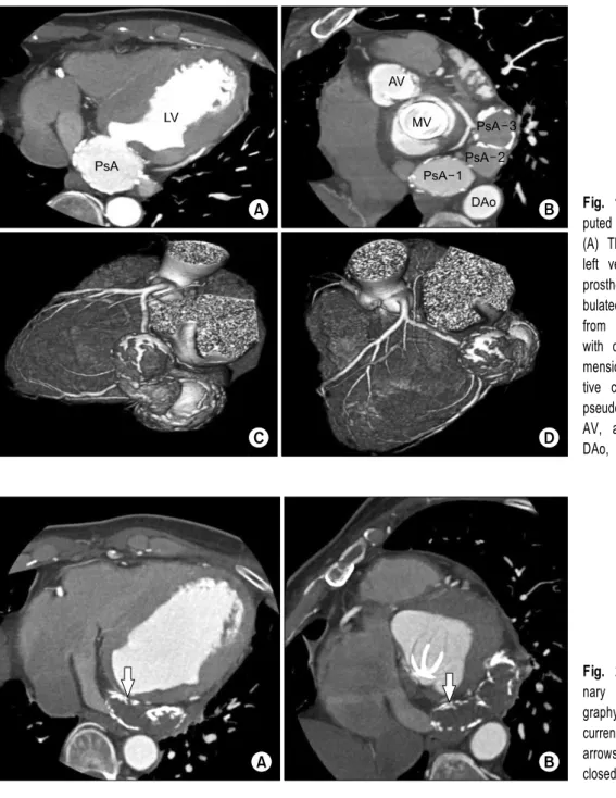
An electrocardiogram revealed myocardial ischemia in the inferior wall. However, no abnormalities were observed in laboratory findings, including cardiac markers. Echocardio- graphy revealed normal motion and function of the mitral and aortic valve prostheses, and coronary angiography findings were also normal. However, coronary computed tomographic angiography revealed three large lobulated aneurysms with calcified walls bulging from the base of the left ventricle

(Fig. 1). The leak in the left ventricle was located in the sub-mitral valve prosthesis area. Based on this finding, we di- agnosed the patient with a pseudoaneurysm of the left ven- tricle, and surgical treatment was planned.

Surgery was performed under general anesthesia using dou- ble-lumen endotracheal intubation. A left lateral thoracotomy was performed via the fifth intercostal space. Arterial cannula- tion via the left femoral artery and venous cannulation via the left femoral vein and left pulmonary artery were performed.

Three communicating aneurysmal sacs were observed in a single plane, identical to that observed in coronary computed tomographic angiography. The neck of the first sac was lo- cated at the base of the left ventricle. Under cardiopulmonary bypass and fibrillation, the aneurysmal sacs were incised and the thrombi in the sacs were extirpated. A defect measuring 15 mm in diameter was identified in the sub-mitral left ven- tricular wall with calcification but with no infection. The

Korean J Thorac Cardiovasc Surg 2015;48:63-66 □ Case Report □

http://dx.doi.org/10.5090/kjtcs.2015.48.1.63

(2)

Jun Ho Lee, et al

− 64 −

Fig. 1. Preoperative coronary com- puted tomography (CT) angiography.

(A) The location of the leak in the left ventricle is the sub-mitral valve prosthesis area. (B) Three large lo- bulated aneurysms are seen bulging from the base of the left ventricle with calcified walls. (C, D) Three-di- mensional reconstruction of preopera- tive coronary CT angiography. PsA, pseudoaneurysm; LV, left ventricle;

AV, aortic valve; MV, mitral valve;

DAo, descending aorta.

Fig. 2. (A, B) Postoperative coro- nary computed tomographic angio- graphy. There is no evidence of re- currence of the pseudoaneurysm. The arrows show no leakage at the closed aneurysmal neck.

pseudoaneurysm of the left ventricle was determined to be caused by myocardial discontinuity. Closure of the defect was performed with a Supple Peri-Guard Pericardium Patch with APEX Processing (Synovis, St. Paul, MN, USA) using pledg- eted horizontal mattress sutures. The walls of the aneurysmal sacs were closed, and the patient was weaned from car- diopulmonary bypass without difficulty. The total cardiopulmo- nary bypass time was 70 minutes.

The vital signs of the patient remained stable, and she was

extubated six hours after the operation. Follow-up echo-

cardiography was performed on postoperative day eight, and

no abnormal findings, including cardiac wall motion, were

noted. The patient underwent coronary computed tomographic

angiography on postoperative day 14. No leakage at the

closed aneurysmal neck was observed, and there was no evi-

dence of recurrence of the pseudoaneurysm (Fig. 2). The pa-

tient was discharged on postoperative day 16, without any

complications. She has remained disease-free for six years

(3)

Left Ventricular Pseudoaneurysm

− 65 − postoperatively, with regular Coumadin anticoagulation therapy due to the presence of the prosthetic aortic and mitral valves.

DISCUSSION

A left ventricular (LV) pseudoaneurysm, or false aneurysm, is defined as a contained rupture or perforation of the my- ocardium [1]. The rupture of the myocardium occurs rarely in clinical practice; it is most often associated with myocardial infarction or cardiac surgery, such as mitral valve replacement (MVR), and is usually fatal [2-4].

In 1980, Cobb et al. [5] described the transverse mid- ventricular disruption of the LV free wall after MVR. They proposed that an incomplete rupture could be the cause of a false aneurysm. Roberts and Morrow [6] suggested that in- advertent invasion into the LV free wall is possible during excision of the mitral valve if there is poor visualization of the operating field when using the tips of the scissors. Other causes of complete or incomplete rupture of the LV free wall include an oversized prosthetic valve, excessive extirpation of calcium in the mitral annulus, myocardial erosion caused by the struts of the prosthetic valve, the untethering of the fi- brous structures of the left ventricle during resection of mitral leaflets, an increase in LV contractility after aortic cross- clamping, enhanced LV wall stress with the support of in- otropic agents, and other mechanical trauma between the free wall and the papillary muscles, such as rubber catheter wedg- ing or metal pump suction during valve replacement [7].

Moreover, perioperative rupture of the myocardium is known to be a lethal complication [4].

The wall of a false aneurysm comprises the fibrous ob- literation of the pericardial sac, resulting from adhesion between the parietal and visceral layer of the pericardium. This can pre- vent the complete and fatal rupture of the LV free wall [7].

While a true aneurysm has nonrestrictive continuity with the LV cavity, a pseudoaneurysm has a defect in the my- ocardial continuity and a well-defined neck, representing a history of LV wall perforation [1,2]. Thus, such pseudoaneur- ysms are more likely to undergo rapid enlargement and rup- ture than true aneurysms [1]. Therefore, surgical correction, including the resection of the aneurysmal sac and patch repair or primary closure of the aneurysmal neck, is indicated and

recommended for pseudoaneurysms [1,2].

In the present case, we diagnosed a LV pseudoaneurysm using coronary computed tomographic angiography. Myocar- dial infarction was excluded as a cause of the LV pseudoa- neurysm because coronary angiography revealed normal find- ings and cardiac markers were in the normal range, although electrocardiography revealed myocardial ischemia in the in- ferior wall. The cause of LV pseudoaneurysm in our patient was a late-term complication of MVR. Echocardiography re- vealed normal function of the mitral valve prosthesis; there- fore, mitral reoperation was not indicated.

In such patients, if a repeat sternotomy for entering the op- erative field is considered, it may be difficult to approach the heart due to massive pericardial adhesions, and the possibility of injury or rupture of the pseudoaneurysm during dissection may be higher [4]. A direct approach to the operative field via a left lateral thoracotomy can avoid these complications and preserve the previously implanted prosthetic valves.

Further, it has other advantages, such as making it easier to establish cardiopulmonary bypass and obviating the need to cross-clamp the aorta, in contrast to the sternotomy approach [4]. In our operation, a fibrillator was used and the exposure of the operative field was excellent.

In the operative field, we found three large lobulated pseu- doaneurysms with calcified walls in a line, bulging from the base of the left ventricle. The configuration of the pseudoa- neurysms suggested a previous episode of LV rupture, which had developed over time.

In patients with LV pseudoaneurysms, there is a well-de- fined approach to surgical correction. The neck of the pseu- doaneurysm is dissected, and the perforated site can be closed by patch repair or primarily. The mortality rate of this sur- gery is approximately 10% [8]. However, no complications occurred in our case.

In conclusion, we have reported a case of LV pseudoaneur-

ysm that occurred as a late complication after MVR. We per-

formed a left lateral thoracotomy via the fifth intercostal

space to approach the operative field directly and successfully

repaired the sub-mitral LV pseudoaneurysm using a prosthetic

patch. A few cases of LV pseudoaneurysms have been pre-

viously reported; however, no case reports have previously

been presented of Korean patients with an LV pseudoaneur-

(4)

Jun Ho Lee, et al

− 66 − ysm following MVR who were treated with an operative tech- nique employing a left lateral thoracotomy. We consider the approach described here to be feasible, safe, and reproducible.

CONFLICT OF INTEREST

No potential conflict of interest relevant to this article was reported.

REFERENCES

1. Kollar A, Byrd BF 3rd, Lui HK, Drinkwater DC Jr. Mitral valve replacement and endocavitary patch repair for a giant left ventricular pseudoaneurysm. Ann Thorac Surg 2001;71:

2020-2.

2. Kim HS, Kim KH, Hwang HY. Left ventricular pseu- do-pseudoaneurysm with hemopericardium. Korean J Thorac Cardiovasc Surg 2011;44:247-9.

3. Choi JB, Choi SH, Oh SK, Kim NH. Left ventricular pseu- doaneurysm after coronary artery bypass and valve replace- ment for post-infarction mitral regurgitation. Tex Heart Inst J 2006;33:505-7.

4. Suda H, Ikeda K, Doi K, Shiraishi R, Furukawa K, Ito T.

Successful repair of left ventricular pseudoaneurysm after mitral reoperation under hypothermic circulatory arrest. Jpn J Thorac Cardiovasc Surg 2003;51:18-20.

5. Cobbs BW Jr, Hatcher CR Jr, Craver JM, Jones EL, Sewell CW. Transverse midventricular disruption after mitral valve replacement. Am Heart J 1980;99:33-50.

6. Roberts WC, Morrow AG. Causes of early postoperative death following cardiac valve replacement: clinico-pathologic correlations in 64 patients studied at necropsy. J Thorac Cardiovasc Surg 1967;54:422-37.

7. Waller BF, Taliercio CP, Clark M, Pless JE. Rupture of the left ventricular free wall following mitral valve replacement for mitral stenosis: a cause of complete (fatal) or contained (false aneurysm) cardiac rupture. Clin Cardiol 1991;14:341-5.

8. Frances C, Romero A, Grady D. Left ventricular

pseudoaneurysm. J Am Coll Cardiol 1998;32:557-61.

수치

Fig. 1. Preoperative coronary com- com-puted tomography (CT) angiography.

참조

관련 문서

systemic circulation, in the right ventricle and oxygenated blood from the lungs, or pulmonary circulation, in the left ventricle, as in birds and mammals.. Two vessels,

Sizes of left and right pupils Intensities of left and right pupils Ellipse ratios of left and right

1 John Owen, Justification by Faith Alone, in The Works of John Owen, ed. John Bolt, trans. Scott Clark, "Do This and Live: Christ's Active Obedience as the

ㅇ 미국 등 많은 OECD국가에서 사람들이 사회적 접촉을 꺼리는 현상이 발생하는 것도 사회적 소외와 뒤쳐짐을 악화시키는 요인임.. - 기존 유럽 사회는 교회,

Based on the results of root cause analysis, the following corrective action to mitigate the erosion and corrosion damage at hot leg check valve were

core exercise program showed not only increasing peak torque of the right leg, left leg in Hip Extension of exercise group but also increasing peak torque of the right leg, left

[r]

5 ) Stop Light Switch (Mechanical) 6 ) Front/Rear Balance Valve 6 ) Front/Rear Balance Valve 7 ) Pressure Differentiavl Valve 8 ) Brake Warning Lamp.. 9 ) Brake Fluid